Is the incandescent filament lamp 'in the dock' in the early 21st century? This inefficient, heat generating light source in the domestic environment is slowly being phased out worldwide—shops in the European Union are not permitted to restock with the typical 25-100W household mains lamps—other lamps such as mains halogen spots are to follow. (See e.g. Wikipedia entry Phase-out of incandescent light bulbs). Is their continued use in microscopy also rather bleak? Fortunately, low voltage variants commonly used don't seem to be on the current list of future planned phase outs, but many new microscope models have LED transmitted illumination, conversion of older stands from tungsten to LED is being widely embraced by many enthusiasts and commercial LED retrofits are available for some stands.
Perhaps we will need to write an epitath for the tungsten lamp in microscopy in the not too distant future. The 'case for the prosecution' has been widely aired and the key benefits of LEDs over tungsten are well known—near constant white balance at variable intensities, very little thermal IR directed into the optics train (local heat dissipation is necessary for higher wattage models), essentially no near UV and typically little near IR to filter*, relatively cheap, compact size, very long life, stable white, low voltage/amperage supplies and lower powered designs enable battery operation if field use is desired ... so what's not to like? If the tungsten microscope lamp is to be sentenced in the future by the judge wearing his black cap, perhaps it deserves a case to be presented in its defence.
(*This generally applies to the common white LED lamps where a blue LED is coated with a phosphor—some white LEDs use a near UV source—the makers' data sheets can be checked if uncertain. Warm whites have a typically higher near IR 'tail'. LED based lasers (e.g. pointers) may operate differently and must be checked for safe use on many grounds—some green lasers use frequency doubling of a near IR LED where it is widely documented that if insufficiently filtered, they can emit potentially unsafe levels of near IR.)
Below would be the case I would offer on behalf of the tungsten filament microscope lamp if appointed as its defence attorney. I should add that I'm not a die-hard tungsten lamp user over LED, I have a foot in both camps, there's some microscopes I would be very reluctant to convert, others my brother Ian and I have and/or would readily if needed.
(Fluorescence work with mercury HBO lamps is a special case and not under consideration here as an arc lamp. Using LEDs as an alternative in the home lab are very persuasive on cost, safety and convenience grounds and has considerably broadened the technique to enthusiasts; all my autofluorescence studies uses high power LEDs. A number of modern microscopes systems now adopt LEDs, e.g. the elegant Zeiss Colibri system.)
Using a classic microscope exactly as its designers intended
How strong this argument is depends heavily on each user's 'philosophy' and to a large extent the microscope in question. My main compound microscope for transmitted work is a Zeiss Photomicroscope III with an external sophisticated lamphouse using a 12V 100W tungsten-halogen bulb and for me using this stand, this argument is compelling. Unless the lamps become unavailable, its advantages with a tungsten source outweigh the potential benefits of converting to LED.
When this now classic research stand was designed, a lot of thought went into the mechanical design of the lamphouse and the optical train to optimise the bulb for Köhler illumination. Part of the appeal of using such a classic stand is setting it up as described in the manuals and knowing it is being used exactly as the designers intended; this can be particularly key if enjoy using the microscope at the limit e.g. for tough 'diatom dotting'. Aligning the lamp also has a certain satisfaction and on completing alignment, if choose not to swing in the diffuser in base and on lamp, the classic Köhler setup can be checked and appreciated in the back focal plane—a centred, focussed, even image of the lamp filament spanning the full aperture—bliss for the user of classic microscopes!
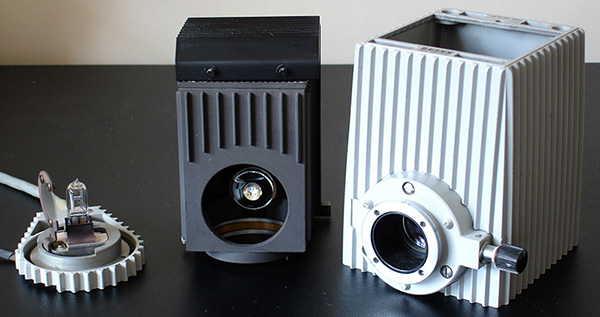
The Zeiss 12V 100W lamphouse is a typical design—similar were being used by most major makers for their higher specified laboratory and research level stands up until the late 20th century at least, and the sort of stands that may increasingly be in the hands of enthusiasts. It is a finely crafted piece of mechanical and optical engineering designed to optimise the use of the widely available tungsten-halogen flat filament bulb
(e.g.
Osram Xenophot) which costs less than £2 in the UK. It has an optional swing-in diffuser on the bulb holder, a concave mirror to double the filament
size, focussing field optics and five screws on rear of housing to align and focus the primary and mirror bulb image. Heat is safely and optimally dissipated using the lamphouse's finned design and air circulation.
Lower wattage small envelope LEDs can be retrofitted into the GY6.35 lamp base (and have done it for the Nikon Labophot 1 small simple external lamphouse) but becomes more complex in lamphouses of the above sophistication for the larger higher powered LEDs requiring heat dissipation. The bulb focus would be retained with an LED conversion and primary bulb centering but image doubling with the mirror and associated
alignment controls become superfluous.
If near UV or near IR studies are of interest, the LED would need changing—non-trivial compared with the simple addition of filters on field iris housing for a tungsten source (see below).
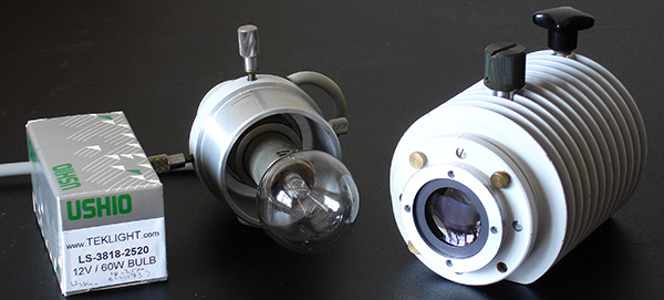
The 12V 60W lamphouse using a tungsten bulb with large flat filament. These bulbs are a little more expensive but quite good value ($15+ in US). These can be more sensitive to mechanical breakage but with care can last years, this example is over 5 years old. Again the lamphouse is carefully crafted with centering and focussing for optimal Köhler. The small 12V 100W tungsten-halogen bulbs are cheap, work but are rather characterless. These larger bulbs with their large spherical envelopes have a certain appeal in the same way that valve audio amplifiers do.
This 'using as the maker intended' argument is less persuasive for stands with simpler lamphouses, either internal or external as illustrated below. The 6V/15W barrel assembly for the Zeiss Standard variants for example are readily amenable to LED conversion (the Photomic also came with this option although rather underspecified in this form for 'light guzzling' work).
The argument also fails at the first hurdle if the lamps are so specialised that they are now either very expensive and/or near unobtainable. Thankfully the lamp used for the 12V 100W is a widely used and available domestic grade, very cheap and hopefully not to be earmarked for phasing out (if it is I'll be stocking up!). The bulbs for the 12V 60W lamphouses although more specialised are still made, widely available and modestly priced if shop around.
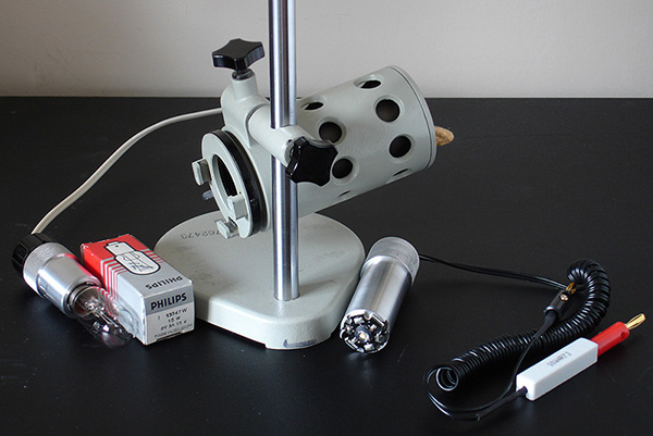
The external Russian LOMO OI-19 Köhler lamp shown above uses a simple metal barrel fitting for the bulb holder and is eminently suitable for LED conversion without any changes to the lamphouse. A number of stands such as some Zeiss Standards have similar internal lamps.
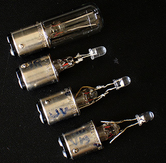 The original tungsten lamp fitting is shown left and the conversion using a 1W white LumiLED shown right (lamp fitting kindly made by Robert Pavlis). Electrical fittings with in-line resistor by Ian Walker. The original power supply cannot of course be used as AC not DC and ideally should
be current limited.
Another popular method of converting suitable lamps to low powered LEDs (i.e. designs requiring little to no heat dissipation) is to mount the LED on the stub of an old bulb. The bulb's original mounting and centering can then be used and LED junction is at or near the original filament plane to mimic Köhler.
Shown right are my own conversions when first starting out with LED trials some years ago to allow a Russian LOMO lamp to use a 5 mm white LED, near IR or near UV.
The major disadvantage of having plug-in multi-spectral LEDs cf filtering on an original tungsten-halogen lamp as described below is that critical lamp alignment is potentially lost each time and does not allow quick interchange of lighting for comparison purposes. Lamp setup for near UV and near IR has to be carried out without the aid of the eye if the lamphouse is inadvertently moved while interchanging bulbs.
The key aspects for me when considering an LED conversion is whether 1) it is non-destructive to the lamphouse, 2) the original mode of operation is significantly being changed and 3) other features are lost in the process (e.g immediate access to near UV/IR lighting). For all three of these reasons I would not convert the 12V 100W or 12V 60W lamphouse for the Zeiss Photomicroscope.
Instant interchange between Köhler lighting in the visible, near UV and near IR
One of the cited benefits of white LEDs based on blue LEDs with excited phosphor over tungsten is the negligible emission of thermal / near infrared and near UV into the optics train. Damage caused by thermal IR has been the bane of many a microscopist including myself when buying used (Zeiss) equipment and faced with evidence of 'cooked' optics and/or mechanics. With the modern sensitive video and still cameras now available and affordable it's arguable whether lighting above 100W is now required
for transmitted
studies; 100W lamps and lower are readily controlled for heat with a thermal IR filters (often fitted as standard, place another on field iris if wish to be certain)—both absorption types such as the Schott KG series or interference hot or cold mirrors are available.
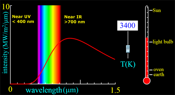
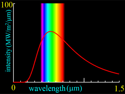
Above: The spectrum of a black body radiating at 3400K i.e. the cited colour temperature of a tungsten lamp operating at 12V 100W (Zeiss Photomicroscope manual). Created using the Java black body spectrum simulation (an interactive web page). Credit: PhET Interactive Simulations, University of Colorado http://phet.colorado.edu
A 12V 100W tungsten-halogen operating at ca. 3400K has a dominant near IR spectral content and small but usable near UV content (latter if bulb envelope not treated to block near UV) both which can be turned around from a cited disadvantage to the microscopist's benefit when required. Placing a long-pass filter on the lamphouse and using a monochrome sensor camera which are usually sensitive to this band, the benefits of near IR are immediately on hand. Near IR is useful for studying subjects such as the denser insect exoskeleton thin mounts where it transmits more readily than visible allowing studies of the structures better. Depth of field is also improved. For the type of subjects where this method is useful, the decrease in resolution because of the longer wavelengths is not usually detrimental.
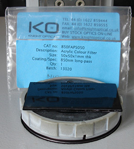
Left: An 850 nm long pass filter for near IR studies, this is an acrylic filter with modest cost. This example is quite some way into the near IR as wished to make studies essentially free from visible light, but grades from 700 nm are available. A high quality camera lens grade filter is not required if placed at this position.
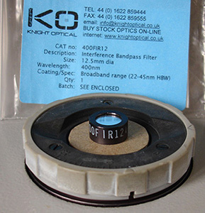
Right: An interference 400 nm filter with 45 nm half bandwidth. As these studies are typically made at higher mags e.g. 40X and higher, the filter only has to be large enough to fill the field iris aperture set for the lowest mag chosen, thus allowing the smaller cheaper grades to be bought. A card annulus blocks any residual light from the edges.
I prefer not to go further into the near UV than 400 nm so as to maximise light available from the 100 W tungsten-halogen lamp
(an Osram Xenophot does not have a UV block on the bulb envelope, some may). There is a marked improvement in resolution going from 500 nm to 400 nm, the improvements plateau out as eke the last tens of nm and as long as don't go too far into the near UV (365 nm is about the max for normal objectives) and avoid unnecessarily narrow filter bandwidths, there is plenty of near UV from 100W lamps at 12V for demanding diatom dotting, e.g. at highest powers with annular illumination. I use
a cheap monochrome C mount camera used for telescope guidance.
Near UV Examples
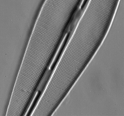
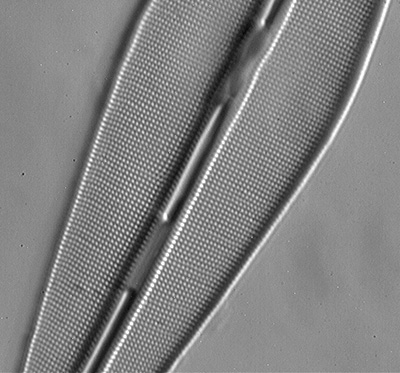
Diatom, slide label "Frustulia rhomboides", 8 form test slide, but correctly Frustulia saxonica Rabenhorst (see Frithjof Sterrenburg's article which discusses the history of both the misidentification and incorrect nomenclature of this popular test diatom). Zeiss 63X/1.4 oblique using offset brightfield port in phase condenser. The diatom long axis is ca. 45 degrees to direction of oblique. Single shot using Opticstar PL-130M 1.3 Mpixel monochrome C-mount camera, MICAM image capture software.
Lee dark blue acetate filter ca. 440 nm (left) the punctae are resolved, but decreasing the wavelength to 400 nm (right) and tightening the bandwidth using the above interference filter, the punctae are better resolved and image more contrasty. The latter can be placed over the field iris in seconds to compare result without disturbing Köhler. A near UV LED exchange from a white LED would be less convenient and may upset any critical lighting.
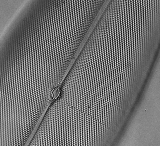
Above: Pleurosigma angulatum, 400 nm interference filter, Zeiss 63x oblique using offset brightfield port in phase condenser. Nikon D5000 DSLR with Zeiss 10X Kpl W eyepiece on short collar as relay lens. IS0 200, 2 secs, crop of frame.
Another benefit of an interference filter just in the visible is that consumer digital SLRs can be better behaved, I've had rather odd results when going out of the visible.
Images and further details from the Micscape December 2008 article: Fun with filters: Assessing a 400 nm bandpass interference filter on a tungsten lamp to improve resolution as an alternative to an LED.
Near IR example
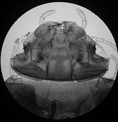
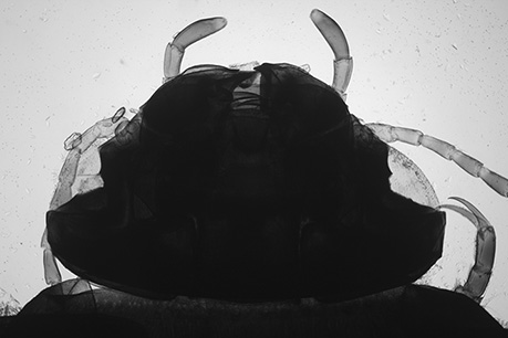
Above: Diving beetle Agabus sp. prepared by author on NBS slide making course taught by the late Eric Marson. Zeiss 2.5X objective. 12V 100W tungsten-halogen lamp.
Left: Near IR with 760 nm filter on field iris, using a home modified old Sony S75 consumer digicam for solely near IR work (microscopy or landscape) described in this Micscape article. The camera is able to manual white balance avoiding the red tint of near IR imagery. LM-Scope relay lens used.
Right: Visible light brightfield image with Canon 600D DSLR body, Zeiss 10X Kpl W on short collar as projection lens. Visible light barely penetrates and photography is difficult because of the extreme contrast.
Near IR can penetrate denser areas of insect exoskeletons and can be useful
for showing detail. Out of camera images can be tonally flat so tonal balance is
required.
By retaining the tungsten lamphouse, near IR, visible and near UV are instantly available by filter interchange on the field iris, essentially maintaining Köhler (slight refocusing of objective and possibly condenser iris required if critical work). Converting the lamphouse to a white LED loses this facility and requires stopping work and fitting a near IR or near UV LED as appropriate. Checking lamp alignment can also no longer be done by eye.
Incandescent lamp spectral qualities extensively documented for visual, film and digital photography cf atypical white LED spectra (Also see Footnote.)
In the pre-digital days when film photomicrography was the sole method of recording colour images, 'nailing' the colour balance corrections with filters and/or lamp intensity control was vital if using positive slide film for direct projection. The slightest mismatch was all too clear as a typical pale red or blue tint if the lamp colour temperature was under or over corrected. Ageing of microscope slide mountants, bulbs, filters,
optics further created the need for colour correction requirements beyond those published for various film / lamp combinations—as I know all too well with the many examples of slide / print films which I have where I didn't quite 'nail' it. If the colour film (positive or negative) was to be printed, additional corrections could be made at the enlarger stage if performing own colour developing but there was no control if commercially printed. Large sections of textbooks on photomicrography
were devoted to colour correction.
Cue the digital camera era—this potentially awkward aspect of tungsten lamps has essentially gone. The typical modern consumer digital camera, e.g. the Sony NEX 5N camera body I have in front of me has a number of approaches to correct the colour balance before pressing the shutter:
- Auto white balance (AWB) - this can often cope with a wide range of colour temps automatically.
- Set the colour temp in Kelvin if known (makers often supply a voltage v. colour temp graph for given bulbs) - for the NEX 5N the user can dial in a temp from 2500 to 3500K manually, with additional colour wheel for fine tuning.
- Set the tungsten lamp setting if a good match to what is being used.
- Always use RAW setting, as allows post capture correction in software if incorrectly set.
- Custom white balance - most cameras allow this to be set and often one or more custom settings can be saved to memory.
- And if any of the above don't get it quite right, or the microscopist set jpeg and forgot entirely to check the colour, all is not lost. If there was an area in the subject that should be white e.g. the background in a brightfield subject, use the eye dropper in the photo editing software to set that for white and voilà, perfect white balance!
Incidentally, option 6 has saved the not quite correct slide film photomicrographs which I have. Scan them, then set the white balance. Dedicated digital cameras for microscopy will also have features to correct the white balance from tungsten sources.
|
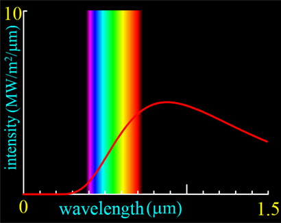
Credit: PhET Interactive Simulations, University of Colorado http://phet.colorado.edu |
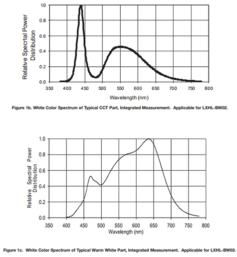
Upper right is the typical spectrum of a white LED, cited as having a 'typical' colour temp. of 5500K. The equivalent black body temp. is shown upper left. Lower right is the same maker's 'warm white' with a markedly lower blue spike, 'typical' color temp. cited as 3300K. The equivalent black body temp. is shown lower left. Source: Philips Lumileds Luxeon Emitter Technical Datasheet DS25. Note that these are not necessarily normalised for same y axis units, but are illustrative. The overlain normalised spectra presented e.g in the Olympus BX46 brochure below are similar. |
There's an argument that the well documented tungsten-halogen lamp spectral output has a benefit over the typical white LED for critical work e.g. quantitative polarisation studies and critical colour assessment in stained specimens—the white LED's spectra is atypical as a consequence of the exciting blue LED's output mixed with that of the phosphor's emission. The standard white's have a marked blue spike, the warm white's less so but the latter at the penalty of a much lower colour temp—lower than a 12V 100W tungsten-halogen.
Some manufacturers offer microscopes with LED lamps based on RGB LED sources to give a more balanced light output for critical work. Olympus state they are using 'mixed-matrix brightfield LED technology' in some stands e.g. the BX46 which they call 'True Color LED'.
Quote from the web page linked to above: "True Color LED. The most advanced mixed-matrix brightfield LED technology in the BX46 provides a colour rendering index very similar to that of halogen bulbs with daylight filters, allowing clear differentiation between similar colours. Such clarity cannot be provided by standard LEDs, in which case diagnostic imaging becomes difficult. The advanced colour rendering technology in the BX46 provides a wavelength range ideal for the most commonly used stain colours - purple, blue and red, for example haematoxylin and eosin (HE) or Papanicolaou stain (Pap)."
A background article on the Olympus system 'Don't fight to get the light right' shows three photomicrographs taken with tungsten, white LED and mixed matrix. In the BX46 brochure they have a graph which overlaps the visible spectrum of halogen with blue filter, white LED with marked blue spike and their own LED system. Their's gives a much better approximation to black body emission.
The spectral properties of incandescent filament lamps are well documented, the human eye evolved for such spectra (the sun). When the near IR and near UV is undesirable from a filament lamp, these bands are outside of the visible range and readily filtered. The atypical features of a white LED are within the visible range, any corrective measures e.g. tunable interference filter or weak yellow filter to suppress the blue spike may affect the white balance for critical applications.
Hot lamphouses can be useful!
All my microscopy has to be carried out on a modest sized sturdy writing desk, there's no room for a dedicated wet area or home lab—space is key. I don't make many permanent mounts but they tend to be Balsam based when I do which require a gentle heat to accelerate drying. In the absence of room for a small oven or hotplate, the lamphouse's flat ventilated top plate is a very effective area for heating the occasional slide under controlled conditions. The lamp voltage allows the lamphouse to be
calibrated with a
thermocouple for a known heat setting. It is safe to be left on
overnight.
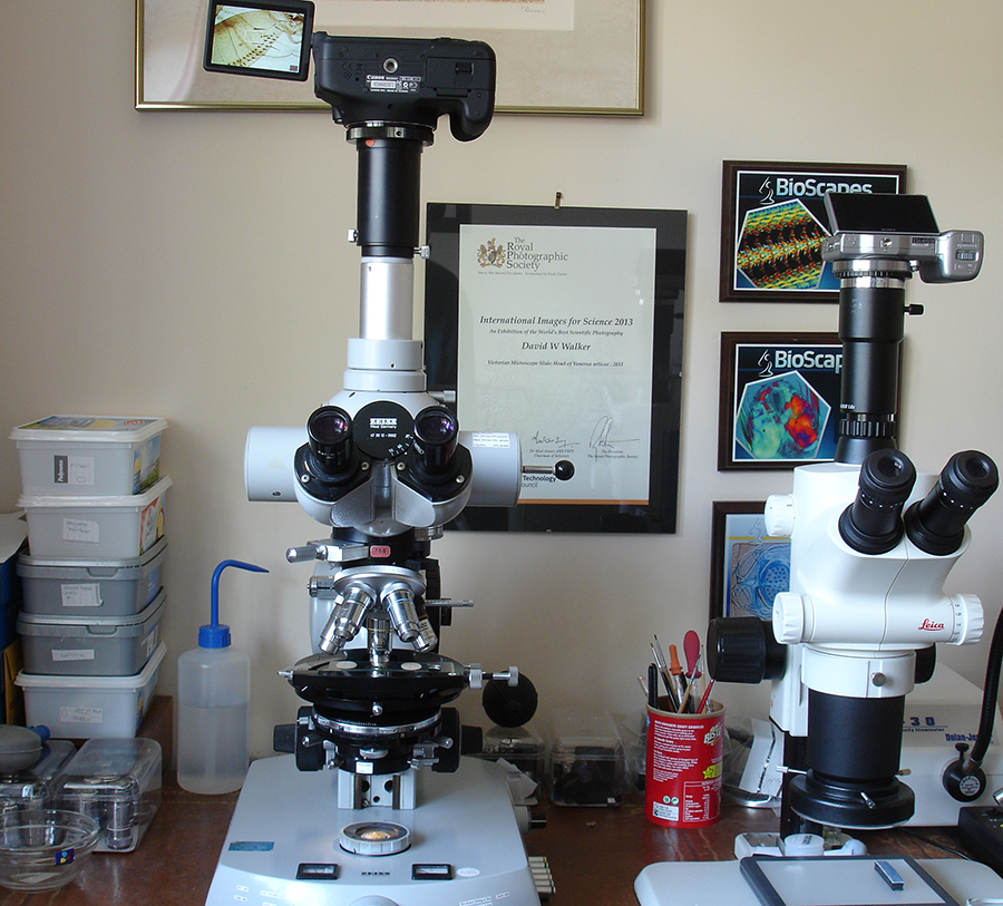
A cluttered work area doesn't leave any room for a hotplate.
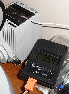
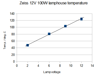
An industry standard type K thermocouple with digital readout is useful for calibrations where the temp. may exceed the range of a mercury / alcohol thermometer. The example shown with thermocouple and digital monitor was typically ca. £7 incl. postage on eBay UK (PP9 9V battery required). The graph shows the plot with thermocouple sitting under a slide on the lamphouse (temp. allowed to stabilise at each voltage); a useful range of temps. are available.
****************************************
My case rests m'lud for the defence of the continued use, when appropriate, of the tungsten lamp for microscopy further into the 21st century and I await the verdict of the jury.
Comments to the author David Walker are welcomed.
Footnote: 'Blue light hazard' of some white LEDs? There are widespread debates at present as to whether the marked blue spike in the spectral output of the typical neutral white LED poses a potential health hazard to the human eye. The interested reader unfamiliar with this debate is invited to type in 'blue light hazard' with 'white LED' into a search engine and to explore the commentaries and documents from various sources. Two documents that I found useful were the 'Optical Safety of LED Lighting' (European Lamp Companies Federation, July 2011, 18pp pdf) and 'Health Effects of Artificial Light' (European Commission - Scientific Committee on Emerging and Newly Identified Health Risks, March 2012, 118pp pdf). To date I haven't found resources that specifically address their safety in use in transmitted light microscopy, i.e. where the user is staring into the source for possibly long periods albeit diffused and lower intensity (I would be interested in hearing of such documents.)
