Like many hobbyists I enjoy using the affordable range of short wavelength LEDs (deep blue, violet, near UV) for improving the resolution of subjects like diatom frustules. Various LED lighting designs have been described for either modifying existing lamps or for building dedicated lamps (e.g. see the Micscape Library where contributors have shared various designs).
A potential disadvantage of using LEDs is that the beam properties don't necessarily match a microscope's field lenses and/or substage optics to fill the aperture of high NA objectives, which can be vital for more demanding studies. The cheap UV LEDs can be underpowered as well for techniques like oblique, unless a sensitive enough camera is used.
The emission spectrum of a tungsten microscope lamp peaks in the near IR but there is a finite emission tailing into the violet - near UV*, which increases with the filament temp. So another approach for studies with short wavelength visible light is to use appropriate narrow bandpass filters on the existing microscope lamp. This is nothing new of course but was an approach I hadn't tried. With modern sensitive digital cameras this limited emission may be exploited. (*This McGraw Hill Java applet, allows 3400K, the stated colour temp of a Zeiss 12V 100W quartz halogen lamp or other lamp temp., to be entered to examine spectrum. Actually for stars but lamp filaments are essentially black body radiators.)
Below are some comments on a short wavelength filter I've tried compared to a typical deep blue filter. It's very much a 'try it and see' approach (my favorite type of microscopy!).
Update: Thank you to a reader who tells me that tungsten halogen bulbs with a near UV block in the envelope are available, depending on wattage and rating. These would not be suitable for the trials below. Consult manufacturer's data sheets for details of extent of near UV protection.
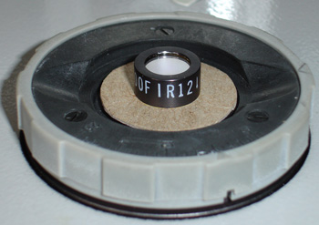
Knight Optical interference filter '400FIR12' - 400 nm bandpass with 40 nm half bandwidth. Interference filters are available in a wide range of wavelengths and bandwidths. I picked a 400 nm filter which is just on the border of visible and near UV as I liked the challenge of photomicrography just in the visible. I was unsure how much light at 400 nm would be available from a 100W microscope lamp for my camera so chose a relatively broad band of 40 nm at 50% transmittance; filters with 20, 10 nm and lower bandwidths are available. A typical blue LED has a quoted half bandwidth of ca. 25 nm.
Large diameter interference filters can be expensive so chose one
10 mm wide for ca. £20. High resolution studies are likely to be using a 40x
objective and higher where the field iris when set to just clear the
objective's field of view is much less than 10 mm on my Zeiss stand.
Safety note - such a filter is for camera use only. The eye is insensitive to and could be damaged by these wavelengths and the filter will be transmitting near UV as well. Most monochrome cameras eg for security use are sensitive to this wavelength for live view focussing. The microscope can be safely set up with a normal dyed blue filter then replaced with the interference filter for camera use.
|
Right: The 10 mm diameter interference filter sitting on the field iris of a Zeiss stand. It covers the field iris diameter for objectives 40x and higher. See eg this link for a description of how they work, to the eye they look like opaque mirrors. A card stop is used outside the filter diameter because although the iris is stopped down, the filter reflects white light back onto the iris, some of which may be reflected up. Blocking all extraneous light from the lamp outside the filter's transmission band is vital. I tried projecting the light onto white paper but the camera doesn't show its very deep violet colour correctly. |
|
Lee acetate filter - '119 dark blue'.
This a typical deep blue filter to compare with the above. I preferred this example as Lee publish
its spectra; its peak emission is ca. 440 nm
and half bandwidth of ca. 120 nm. It comes
in some of Lee's 'Colour Magic' filter packs*.
To my eyes this is as deep a blue that can be used for visual work although
fine detail like diatom punctae I'd struggle to see, this is why cameras
are vital.
This filter also has a marked near IR transmission so was
used with a near IR blocking filter.
(*See
Ian
Walker's Micscape article on Lee's 'Colour
Magic - Complementary'
filter set, the 'Saturates' set may also be of interest. In UK available
from e.g. Speed
Graphic for ca. £15).
Images
Single
captures from an Opticstar PL-130M 1.3 Mpixel monochrome C-mount camera using Micam
capture software.
(This has a Micron MT9M001
chip, quoted responsivity 2.1 V/lux-sec sold as an Astro imaging / guide
camera,
but with programmes like Marien van Westen's Micam freeware
programme,
it works well for microscopy.)
(Diatoms on Klaus Kemp's invaluable '8 form' test slide, Hyrax).
Frustulia rhomboides, Zeiss
40X NA0.75 Neofluar (ph) - oblique using offset brightfield port in
phase condenser.
The diatom long axis is ca. 45 degrees to direction
of oblique.
The camera isn't very sensitive but
only needed 7V (with oblique) on a 12V 100W lamp to expose with the interference
filter (dark
blue used 4V).
(At lower voltages, the near UV component of the filament
lamp within the 380-420 nm bandwidth of the filter emission will decrease.)
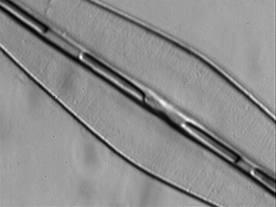
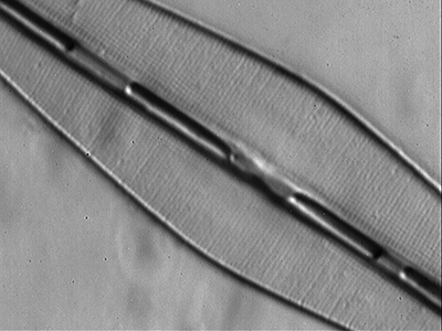
Left
above - Green filter ca. 550 nm (internal filter on Zeiss Photomicroscope
III) - a hint of frustule detail.
Right above - Lee dark blue filter 440
nm
- some evidence of punctae and 'beaded striae'.
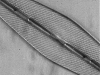
Above - 400 nm interference filter, punctae clearer over whole frustule cf the deep blue.
Amphipleura pellucida,
Zeiss 40X NA1.0 oil apo, dry condenser - oblique using
offset brightfield port in phase condenser.
The diatom long axis is parallel to
direction of oblique.

Lee
dark blue filter - very slight hint of 'striae' at left.

400
nm interference filter - 'striae' quite clearly shown.
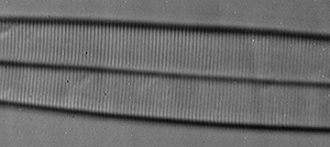
Above:
The LOMO 40X NA0.95 dry apo is also able to show the striae in oblique with
the 400 nm filter.
'Striae' are the accepted appearance of unresolved punctae under the optical microscope, they are not real features on the frustule. For Amphipleura pellucida they are typically reported to be 0.28 µm apart, and thus resolvable at 400 nm by an NA 0.95 objective from the Raleigh equation (Resolution = 0.61 l / NA ).
Frustulia rhomboides, Zeiss
63X - slight oblique using offset brightfield port in
phase condenser.
The diatom long axis is ca. 45 degrees to direction
of oblique.
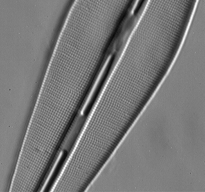
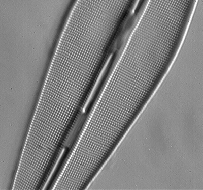
Left: Lee dark blue filter ca. 440 nm the punctae are resolved, but decreasing the wavelength to 400 nm (right) the image look crisper and more contrasty.
Nikon DSLR sensitivity to '400 nm' interference filter
If my Nikon DSLR with CMOS sensor is
typical, a DSLR seems sufficiently sensitive to this short
wavelength and narrow band output from a 100W microscope lamp. Apart from
extreme oblique there was plenty of light for Live View to compose
and focus. With 100W lamp at 7V, a 2 second exposure at ISO 200
was needed, the sort of exposure I'd use anyway at high mags to avoid camera
vibration. As with the earlier images, diatoms tend to have flat
tonality so the histogram needs adjusting. Images were best converted to
monochrome as in colour they were a pale looking violet.
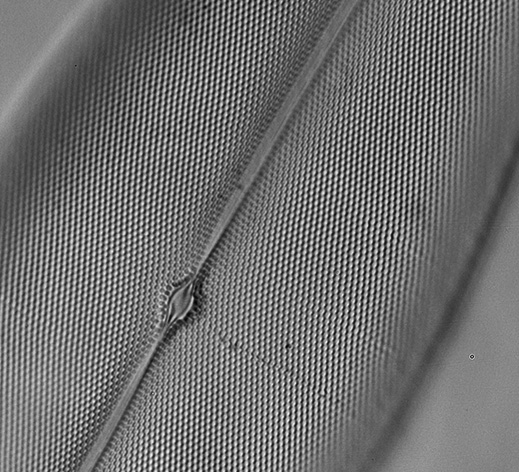
Pleurosigma
angulatum, 400 nm interference filter, Zeiss 63x oblique using
offset brightfield port in phase condenser.
IS0 200 2 secs, crop of
frame.
