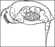
 Life is every bit as tough on the microscopic scale as on the
plains of Africa. There are the hunters, the hunted and the scavengers all playing their
role in the microscopic world of a freshwater pond.
The following real life story is a summary of an
extraordinary piece of video footage captured by Ken Jones, a well known UK amateur
microscopist specialising in video microscopy. This article, illustrated by stills from
the video, tells a no-holds barred story of birth, life and death of a female rotifer
(shown right) and her offspring.
Why this video sequence is so unusual
The Characters
The Good
The innocent party of this story is a female rotifer of the genus Colurella (shown
above) carrying an egg. Rotifers are multicellular microscopic organisms which are
typically 0.1 to 1mm long and are found in a variety of aquatic habitats. They are a
popular subject for the amateur microscopist because of their wonderful variety of form,
habits and life cycle.
The Bad
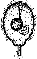
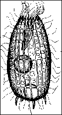 There are two
villains in this story, both of them protozoa. The protozoa are aquatic single-celled
organisms which are usually much less than a millimetre long.
The first is a member of the genus Coleps
and is typically 80 microns in length (shown on the left). It is the armoured tank of the
protozoa world because its body is protected by many small plates which often bear spines.
The second and real villain of the story
is a diminutive protozoan thought to be a member of the genus Urotricha (shown on
the right). It is almost spherical in shape and this individual is barely 15 microns in
length.
The Ugly
The ugly member of this story is a fungus called Zoophagus insidians. The fruiting
bodies of the larger terrestial fungi such as mushrooms are familiar to all. However, Zoophagus
is an aquatic fungi which predates on other organisms such as rotifers, protozoa and
nematodes (roundworms). The organisms are trapped on hyphae which are coated with a highly
effective adhesive substance. Hyphae are a characteristic of fungi, and are tubular
filaments which branch repeatedly to form a network called the mycelium, which extracts
nourishment from the surrounding medium. The mycelium normally obtains its food from dead
and decaying organic matter, but in this case the hyphae actually captures food.
The Tragedy Unfolds
The following stills from the video are complemented by captions, which illustrate and
relate the story as it really happened. The black bar on the first image represents 50
microns or 1/20th of a millimetre.
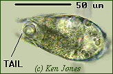 Female rotifer trapped by the fungus
The rotifer is being viewed from 'above' and her tail is pointing into the screen. The
fungal hypha is the vertical out of focus band on the right of the image.
The rotifer has been trapped by the sticky fungal hypha for some time and has been
struggling to free herself without success. Death is inevitable after the fungus captures
its prey. A penetration tube grows out of the hypha and enters the rotifer's soft body.
Enzymes are secreted which quickly kill the animal, and the rotifer's cytoplasm is
devoured by the fungus.
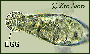 Out of
death comes life
The female rotifer in its death throes releases an amictic egg, which was probably due to
be laid regardless of whether she had been trapped by the fungus. Amictic eggs are eggs
which have not been fertilised by a male, and will develop into another female.
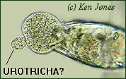 The tiny
attacker
Shortly after egg laying commences, the tiny protozoan (possibly Urotricha genus)
attacks the egg. Despite its size, the protozoan apppears to attach itself quite firmly
and pushes and pulls on the egg membrane. The image left showing the distorted egg
illustrates the force that the tiny attacker is exerting.
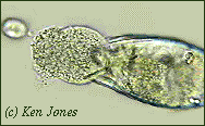 The egg
is destroyed
The protozoan does not succeed in the first two attacks on the egg, but in the third
attempt the egg is ruptured and disgorges its contents into the surrounding water. The
protozoan can be seen moving away out of the plane of focus of the video still.
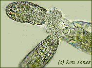 The
scavengers move in
Like hyenas on an African plain, there are scavengers in the freshwater world eager to
feed on the dead and dying. Barely thirty seconds after the egg is first ruptured, two
larger protozoa of the genus Coleps eagerly dive onto the granular contents of the
egg.
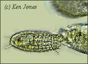 The end
of a mother and her young
Less than seventeen seconds after the Coleps' first arrived, the egg had been
completely consumed. The image shows one of the Coleps eating the last vestiges of
the egg yolk, with its partner in crime leaving the plane of focus on the right having had
a tasty meal.
The mother, having failed in an attempt to give birth to her potential offspring, is still
left in the clutches of the fungus, which will make a meal of her. Life is tough in the
microscopic world!
Why this video sequence is so unusual
The video sequence recorded by Ken Jones was studied by Dr Alan Warren of the Natural
History Museum, Microbiology Group. Dr Warren remarked that he and his colleagues have not
previously observed ciliated protozoa thought to be of the Urotricha genus
attacking and rupturing rotifer eggs. He was also not aware of similar instances reported
in the literature.
This article shows that it is possible
for the amateur microscopist to make a valuable scientific contribution to our knowledge
of the behaviour of freshwater organisms.
Postscript
After Ken Jones and Eric Hollowday originally published their article on this video
sequence, they found out that a microscopist in 1859 or earlier had made a similar
observation! In his classic book 'Evenings at the Microscope' P. H. Gosse described how a
group of Coleps attacked and killed a rotifer hatching from its egg. This was later
followed by an attack by a group of ciliated protozoa.
This postscript illustrates an
interesting point which Jones and Hollowday eloquently state in their letter to
'Microscopy' ie that Gosse's observations 'are not only of great interest for their
relevance to our paper over a century later, but also serve to demonstrate the
scientifically valuable information pertinent to modern studies which one can so often
find in old works which might otherwise be thought obsolete and unwisely discarded.'
Go to top of this
article
Other illustrated articles in Micscape on protozoa and
rotifers
Protozoa
Paramecium
Vorticella
Stentor
Rotifers
Rotifers in a bird-bath
Acknowledgments
The Micscape editors would like to thank Ken Jones and Eric Hollowday for allowing the
video sequence and their original paper on this work to be used (in a more dramatic
style!) for Micscape.
The Micscape editors also thank the Quekett Microscopical
Club for permission to use the above authors paper for this article.
The original paper entitled 'A Note on the Consumption of a
Rotifer egg by Ciliated Protozoa (Ciliophora)' by K R Jones and E D Hollowday was
published in Microscopy, vol 36, p718-720, 1992.
The letter by the above authors drawing attention to the observations by P.H.Gosse was
published in Microscopy, vol 37, p80, 1993.
Microscopy is the Journal of the Quekett
Microscopical Club.
The Micscape editors also thank the Natural Environment Research Council (NERC) and the Freshwater Biological Association for permitting the line
drawings in this article to be copied from the following publications.
Rotifer Colurella from figure 19 page 33 of:
A Key to the British Freshwater Planktonic Rotifera by R M Pontin. Freshwater
Biological Association (FBA) Publication No 38, 1978. 178 pages. (Available from The
Librarian, FBA, Ferry House, Ambleside, Cumbria LA22 0LP, UK). SBN 900386 33 9, ISSN
0367-1887. A well-illustrated guide to the identification of the UK planktonic species.
Protozoa Coleps and Urotricha from
figures 74 and 75 page 45 of:
A Beginners Guide to the Collection, Isolation, Cultivation and Identification of
Freshwater Protozoa by B J Finlay, A Rogerson and A J Cowling. Published by the
Freshwater Biological Association, The Ferry House, Ambleside, UK, 1988. (78 pages) ISBN 1
871105 03 X.
Technical Details
Video sequence filmed by Ken Jones using a Panasonic WD130 video camera on a custom-built
photomicrography stand. The stand uses standard microscope objectives to project an image
into the camera.
A good general introduction to video microscopy: 'An
Introduction to Video Recording at the Microscope' by A V Dodge, S V Dodge and K Jones,
Microscopy, 1988, vol 36, p43-53.
All images captured from still frames of the VHS video
using a Creative Video Spigot capture card.
Image manipulation using Photostyler v2.0 software. Note
that the colours of the organisms are similar to, but not necessarily identical to those
seen in real life.
Feedback and comments to the author Dave Walker are welcomed.
© Microscopy UK or their contributors.
Please report any Web problems or
offer general comments to the Micscape Editor,
via the contact on current Micscape Index.
Micscape is the on-line monthly
magazine of the Microscopy UK web
site at Microscopy-UK


Life is every bit as tough on the microscopic scale as on the plains of Africa. There are the hunters, the hunted and the scavengers all playing their role in the microscopic world of a freshwater pond.