Micscape Article: Paramecium
Text by Mike Samworth, images by Mike Morgan. Dec. '95.
Updated by the Micscape
Editor Sept. '99.
Note to readers: since this article was posted in 1995, the
Microscopy UK parent website has uploaded an enormous range of resources on the microscopic world. These include
video clips, 3D images, high quality images of protozoa, articles on pond life and many other topics. Click on
the resources offered on the site's main index to see them. Or search for a particular topic in the on-line library.
Links at base.
Overview
Paramecium is a small unicellular organism that is plentiful in freshwater ponds. It swims, rotating slowly,
and often changing its direction. Under suitable conditions it may reproduce by dividing two or three times a day
and so large numbers may build up. It belongs to the group of organisms called Protozoa. Most of these are microscopic
with over 80,000 different species identified.
Protozoa
The cells of fossil protozoa are often found in enormous numbers. They can help in tracing oil-bearing rocks and
sediments. Many are also of considerable importance as they are responsible for various diseases and infections.
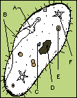
Paramecium Structure
Although just visible to the naked eye, to see Paramecium properly, a microscope is needed. You should
be able to recognise most of the processes from the diagram below when observing them for real.
Studying Paramecium
 Although
Paramecium is a small organism, it has a complex structure and a microscope, stains and a degree of skill
are needed to reveal all the details. It is usually best to suspend a drop of water upside down on a cover-slip,
using blobs of Vaseline to support the cover-slip. Remember this is a diagram that has been drawn from a living
specimen under a powerful microscope by an expert. You are more likely to see less features than this, but look
carefully for each of the structures shown. I have labelled some of the more important ones: (A) = gullet, (B)
= oral groove, (C) = micronucleus,
Although
Paramecium is a small organism, it has a complex structure and a microscope, stains and a degree of skill
are needed to reveal all the details. It is usually best to suspend a drop of water upside down on a cover-slip,
using blobs of Vaseline to support the cover-slip. Remember this is a diagram that has been drawn from a living
specimen under a powerful microscope by an expert. You are more likely to see less features than this, but look
carefully for each of the structures shown. I have labelled some of the more important ones: (A) = gullet, (B)
= oral groove, (C) = micronucleus,
(D) = macronucleus, (E) = food vacuoles, (F) = contractile vacuoles ; and the small dots are plasmasol. The function
of each of these should become clear as we consider the organism in more detail.
Being unicellular, this one cell must contain everything needed for survival.
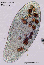
A photograph of Paramecium is shown right below.
 Movement
The outer surface of the cell is covered with many hundreds of tiny hair-like structures called cilia. These act
like microscopic oars to push through the water, enabling the organism to swim. The speed of motion is about four
times its own length per second. It moves so quickly that microscopists have to add a thickening agent to the water
to slow it down to study it.
As it moves through the water it rotates on its axis and small particles of debris and food are collected and
swept into the gullet. If Paramecium comes across an obstacle, it stops, reverses the beating of the cilia,
swims backwards, turns through an angle and moves forward again on a slightly different course.
Feeding
Paramecium has a permanent feeding mechanism, consisting of a funnel-shaped gullet into which food is
drawn by the combined action of cilia which cover the body and other cilia lining the gullet. They feed on small
organisms such as bacteria and even other smaller protozoa.
Reproduction
Paramecium has two means of reproduction, simple division and conjugation.
A. Division. In favourable conditions the cell divides in two by a process called binary fission.
This forms two new cells, each of which rapidly grows any new structures required and increases in size. This whole
process make take place two or three times a day if conditions were right.
B. Conjugation. This is a more complicated method. It involves two cells coming together to
exchange nuclear material. The two cells then separate and continue to reproduce by simple division. It is similar
in some ways to sexual reproduction in more complex animals.
More observations
Here are a few things to think of when studying Paramecium under a microscope. Observe the way in which
Paramecium moves. Can you see the cilia beating? Notice the reaction of the animal when it hits an obstacle.
Can it swim backwards? Are all the Paramecia the same size? Estimate the speed of movement. Slow down
the motion of the organisms with something. Can you now see the cilia beating with a wave-like motion? Place a
drop of neutral red underneath a cover-slip. The food vacuoles become pink and the route taken by the vacuoles
as they travel through the cell may be seen. There are vacuoles in Paramecium that do not contain food,
usually one at each end. These are used to remove excess water. You may see these fill up and then empty. Add a
drop of yeast culture stained with Congo Red stain. this will allow you to see food particles being swept into
the gullet. Can you see any Paramecia dividing on your slide?
Other points of interest on Paramecium and other specific Microscopy UK resources
-
Paramecium is not one species but the name of the genus to which a number of species belong e.g.
Paramecium caudatum which is elongated and streamlined, and Paramecium bursaria which resembles a
footprint. There are at least eight well-defined species.
-
Click here for a video clip showing the collapsing contractile vacuole
in a protozoan (taken by Edward Cowen).
-
Click here for a video clip showing the beating cilia of a protozoan
(taken by Edward Cowen).
-
High quality images of Paramecium by James Evarts showing many of their features can be found here
and here.
-
An excellent introduction to the types of microscopic life found in ponds with stunning images can be found
on Wim van Egmond's Smallest Page on the Web.
© Micscape and/or their contributors.
Micscape Magazine, the official web magazine of Microscopy UK
© Onview.net Ltd, Microscopy-UK, and all contributors 1995 onwards. All rights
reserved. Main site is at www.microscopy-uk.org.uk with full mirror at www.microscopy-uk.net.