 Stentor are one of the largest protozoa found in water, and
is therefore one of the easiest for the microscopist to study. This article describes
where you can find the Stentor and how different types of illumination under the
microscope can be used to show the characteristic features of a protozoan.
What are protozoa and where to find the Stentor
Protozoa are single-celled organisms and are one of the simplest forms of life. In the
modern classification of organisms, the protozoa belong to the kingdom Protista along with
the algae and lower fungi. The Protozoa are unique in that the single cell performs all
the functions of feeding, digestion, excretion, respiration and reproduction.
Although most protozoans are microscopic, the Stentor is the exception to this rule and can be up to 2mm long! It is therefore larger than many of the multi-cellular organisms found in freshwater such as rotifers and water-fleas, and has been known to eat the smaller members of these groups.
Stentor are usually present in most freshwater ponds. Take a few pieces of the stem of any floating plant eg Potamogeton, which has spear-shaped leaves. Place the stems in some water in a shallow dish and allow a few minutes for the water to settle. Examine the stems with a 10x hand lens - any Stentor present on the stem will be visible as long trumpet-shaped organisms. Stentor may also be swimming freely, when they become more oval in shape with a narrowing at their posterior.
Autumn (the Fall) is a good time of year to find Stentor because leaves falling into a pond increase the bacteria population feeding on the decaying vegetation. This leads in turn to an increase in the population of protozoa such as Stentor which feed on the bacteria.
Colonies of Stentor often occur, in which the lower parts of the animals are covered with a mucilaginous substance which they secrete.
Stentor is remarkable for its regenerative powers; a small fragment one-hundredth the volume of an adult can grow back to a complete organism. This capability has made Stentor a favourite subject amongst biologists for studies of regeneration in protozoans.
General features of the Stentor
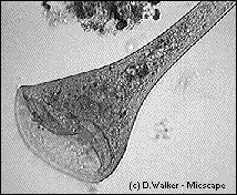
 The image at the top of this page shows
a typical view of a Stentor using bright field illumination. When attached to a
substrate, the Stentor has a trumpet shape. Any disturbance makes the whole body
contract to become a blob of protoplasm. The image to the left shows Stentor when
swimming where it adopts an oval or pear shape.
The rim of the trumpet is surrounded by a crown of cilia (vibrating hairs) which beat to
produce a current that draws particles of food down into the gullet.To find new feeding
grounds, the animal swims by means of cilia that cover the body.
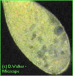
 Stentor can be very colourful -
there are green, blue and amethyst coloured species. The figure on the left shows a green
form using dark field illumination. The green colour is a result of the microscopic green
algae which Stentor ingests. The algae live in symbiosis with the Stentor, ie the
algae and Stentor mutually benefit from the close association. The algae uses
photosynthesis to convert Stentor's waste products to useful nutrients.
Observing features using phase contrast
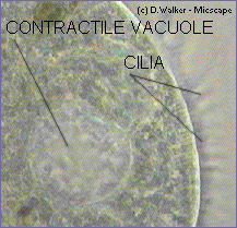
To observe the cilia of Stentor, a special type of illumination called phase contrast can
be very useful. The figure on the right below shows the cilia, which are not sharply
focussed because they are in constant motion.
Although not clearly seen in the photographs, Stentor has a macronucleus in the form of a string of beads along the body length.
 A second feature of Stentor as a typical
protozoan is the contractile vacuole, which is also seen in the right hand figure. The
concentration of salts in the bodies of freshwater protozoans is higher than in the
surrounding water, so the water continually enters the animal through its external
membrane by osmosis. The excess water collects in the contractile vacuole, which at
intervals discharges it to the outside. A few minutes studying Stentor should show
one of them with a collapsing vacuole.
When examining microscopic subjects, particularly the Protozoa, it is worth trying the various forms of illumination available to you. Bright field is the standard method of illumination, but dark field, oblique and Rheinberg illumination are worth trying and well within the scope of the amateur. Phase contrast illumination is a more specialised form of microscopy which requires special objectives and condensers. Phase contrast is particularly valuable for highlighting structures such as cilia and flagella of Protozoa.
Equipment used
All images captured from still frames of VHS videos using a Creative Video Spigot capture
card.
Videos originate from Maurice Smith (first image) and David Walker (other images).
Primary microscope magnifications in the range 200-400x before projection into a CCD video camera.
Phase contrast images taken using Cooke, Troughton and Simms phase contrast microscope.Other microscope images taken using Russian Biolam microscope.
Image manipulation using Photostyler software.
Comments to the author Dave Walker welcomed. But check refs. below for detailed info' which the author is not able to provide.
References and sources of information:-
1) A Beginners Guide to the Collection, Isolation, Cultivation and Identification of
Freshwater Protozoa by B J Finlay, A Rogerson and A J Cowling. Published by the Freshwater
Biological Association, The Ferry House, Ambleside, UK, 1988. (78 pages) ISBN 1 871105 03
X.
2) Animal Life in Freshwater by H Mellanby. Published by Chapman and Hall 6th Edition 1971. (308 pages) ISBN 0 412 21360 5.
3) Encyclopaedia Britannica 15th Edition 1993. If you can't find a specialised book on protozoan biology or invertebrate zoology in a local library, don't overlook the large encyclopaedias such as Britannica, which has twenty pages in the Macropaedia (Volume 26) on Protists and Protozoa. Or read Britannica's on-line article on Protozoa here, which covers aspects like cell structure, reproduction, excretion etc.
4) In depth info' on Stentor can be found in 'The Biology of Stentor', by Vance Tartar, Pergamon Press, 1961, Library of Congress # 60-15714. Ask your local main library for obtaining this on their inter-library loan scheme. (The author thanks William H Amos for providing this reference).
Also see these other Micscape articles on Stentor.
