by Mike Dingley, Australia
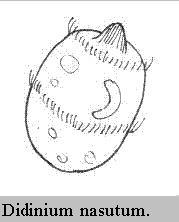
Didinium nasutum is an oval ciliate ranging in
size from 60um to 200um, lives in freshwater habitats and is frequently
seen in pond samples. It has two bands or girdles of cilia (pectinellae);
one round the front pointy end and one band just below the middle. The
pointy end, snout, nose or proboscis gives it its species name, nasutum.
There is another very similar animal which only has one girdle near the
anterior end which has been placed in the genus Monodinium.
Didinium is a voracious hunter of live food, namely Paramecium. It is found quite commonly in freshwater habitats such as ponds and other still waters. It is in constant motion, swimming around in search of prey. When it bumps into something it seems to hesitate and examine the obstacle before swimming off if it is not edible. When its nose (proboscis) strikes a food source such as Paramecium it extrudes trichocysts (threadlike filaments). These same trichocysts do not release when it encounters other protozoans. My references inform me that it is not known what causes the extrusion of these trichocysts. It is not a collision-stimulated response as it bumps into many other protozoans without releasing them.
When it captures a paramecium a food vacuole begins
to form inside Didinium and the cytostome expands to ingest it.
Of all the images that the author has seen it appears that Didinium
makes contact with Paramecium half way along the Paramecium's
length. It then somehow (using the anterior band of pectinellae?) moves
the Paramecium to one end and then manoeuvres this end into its
cytostome. Paramecium measure about 300um by 200um and so you can
see that it is a great deal larger than Didinium.
Take a look at the images photographed from prepared slides.
Both Didinium and Paramecium have been stained with a red
and blue dye. Notice the blue bands of cilia.
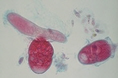
Image 1.
This shows two Didinium (didinia?), one of which
is in contact with the side of a Paramecium.
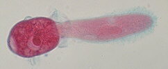
Image 2.
Shows the start of feeding. Didinium has manoeuvred
the Paramecium to its anterior end whereupon it has started to ingest
it.
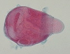
Image 3.
The Paramecium has almost been totally engulfed
by Didinium who must be feeling very full at the moment.
In a book by Eric Gravé, Discover the Invisible - A Naturalist's Guide to Using the Microscope.Prentice Hall, 1984. In it he explains in detail this feeding behaviour and illustrates the text with seven photomicrographs.
Another source of information illustrated with scanning electron micrographs is The Protozoa. Introduction to Protozoology by John N. Farmer. Published by the C.V. Mosby Company, 1980, 732 pages.
© Mike Dingley.
Editor's note: An article on Paramecium is in our Articles Library. See also an article about Protozoa attacking a rotifer's egg.
Micscape is the on-line monthly magazine of the Microscopy
UK web
site at Microscopy-UK