INTRODUCTION
There have been many new technologies in the past ten years or more with the most recent capturing the headlines and news channels being the advent of A.I. (Artificial Intelligence). How many of these might help the microscopist or in a wider sense, to study our world and ourselves. I am a person suffering from an end-of-life lung disease triggered by smoking; one of many such victims born in the era of 1940 to 1960. In such times, adverts for smoking were thrown at us from every
direction. In a recent medical quest to look at my lungs I had an X-ray. The range of lung diseases are collectively termed COPD (Chronic Obstructive Pulminary Disorder or Disease). My particular one is emphysema. This is where air sacs are destroyed and it is an irreversible process.
It is below.
X-RAYS
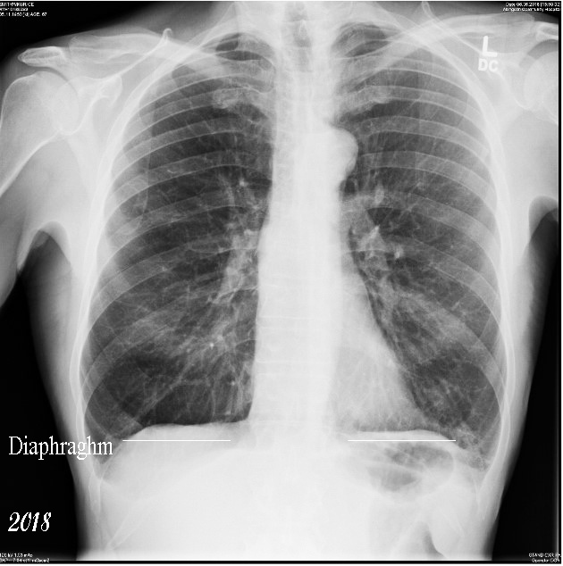
The image is taken as though you were looking straight at me. It was taken towards the end of 2024. It looks hardly any different to one taken of me 6 years ago, but in fact my capacity to breathe has declined on an exponential scale in those 6 years. The X-ray shows several things of note. The first is that there is no sign of a tumour/cancer. One blessing, I guess. The other is an increased lowering of the diaphragm compared to the one in the image taken in 2018
I am no doctor but as my lung is continually expanding to try and produce as much contact between the air sacs and blood vessels to pass oxygen in and extract CO2 out, my lungs are now pressing down hard on the diaphragm.

This interesting thing about this is that an X-ray cannot show the extent of destroyed air sacs and (in my observations) do not show a worsening of my disease. I can only assume the consultant just wanted to see if I had cancer or not. But indeed, my lung function has declined to such an extent, I can no longer stand for more than thirty seconds and I'm wheelchair bound and nigh on helpless.
The lungs will not stop expanding so ultimately they will crush one of the valves of my heart leading to death by heart attack, a predicted end as told to me. In the UK and the NHS, there is no attempt to measure this condition using MRI scanners. Why bother, you are a dead man walking. If you still vape or smoke, they do not give you oxygen either which would help a little. I do have oxygen concentrators which were purchased by myself and they do help me quite a lot.
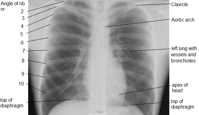
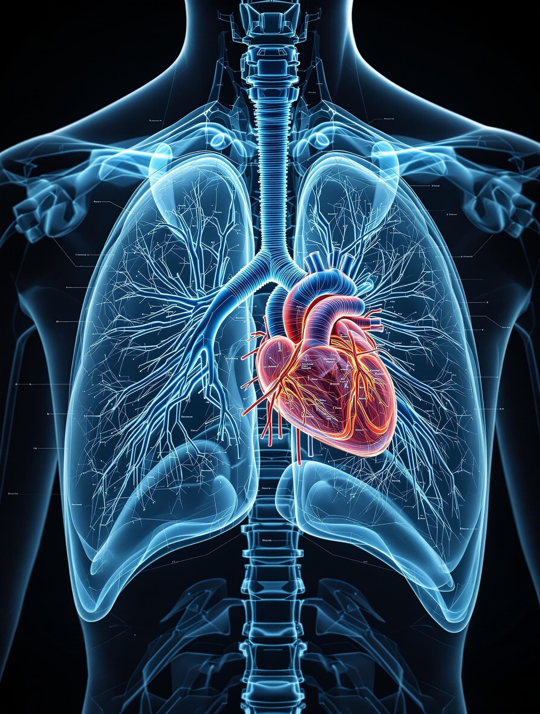
To help me interpret the X-ray, I used the X-ray of a healthy lung from Wikipedia which has been labelled. Used here under their Common licence.
But to understand more, I needed something else...
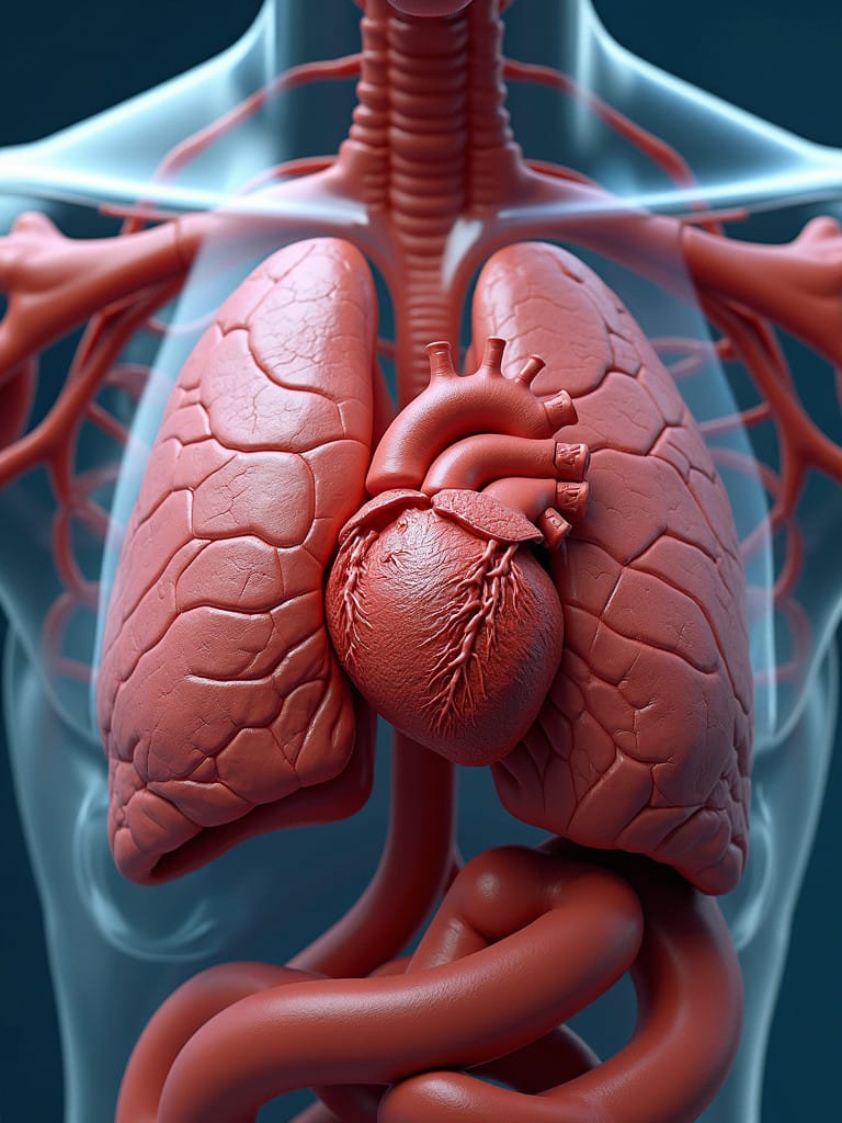
| 3D Models To help further I used a 3D animation I made for an article for Micscape many years ago. This was animated using POSER 3D SOFTWARE. |
|
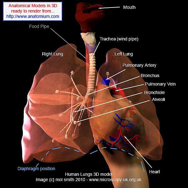
This is a detailed 3D model from Anatomium, a company who has since gone.
I have found using 3D modelling a helpful tool as I'm sure people studying all kind of different subjects do as well.
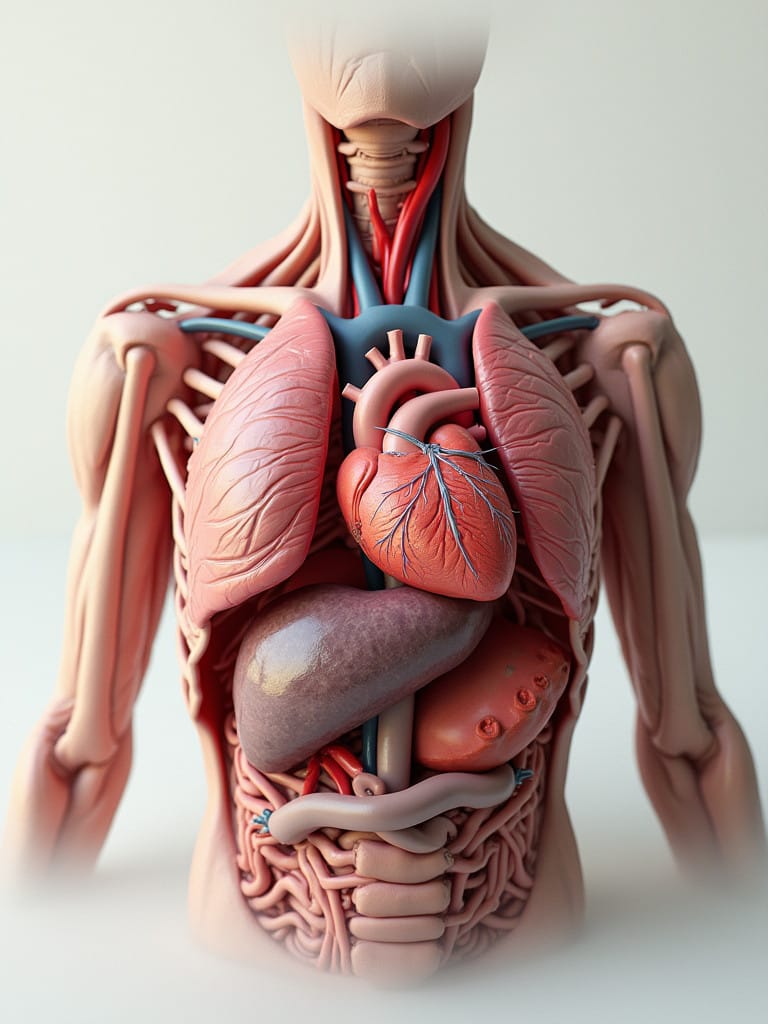
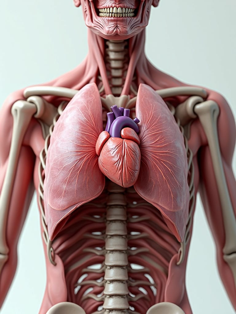
So the next thing to try is A.I. image creation. I use several such site but for the sake of this article, I used Nightcafe.
There are several different A.I. so-called engines you can use. I pay a subscription as I also create books and art. I used several of the best and the more expensive ones for this article.
The results are below.
|
This is my first stab. I used the Flux engine with this comment: |
 To see this larger, click on it. To see this larger, click on it. |
| Give it another go and... |
|
|
One more go but change the instruction... |
|
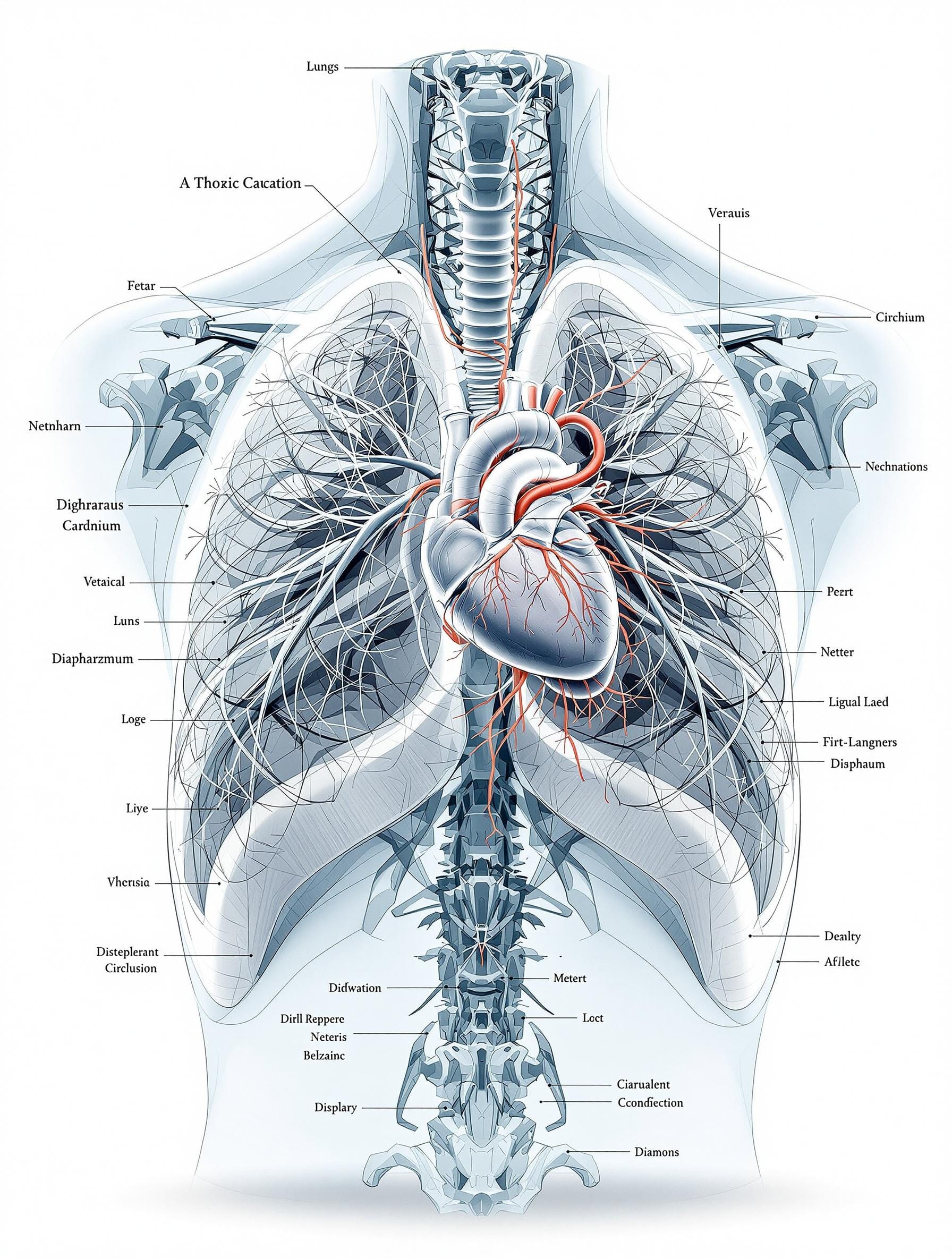
| I decided to try another engine. I tried Flux Pro Ultra... |
|
| Another go... If you enlarge it, you'll see the labels are gobbledigook! |
|
| So far, none of this has really proven of value to microscopical study other than to understand that if you look at the cells of the lung as in this article, where lungs are and thus where the cells being studied are coming from. |
|
|
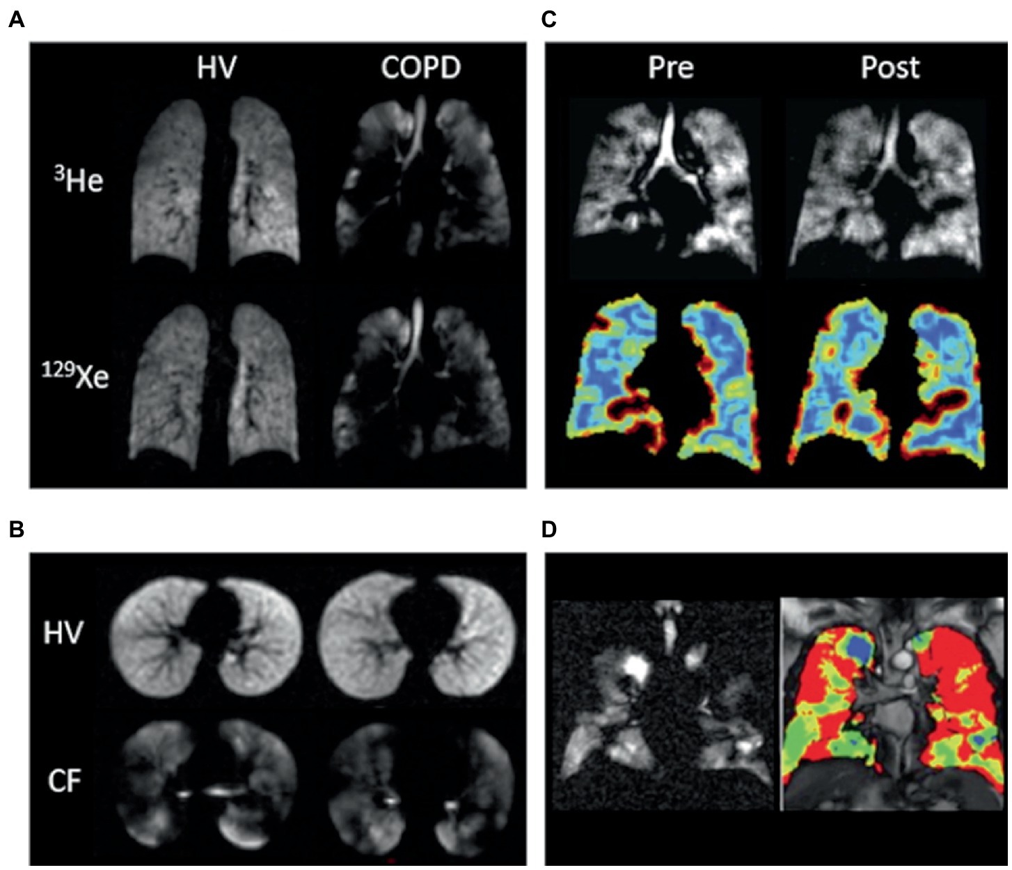
Now an MRI scan is a winner when showing COPD as this article shows.
|
|
| |
|
|
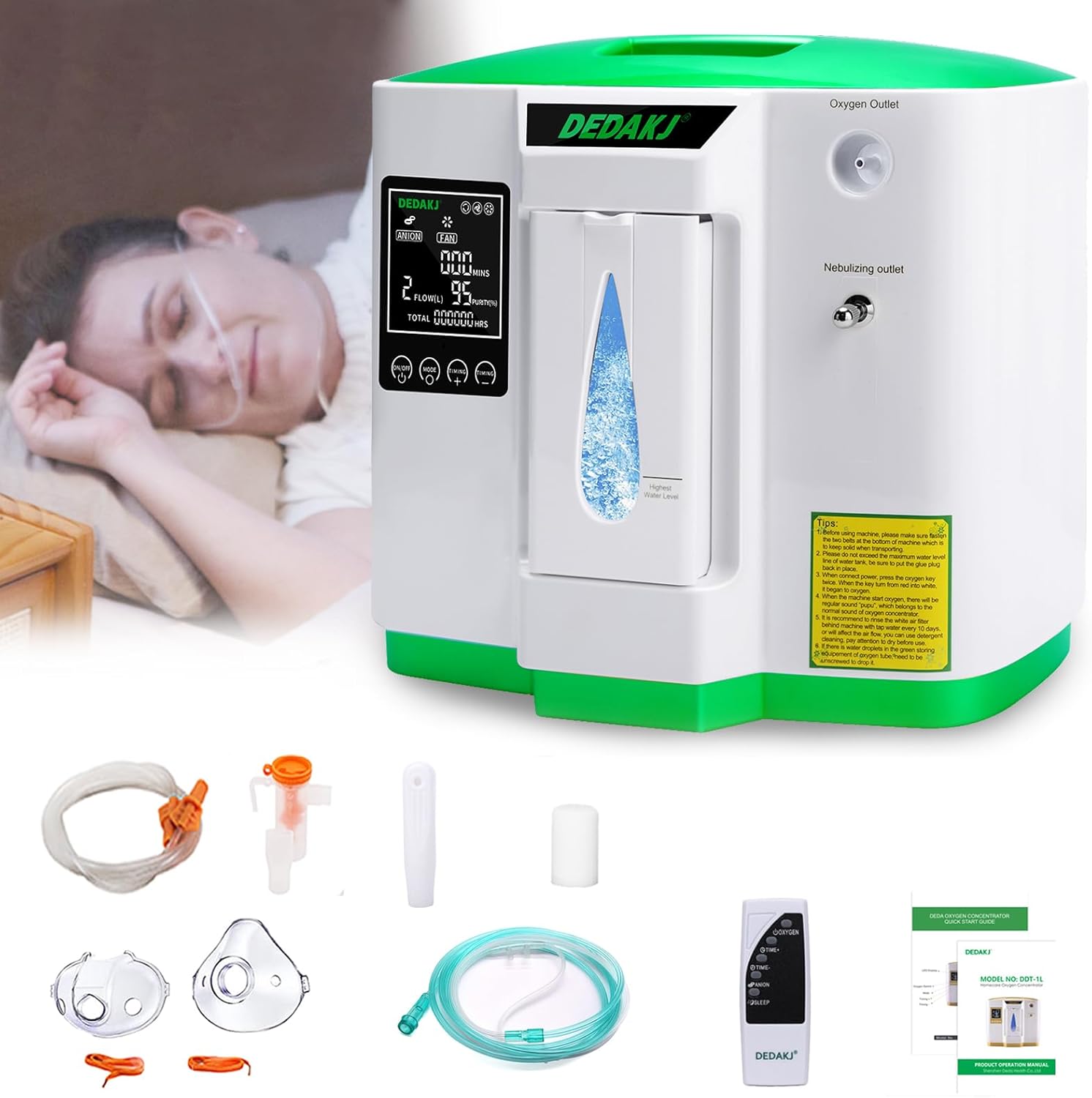
In conclusion, possibly some Internet technologies can help one to understand about
your body and ill-health. Coupled with studying real specimen slides completes the picture. But if any reader has COPD, then this is the oxygen concentrator that I find is the most helpful. I have tried others but this is the best. |
|
| |
|
Microscopy UK Front Page
Micscape Magazine
ArticleLibrary
Published in the Feb 2025 edition of Micscape.
Please report any errors to the Micscape Magazine Editor
via the contact on current magazine index.
Micscape Magazine is the free monthly web magazine of Microscopy-UK.
© Onview.net Ltd, Microscopy-UK, and all contributors 1995 onwards. All rightsreserved.