One of the appealing aspects of microscopy for me is exploring subjects at both near IR and near UV wavelengths. The former is useful for dense insect exoskeletons where near IR has better transmittance revealing more detailing and the smaller contrast range allowing imagery. A bonus is increased depth of field but resolution is decreased. Near UV is a popular method of increasing the resolution for subjects such as diatom frustules with fine details.
One of the benefits of retaining the original maker's 100W quartz halogen lamp if the bulbs remain available and affordable is that both near IR and near UV is readily available with an appropriate filter on the field lens. A monochrome camera is required which do not usually have a NIR blocking filter. The NIR band from a hot body is of course substantial. There is a finite near UV tail if the bulb is not UV blocked but requires a sensitive camera especially when combined with techniques such as phase and DIC.
For microscopes converted to a modular white LED system, two LED cassettes for near IR and near UV would be required, the latter can be more powerful of course than exploiting the near UV tail of a halogen bulb.
To date, I hadn't explored using either phase or DIC with either near IR or near UV ... so why not 'try it and see'. With an admittedly limited knowledge of the principles behind phase and DIC it also prompted some reading to gain an insight into whether one or both techniques should still be effective. This is part 1 of a four part series of short notes to share these explorations.
DIC at near IR wavelengths
A search online was valuable to assess if it was a technique that was used, if so, it immediately suggested that DIC did work at the extremes of visible (assuming standard DIC systems were in use). One paper published in 1990 was quickly found Visualizing unstained neurons in living brain slices by infrared DIC-video microscopy by Hans-Ulrich Dodt, Walter Zieglgänsberger. Only the abstract was available for non-affiliated readers like myself but revealed that NIR had some benefits. The improved transmittance was exploited and the increased depth of field with the DIC revealing details. Objectives of 20-40X were useful as well as a 63/1.4 objective the latter which presumably would go some way to recovering the resolution desired. Video enhancement techniques were also used.
Image trials.
Zeiss 16/0.35 planachro' for the cheek cells, this is the correct objective for the number I prism of condenser owned. Serial number not optimal so not an evenly toned background.
Zeiss 6.3/0.16 for the insects. An August 2024 note describes the DIC setup used which although not the recommended one gave good DIC imagery.
Visible - full spectrum, near IR blocking filter.
Near IR - Hoya R72 filter, 720 nm long pass. Not quite true near IR but a readily available filter.
Camera ZWO ASI178MM monochrome camera. This small sensor used without an eyepiece or relay lens only captures about a third of the central visual field. As planachromatics may not be well corrected for spherical aberration at wavelengths near edge of the visible, this central image capture should go some way to minimise any uncorrected aberration of this type.
Image work up. The tonal range can vary widely when changing wavelengths so the highlights and shadows were adjusted but tonal mid-range left.
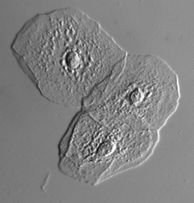
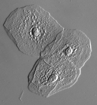
Fresh unstained epithelial cheek cells mounted in water. Zeiss 16/0.35 planachromatic. This is a useful subject to assess a contrast enhancement technique as they are near transparent in on-axis brightfield.
Lefthand image, visible DIC. Cell details are clearly presented in DIC.
Righthand image, NIR DIC. The DIC effect is clearly still present providing a hands-on demonstration that DIC is active in near IR.
For transparent subjects like these there is no merit in using near IR with DIC. Resolution is lost and for this flat subject little evident of improved depth of field. The tonal range despite correction is also flatter.
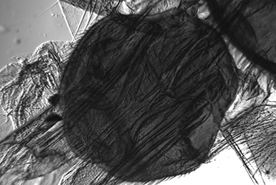
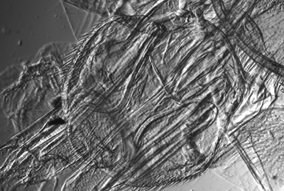
Water gnat 'Hydrometra argentata' abdomen extremity, Victorian whole mount in balsam labelled A. N. Tabor and dated 1894.
Lefthand image, visible DIC. The keratin transmittance is low in visible light making DIC impractical. The camera also struggles with the large contrast range.
Righthand image, NIR DIC. With improved transmittance, DIC becomes more practical and usefully showing detail in the formerly dense areas. The tonal balance is also lower which the camera can cope with. Depth of field which increases with increasing wavelength for a thick subject is also good.
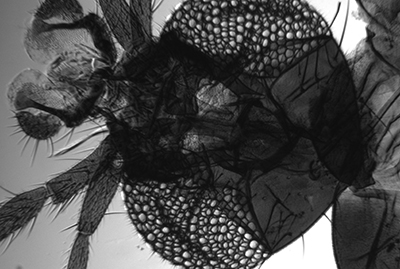
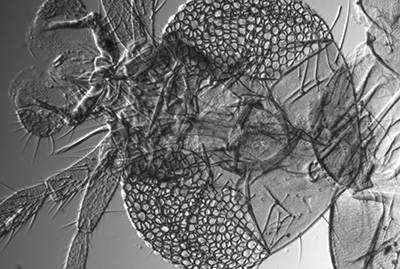
Winter gnat 'Trichocera haemalis' Victorian whole mount in balsam, unnamed, no date, diamond inscribed label.
Lefthand image, visible DIC.
Righthand image, NIR DIC.
Similar comments to the previous image.
Comment. There are not many subjects in my own prepared slide collection where NIR with DIC excels but the main aim of the study was to explore the combination of techniques at the extremes of visible light. As remarked, at least one paper referenced suggests with suitable subjects the technique does have value.
Postscript. Thinking aloud on why DIC works at near IR (or near UV) wavelengths. With my admittedly limited grasp of theory the thoughts shared are speculative and welcome comments. The Molecular Expressions site is invaluable for describing the fundamentals of DIC and reference 1.
The Nomarski prism below the condenser splits the plane polarised incoming light into an ordinary and extraordinary ray. This ability (and for the prism above the objective recombining rays) is not wavelength dependent for the technique to work at the near IR or near UV wavelengths.
The DIC technique "transforms local gradients in optical path length into regions of contrast in the object image" (ref. 1).
Optical path length = refractive index x thickness (between the two local points on that path) (ref. 1).
The variation of RI from near IR through visible to near UV for water in say a water based organism is quite small (see this graph). The difference in RI between the ordinary and extraordinary ray also remains constant for e.g. sapphire (see this graph). So presumably there would be little evidence of a change in the 'strength' of the DIC effect as change the wavelength but details resolved would be affected by the change in resolution.
Reference
1 - Fundamentals of Light Microscopy and Optical Imaging by Douglas B. Murphy and Michael W. Davidson, pub. Wiley-Blackwell 2013, p.173ff.
Comments to the author David Walker are welcomed.