Plants
|
RODOPHYTAE |
By JM CAVANIHAC FRANCE |
|
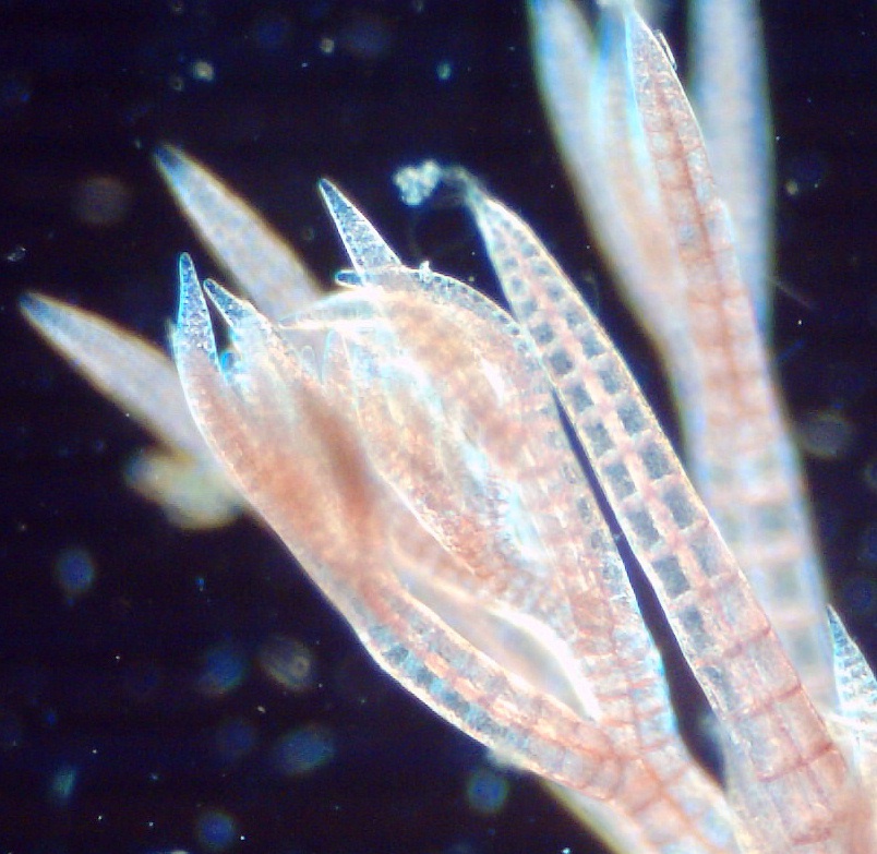
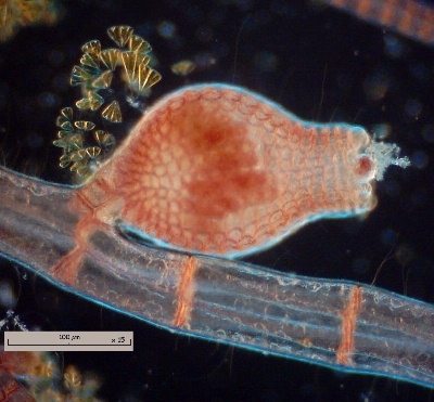
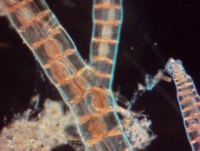
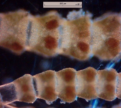
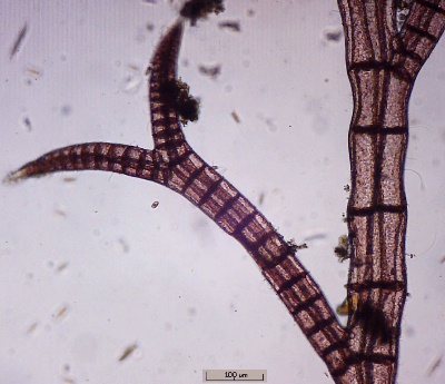
The name a little confusingly includes all the varieties of red algae (but some of them could be blue or even green, according to the proportion of phycocyanine or chlorophyll they contain). However, they all possess the pigment phycoerythrine which absorbs the blue spectrum radiation and reflects the reds. This adaptation allows them to use light to great depths in the sea. They stock this energy in a particular kind of starch (for their group) called floridean created from fifteen by-products of glucose. More than 5000 species exist and appeared up to 600 million years before our era and maybe earlier! We are going to look at two current species, which have very complex cycles of reproduction. To the naked eye, their appearance could be rather similar, but examination under the microscope will allow us to differentiate them. These plants possess a rather special reproduction cycle increasing the chromosomes from N to 2 N: it is a looped cycle: it could be analyzed from any point. This cycle is said to be "trigenetic" because they produce three successive generations: The first generation (1) is composed of a gametophyte, (literally: plants giving gametes) i.e. male and females presenting N chromosomes: the gametes produced by each one give a zygote by fusion, which goes on to develop parasitically on the female gametophyte. The zygote gives birth to some carposporophytes with 2N chromosomes (2) producing then the carpospores. Germinating carpospores will give the third generation of plants: tetrasporophytes (3) producing tetraspores (*) inside their cells (* which ase their name indicates, each includes 4 spores: 2 male and 2 female to N chromosomes) and a tetraspore will complete the gametophytes (generation 1) and the cycle can restart … Why make it simple when it can be made complicated? Don't linger on this process because it is effectively one of the most complex, but it allows us to define some interesting elements of the plant that we are going to notice in the observations and which can explain the variability of our sample collections. It is rather difficult to determine the type of plant: gametophyte or tetrasporophyte because they look the same, however the presence of the fruitions will us allow to differentiate them . The polysiphonia: These look like bushes of thin reddish filaments which can be up to tens of cm long; they extend to a shallow depth and afix to a substratum: stones or shells. The branches are constituted of four lines of cells (siphons) organized around a central cell and explains their name. The ceramium: Their macroscopic appearance is almost the same as polisyphonia but often the extremities of the branches are hooked and, in particular, you can observe some zones alternately red colored and transparent on the stems. The clear cells are axial and red colored cells are periaxial and cortical. The left-hand column below shows polysiphonia images and the right-hand column, Ceramium images; they were made with a x6,3 objective except the spore which used x 40 objective. All images taken with an Aiptek Pencam SD 2 webcam at 1600 x 1200 pixels resampled to 400 pixels width.
|
POLYSIPHONIA ----------------------------------- CERAMIUM
|
|
|
||
|
Plants |
|
|
|
||
|
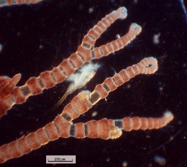
Another image |
Ceramium ciliatum with hair detail. |
||
|
|
|
||
|
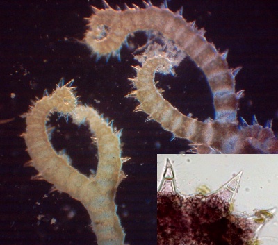
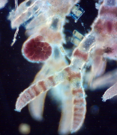
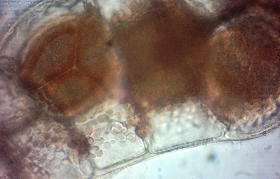
TRICHOBLASTS : they are filamentous extensions carrying male gametes : here they are probably scattered. |
TRICHOGYNES on a ceramium to catch the spermatia released by male gametophytes. Note that male and female gametes are not mobile. |
||
|
|
|
||
|
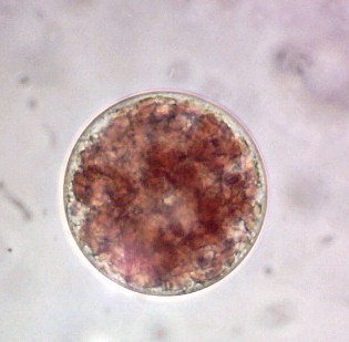
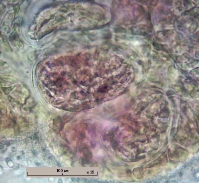
CARPOSPORES on left-hand picture the spores can be seen in transparency. (Rheinberg lighting) |
|||
|
|
|
||
|
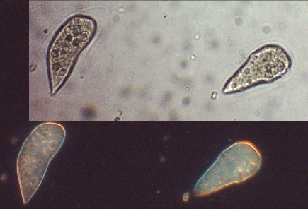
SPORES : each taken with x 40 objective. |
|||
|
|
|
||
|
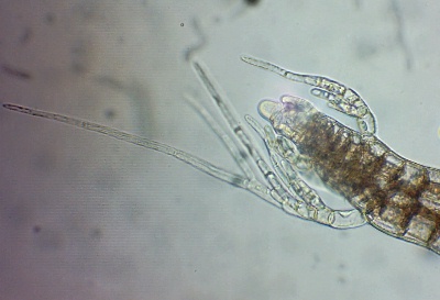
TETRASPOROPHYTES : tetraspores grow inside the cells of plants. |
|||
|
|
|
||
|
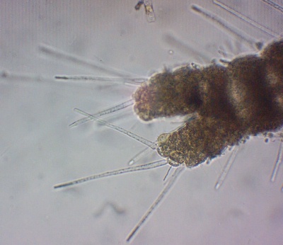
TETRASPORES : in left-hand picture spore division (tetrahedric here) is well seen! |
|||
|
I hope these pictures give a realistic vision on the different phases vital to the life cycle of these red algae. Interesting links - you can find other images at: Comments to the author Jean-Marie Cavanihac are welcomed.
All drawings and photographs © Jean-Marie Cavanihac 2005 Published in the September 2005 edition of Micscape Magazine.
Please report any Web problems or offer
general comments to the
Micscape
Editor, Micscape is the on-line monthly magazine of the Microscopy UK web site at http://www.microscopy-uk.org.uk |