POLYSIPHONIA
a red algae
by Jan Parmentier
POLYSIPHONIA a red algae |
|
||
by Jan Parmentier |
|||
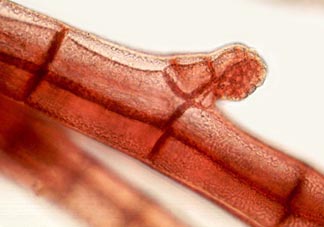
cystocarp beginning to form (see below for explanation) |
Red algae are specialized
plants with peculiar properties and as a consequence they
are studied by specialists. And specialists use a
professional language for the description of red algae,
not always easy for the amateur. The amateur behind the
microscope who wants to study red algae must take some
trouble to learn these terms. It pays, because red algae
are exquisite plants, often with a complex structure and
a life history with phases that are easy to see without
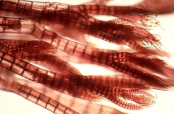
all sort of technical problems. The genus that we will treat here is easy to find; the photographs are from small plants, collected in spring at low tide from brown algae (Fucus species) in the Oosterschelde. Polysiphonia is to be found everywhere along the North Sea coast, on rocks, blocks of concrete, jetties and brown algae. There are 4000 known species of red algae, often present in tropical marine waters; in fresh water we will find only a few species. |
| Their colour is due to the red pigment phycobilin, masking the green colour of chlorophyll. This red pigment is very well suited to absorb the green and blue-green light in somewhat deeper sea water, where we find many of the red algae. Red algae are tough customers. Fragments of the plants, after surviving a winter or buried under sand can grow again into healthy plants if circumstances become better. Red algae are often attached to stones or other plants. The genus Polysiphonia is easily to recognise with a loupe or with the microscope. A "branch" consists of an axis of elongated cells, the central axis. Every cell of this axis is surrounded by a number of cells (ranging from 4-24) with the same length, the periaxial cells. The plants have a segmented appearance. The "branches" often have long hairs (trichoblasts) at the top, which can later on disappear. In the Netherlands we can find about 10, in the UK more than 20 species. Polysiphonia violaceae is very common; we always find it in small pools on other algae. Polysiphonia nigrescens is easily seen, also in winter, on stones at low tide. | |
| The sexual reproduction of Polysiphonia is a complicated process, but we will describe it here because the several phases can be seen very easily in living material. | ||
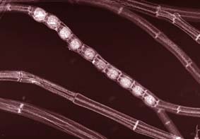
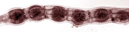
| We start the story with the so-called tetrasporophyte. That is a small Polysiphonia plant with the normal amount of chromosomes (2n), that is forming tetrasporangia, round balls, one at a segment, easily observable in the branches. Most often we see a small row. They are formed between the central cell of a segment and the periaxial cells. These balls divide into four parts; four spores are then formed with half the number of chromosomes (n). From these spores male and female plants are formed. (gametophytes). |
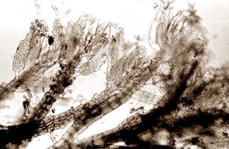
spermatangia |
|
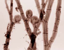
cystocarps |
Tetrasporophyte and gametophytes look identical, apart from the reproductive organs. The male reproductive organs are formed at the top of the branches of the gametophyte. These are the spermatangia, very small cucumbers (length 150 micron) crowded with spermatia. (We don't call them sperm cells because they don't have flagella, these are unknown in red algae). A spermatium, not capable to swim, needs water currents to move to the female reproductive organ, the carpogonium. A spermatium can melt together with the carpogonium. The nucleus of the male cell moves through the tube of the trichogyne (a trichogyne is a hair like receptive protuberance of the carpogonium) to the egg cell and a zygote is the formed, that develops into a cystocarp. |
| In the cystocarp carpospores are formed (with 2n chromosomes), which can escape through a hole at the top and can grow into a tetrasporophyte. From April to September we can find several phases of this reproductive cycle. The tetrasporophyte is easily recognisable; with a 20x objective we see the four spores (most of the time we see only three of them, the fourth one is on the backside). |
tetrasporangia |
 |
And roaming along the sea shore will give us ample opportunities to find other red algae, always very beautiful under the microscope and always with an interesting, complicated life cycle. We will observe diatoms attached to the red algae and many other small organisms and from then on we are lost! |
| References: 1. C.A .Maggs, M.H. Hommersand, Seaweeds of the British Isles ,Vol.1, Rhodophyta, London, The Natural History Museum, HMSO 1993. 2. H.Stegenga, I. Mol, Flora van de Nederlandse zeewieren, KNNV 1983. |
Comments to the author Jan Parmentier are welcomed. Visit Jan's HOME PAGE All Material Copyright: © Jan Parmentier
Microscopy UK Front Page
|