
|
PART 3 of 3 - Other species |
|
||||
| Ceratium, shown on the previous page is only one genus of dinoflagellates. Here is a gallery of species from other genera. Probably, (certainly!), I have made some errors with my tentative identification to sub-species but nobody is perfect, especially in taxonomy; which is a domain for specialists! |
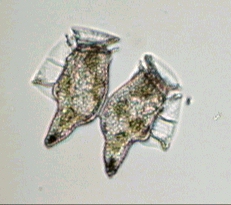
| Dinophysis: A 'rare' specimen, for me: I had to wait for more than 3 years to find one of them! Here is a specimen of Dinophysis caudata. Right hand picture shows reproduction in progress. | |
|
|
|
 |
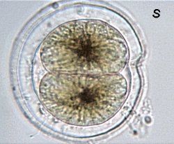
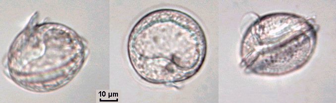
Another dinoflagellate species: Pyrophacus steinii. |
 |
| Some species are a little
more difficult to observe because of their smaller size and particularly
because they move very quickly.
Here too, it's necessary to see specimens at various angles to understand their morphology. Fortunately these specimens were moving and very cooperative in showing themselves on all sides! See video1 (animated gif).(They are a little less cooperative in stopping when you are taking pictures!) See video2 (animated gif). |
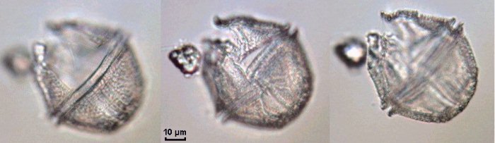
| This perforated specimen is probably a Gaunyolax scrippsae: Three levels of focus. |
 |
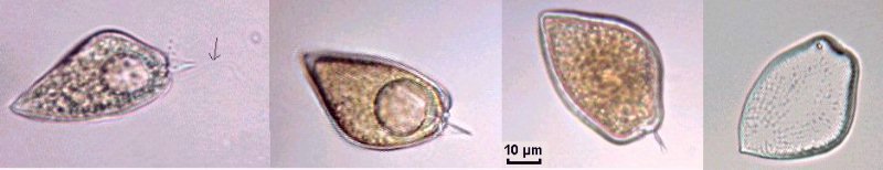
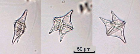
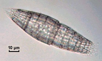
| Maybe a prehistoric arrow tip? No, only Prorocentrum micans: Note flagellum (arrowed) in the left hand picture and perforated theca in the last one. |
 |

Scriptiella, two specimens. |
|

Three images of Gonyaulax: Note the rounded nucleus, well seen in the first and second images of the same specimen. See video (animated gif). |
|
| Maybe Preperidinium: Three views of the same specimen : |
 |
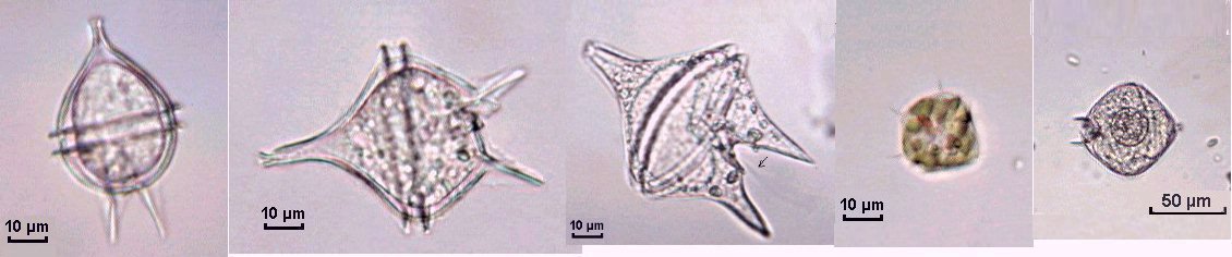
| Protoperidinium: Some sub-species. Three pictures of the same specimen moving on the slide: The first one shows a side view. Video clip (animated gif) shows other views, particularly in bottom view. The tranverse flagellum is arrowed in right hand picture below. | |
 |
 |
| It's often difficult to have in simultaneous focus all parts of the specimen. Some picture montages below (marked with an "S") include images created by summing of two (or more) images made at different depths of focus, using Astrostack software from R.J. Stekelenburg. Some months ago a letter from R. Comer was published on Microscopy-UK to describe this useful freeware. Note that the fourth image below is 'synthetic' but useful to have all the details in the same picture . | |
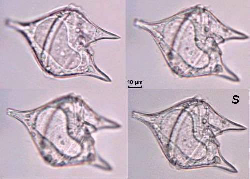 |
Picture marked with 'S' is a 'sum' (in fact a mean + sharpen convolution filter) of the three others taken at three levels of focus. |
| Other sub-species of Protoperidinium: S='sum' of the first two pictures. The second line shows three images of the same specimen moving under the objective. Maybe Protoperidinium depressum (?) |
 |
 |
| Gyrodinium: Three specimens of Gyrodinium spiralis species. The large nucleus is clearly seen. |
 |
| Gymnodinium: Note the large chromatophore containing carotene and the flagellum, which is best seen in the second picture (arrowed). | |
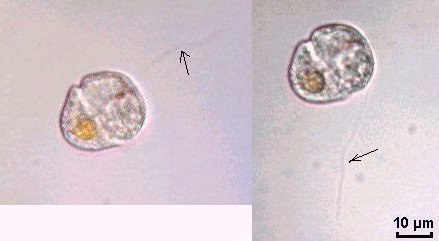 |
Other sub-species are shown below. Note a red spot near sulcus on specimen in the right hand picture. It's probably light sensitive. |
 |
|
| Amphidium
carterae (left hand image below)
and on the right, probably another species of Amphidium with a 'rocket'
shape: Note added March 2002. In fact, the species on the right below
is actually the ciliate Tiarina Fusus - many thanks to Alfred Beran
- Laboratoria di
Biologia Marine -TRIESTE - Italia, for the identification. |
|
 |
 |
 |
||||
|
|
|
|
|
|
| As you can see, the world
of dinoflagellates offers many species. This article shows some of the
more frequently encountered but it's certainly not exhaustive.
Just a note about collecting samples: Until now I used a plankton net, (made from a piece of thin curtain), but the mesh size is ca. around 500 µm and I didn't find the smaller species. I have now changed my method; I use a thin coffee filter (made from soft plastic and with a mesh size around 100 µm) and I pour through this filter, the contents of 3 or 4 buckets of seawater. Then I rinse the filter inside out into the sampling jar. A stereo microscope (x20 enlargement) is useful to sort a sample. I already had some pictures to illustrate this article,
but this year (2001) seems to be favorable to dinoflagellate growth (seawater
temperature was around 24/25°C), and I have caught more than one hundred
of them during this summer. I have found some specimens I had never seen,
but it's possible I was more attentive because I was focused on these species!
To see more dinoflagellate pictures, visit the web sites linked to below: |
|
|
RETURN TO PAGE 2 |
|
Microscopy
UK Front Page
All drawings and photographs © Jean-Marie Cavanihac 2001 Published in the September 2001 edition of Micscape Magazine. Please report any Web problems or offer general comments
to the Micscape
Editor,
Micscape is the on-line monthly magazine of the Microscopy
UK web
|