Microfossil and Nannofossil Image
Gallery
by
Keith Abineri, UK
|
Introduction
Microfossils and nannofossils have
been very much in the news recently. For example, there is still
a hot debate over whether the microscopic features found in an
Antarctic meteorite originating from Mars are evidence of past
life on that planet. Linked to these debates and the search for
extraterrestial life is the question 'how small can a living
organism be?'. Australian researchers who
recently found what are believed to be the smallest living
nannobacteria in rocks deep beneath the earth's surface may force
scientists to review how they define the smallest living
organism.
These debates revolve around
organisms or their fossilised remains visible under the electron
microscope, but many microfossils and nannofossils can be
observed by amateur microscopists using modest optical
microscopes and simple preparation techniques i.e. without the
need to prepare thin rock sections.
To illustrate this, shown below is
an image gallery of a selection of microfossils and nannofossils
that can be found in rocks of the Dorset coast, Southern England.
Many are typical of rocks of this type that can be found
elsewhere. The fossils can provide evidence of the habitat and
climate type that was present when the microscopic organisms died
and slowly fossilised.
The images were taken not of the
rocks but of cellulose peels of prepared rock surfaces, a
technique that is well within the scope of the amateur
microscopist/geologist. Cellulose peels are prepared by painting
the rock surface with a lacquer, that when dry can give a
faithful replica of a rock surface (a 'peel') down to the tiniest
structures, as well as some removal layers of rock material. The
rock can be stained or unstained prior to making the peel to
clearly show certain structure types. Keith Abineri's earlier articles explain the technique in more detail and what can be
deduced from the fossils found.
Image gallery - specimen
locations and image details are in the Appendix.
Terms underlined are
explained in the Glossary.
|
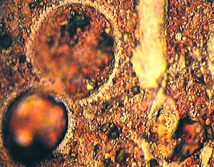
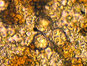
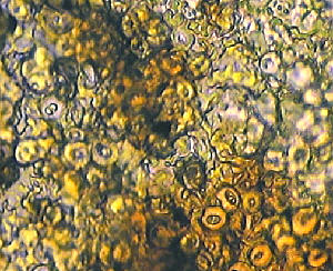
Fig.
1: Globigerinid
forams, other forams, sponge
fragments, calcispheres and nannofossils in chalk.
Stained cellulose lacquer peel, examined under PPL
brightfield illumination. It shows both thin removed
layer images and replica images. The absence
of any blue or mauve staining indicates aerobic
conditions during the deposition of the chalk, in common
with other Upper Chalk microfossils and nannofossils.
Size of the large foram chamber = ca. 93 mm. Limiting
resolution of nannofossils = ca. 2 mm.
|
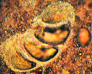
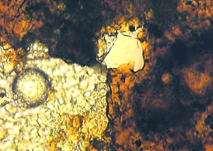
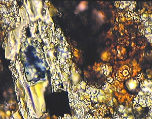
Fig.
2: Large damaged foram section, calcispheres,
chalk nannofossils etc. in chalk. Stained cellulose lacquer peel, examined under PPL
brightfield illumination. It shows both thin removed
layer images and replica images. Length of foram chambers
= ca. 110 mm. Limiting resolution of nannofossils = ca. 2 mm.
|
|
|
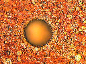
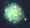
Fig.
3: Chalk calcisphere section on a background of
damaged coccoliths and other nannofossils. Stained cellulose lacquer
peel, examined under PPL illumination. The absence of any
blue or mauve staining indicates again aerobic conditions
during the deposition of the chalk. The diameter of the
calcisphere, shown here, including the complex outer
layer, is ca. 42 mm. The limiting resolution of the coccoliths and
nannofossils is about 1 mm.
|
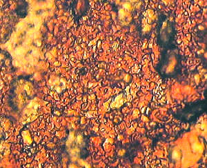
Fig.
4: The fine structure of the chalk is shown
here, including partly buried and damaged coccoliths and
other nannofossils. Stained cellulose lacquer peel, examined under
PPL brightfield illumination. The absence of any blue or
mauve staining indicates once more aerobic conditions
during the deposition of the chalk. The size of the
coccoliths, other nannofossils and fragments range from
<l mm to 9.3 mm in diameter.
|
|
|
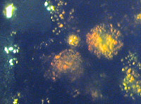
Fig.
5: Coccoliths and coccolithophores,
including some buried in kerogen. Unstained cellulose
lacquer peel, examined under PPL brightfield
illumination. The picture shows both thin removed layer
images and replica images. The three centre
coccolithophores range from ca. 14 to 16 mm in
diameter, and the coccoliths on their surfaces are about
4 to 5 mm in diameter. The numerous free coccoliths in
the field range from about 2 to about 5 mm in
diameter.
|
|
Fig. 6:
Coccoliths and coccolithophores buried in kerogen and
partly not covered. Unstained cellulose lacquer peel,
examined under PPL brightfield illumination. The picture
shows both thin layer images and replica images. The
large uncovered coccolithophore in the lower left-hand
field has a diameter ca. 23 mm. There are
indications of similar features buried in the dark
kerogen in the right-hand field. (See figure (7)). |
| Fig. 7:
This shows features from the field in Figure (6), but
using XPL illumination to indicate the optical figures of
coccoliths and coccolithophores. Note the pale yellow
colour of the objects embedded in kerogen (lower image).
The thick kerogen layer appears very dark. Uncovered
objects show bright white optical figures. The diameter
of the largest buried coccolithophore in the right-hand
field of Fig. 7 is here also ca. 23 mm (upper image).
Note also from comparison between figures (6) and (7)
that some of the coccoliths in the left-hand field are
replica images. |
|
|
Fig. 8: Numerous
coccoliths including some embedded in a layer of kerogen.
Unstained cellulose lacquer peel, examined under PPL
brightfield illumination. The picture shows both thin
removed layer images and replica images. The numerous
coccoliths have diameters in the range from ca. 3.0 to
6.0 mm. The most detailed images appear to be those
coccoliths embedded in the kerogen. |
| Fig. 9: A complex
field including coccoliths and coccolithophores, some of
which are embedded in kerogen, together with some fusain
and pyrites, as well as a replica of part or a
fragment of wood. Partly stained cellulose lacquer peel,
examined under PPL brightfield illumination. The complex
picture shows both thin removed layer images and replica
images. Some of the staining was probably inhibited by
the presence of kerogen. The coccoliths ranged in
diameter from ca. 3.0 to 6.5 mm. Note the small
area of ferroan calcite associated with the wood
fragment. The diameter of the distinct coccolithophore,
embedded in kerogen, at the central area of the
right-hand field is ca. 14 mm. The diameters of
its surface coccoliths are approximately 4.7 mm. |
|
Glossary
Aerobic : in
the presence of oxygen.
Anaerobic : in the absence of oxygen.
Calcareous nannofossils : Nannofossils
largely composed of calcium carbonate. Many forms belong to
the Coccolithophyceae.
Calcispheres : Minute hollow microfossils
and nannofossils of calcareous composition. Found frequently
in chalk and limestone sediments. They have existed in
differing forms since the Devonian Period (circa. 380 million
years ago).
Coccoliths : minute calcium carbonate
platelets secreted by coccolithophores which bear them as
surface plates.
Coccolithophore : a unicellular planktonic
organism of uncertain type (protozoan or algae?). Currently
assigned the phylum Haptomonada.
Ferroan Calcite : crystalline calcium
carbonate with a very small proportion of the calcium
displaced by iron(II) in the crystal lattice. This occurs
under anaerobic environments.
Forams : Foraminifera. A class of protozoa
that form calcareous shells. (See articles in the Micscape
on-line library by Brian Darnton).
Fusain : carbonaceous material derived from
decaying vegetation or wood.
Globigerinid forams : a foram of the genus Globigerina.
Kerogen : A solid complex organic material
which yields petroleum type hydrocarbons under heat and
pressure.
Microfossil : a fossil or fossil fragment
that can only be seen with a microscope.
Nannofossil : fossils of minute planktonic
organisms, especially calcareous unicellular algae.
Period: Jurassic - ca. 218 - 144 million
years ago; Cretaceous - ca. 144 - 65 million years ago.
Pyrites : a most widespread sulphide of
iron. A sure sign of anaerobic sediments.
Removed layer images : actual rock material
removed from rock surface by peel, e.g. kerogen, calcite etc.
Replica images : images fromed by
"printing" by cellulose lacquer; produce no XPL
images.
Staining : in the absence of iron (II) no
Prussian Blue is formed and indicates aerobic conditions.
PPL : plane polarised light (plane
polarising filter in the substage only)
XPL : cross polarised light (plane
polarising filter both in the substage and the eyepiece
aligned at right angles for maximum extinction).
Appendix
Details of the specimen
locations and objective used.
(Objective 40X - N.A. 0.65; objective 100X oil-immersion - NA
1.25).
Figure (1) and Figure (2).
From the Cretaceous, Upper Chalk, Actinocamax Quadratus Zone.
Map Reference SY.851.802. West of Arish Mell on the cliff,
Dorset coast. (Objective 40X).
Figure (3) and Figure (4).
From the Cretaceous, Upper Chalk, Actinocamax Quadratus
Zone Map Reference SY.851.802. West of Arish Mell on the
cliff, Dorset coast. (Objective 100X).
Figure (5).
From the Jurassic, Kimmeridge Clay, Rope Lake Head Stone
Band. Map Reference SY.926.775. Rope Lake Head, West of
Hounstout Cliff, Dorset coast. (Objective 100X).
Figure (6) and Figure (7).
From the Jurassic, Kimmeridge Clay, Maple Ledge Shales. Map
Reference SY. 908.790. East of Gaulter Gap, Kimmeridge Bay,
Dorset coast. (Objective 100X).
Figure (8) and Figure (9).
From the Jurassic, Kimmeridge Clay, Rope Lake Head Stone
Band. Map Reference SY.926.775. Rope Lake Head, West of
Hounstout Cliff, Dorset coast. (Objective 100X).
Editor's note: Some of the quality of
the author's original 35mm slides is lost in the scanned and
compressed web images. Comments to the author are welcomed, who
can be contacted directly at the address below or comments can be
passed on via the Micscape Editor, see magazine index contact..
Keith Abineri. 42 West Borough,
Wimborne, Dorset BH21 1NQ, UK. Tel. 01202 885547
Prepared for the Web by
David Walker. Introduction by David Walker.
© Microscopy UK or their
contributors.
Published in the October 1999
edition of Micscape Magazine.
Article at
http://www.microscopy-uk.net/mag/artoct99/kamast5.html
Please report any Web problems
or offer general comments to the Micscape Editor,
via the contact on current Micscape Index.
Micscape is the on-line monthly
magazine of the Microscopy UK web
site at Microscopy-UK
WIDTH=1
© Onview.net Ltd, Microscopy-UK, and all contributors 1995 onwards. All rights
reserved. Main site is at www.microscopy-uk.org.uk with full mirror at www.microscopy-uk.net.