Diatoms are a popular subject
for many amateur microscopists. Living specimens are particularly attractive
to observe and a number of Micscape articles have described and illustrated
them. (See links in Appendix).
The diatoms are a phylum of algae
(protists) with silica shells which have well defined markings. The markings
aren't always evident in living species but prepared slides of the empty
shells are available where this detail can be clearly seen. Depending on
the species the fineness of the markings varies and some species have markings
close to the resolution limit of the optical microscope. Therefore diatoms
are a favourite subject for assessing various aspects of the optical performance
of a microscope. These include:
-
assessing the relative performance of
objectives e.g. resolution (N.A.), flatness of field, the maker
or type of objective
-
comparing lighting methods e.g. brightfield,
phase
contrast
-
checking the performance of ancillary
equipment like image capture
-
ensuring a microscope has been setup
for optimum performance; diatoms are unforgiving of poorly set up lighting,
incorrect condenser settings etc
-
assessing the fine focus and mechanical
stage controls
-
if planning to buy a microscope using
a set can test most features of the microscope
A number of microscope slide suppliers
can offer either test sets or individual species (see Appendix).
As an illustration, here's how my
two microscopes faired. My main 'scope is a typical budget model - the
Russian Biolam 'R' stand (ca. 1973) with achromatic objectives but
I also own a Cooke, Troughton and Simms
(CTS) M2000 (ca. 1945) with achromatic phase objectives which
is probably comparable to a modern budget to mid-priced stand.
The species names are a bit of a
mouthful but those below are some of the most popular and will be present
in most test sets or available separately.
The images were captured directly
from a cheap B&W security video camera and 'Snappy 2.0' capture card
so the images won't be as crisp as a 35mm slide or visual view. Note that
the green filter (and infra red filter) used gives a rather flat image.
The rough sketches are not to scale
and just show some of the main features.
(An excuse for my poor drawing skills!).
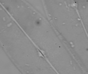
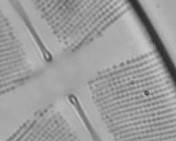
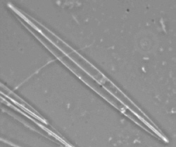
Stauroneis phoenicenteron -
a
good test for low to mid power objectives
Approx. 150 mm
long, lines of dots ca. 0.72 mm
apart.
|
-
a 10x - 20x objective should give a
crisp image of the diatom's main features and show the unresolved dots
as fine lines
-
a 40x dry objective should resolve the
lines into rows of dots
-
if the diatoms are well mounted - good
for checking field flatness and focus with a 40x objective
|
| Right: the CTS 20x
N.A. 0.45 objective was used right but the Russian 20x N.A. 0.40 gives
a comparable view. The camera just captures a hint of the lines from the
clearer visual field. |
|
|
Left: the Russian
dry 40x N.A. 0.65 objective with normal brightfield lighting resolves the
dots. Although not as crisply as the CTS 45x objective. |
Pleurosigma angulatum -
a classic test object for mid powers
Aprox. 150 mm
long, lines of dots ca. 0.53 mm
apart.

|
-
a good subject to show that contrast
as well as resolution is important in microscopy
-
a 40x dry achromatic objective should
resolve the dots but contrast enhancement often needed. Good for assessing
lighting techniques (e.g. oblique or darkfield)
-
with a 60x+ objective the dots should
be clearly resolved
-
poor optics or setups may just show
fine hatchings of lines
|
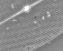
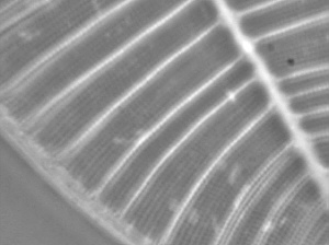
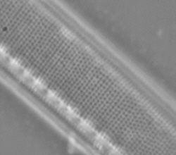
Surirella gemma - a test
for mid to higher powers
Approx. 100 mm
long, lines of dots ca. 0.5 mm
apart.
|
-
a 40x objective should give crisp views
of the diatom and the heavier white borders
-
the finer lines may also be seen
-
a good 40x objective should resolve
the lines to dots, but if not a 60X+ objective should clearly resolve the
dots
|
| The Russian dry 40x
and the CTS 45x oil objective both show the fine lines and the CTS is also
good enough to resolve the lines to dots (shown right, clearer visually
than this image capture). |
|

|
Left: at 60x+ the
dots should be well resolved. The CTS 95x objective does this well. Focusing
in and out gives a black and white contrast reversal effect. |
Nitzschia obtusa - one
of the easier tests for highest powers
Approx. 200 mm
long, lines of dots ca. 0.3 mm
apart.
|
-
a long straight species good for assessing
field flatness at higher powers if the specimens are well mounted
-
a 95x - 100x objective should clearly
resolve the dots
|
| Right: the dots are
clearly resolved with the CTS 95x N.A. 1.3 fluorite. Also shows good field
flatness as the depth of field is minute at this magnification. |
|
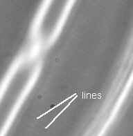
Frustulia rhomboides var.
saxonica
-
the 'going gets tough' for higher
powers
Approx. 50 mm
long, lines of dots ca. 0.3 mm
apart.
Quite a handful as a species name
and quite a handful as a test subject, as this is at the limit of the Russian
and CTS microscopes' capabilities.
Note added 2002: Thank you to
Frithjof Sterrenburg who kindly pointed out the change of taxonomy of F.
rhomboides. Read Frithjof's fascinating Micscape article 'A
second look at some well known test diatoms' where this is discussed.
| Right: with the CTS
95x objective and using the better corrected Russian aplanatic N.A. 1.3
condenser (the CTS phase condenser is only N.A 1.0)
and oiling the slide to the condenser, the lines (perpendicular
to the diatom's long axis) are visible and just
about captured on the CCD camera. The dots aren't resolved visually. |
|
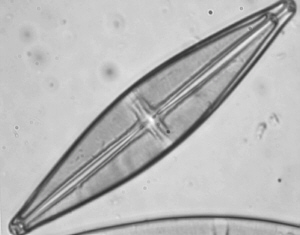
Amphipleura pellucida
-
only
the very best optics will 'crack' this one
Approx. 80-140 mm
long, dots ca. 0.27 mm
apart.
| Right: general view
of this diatom with the 45x CTS objective under phase contrast. Fine lines
perpendicular to the long axis are resolvable with the best 100x objectives.
Resolving these lines to dots pushes the finest optics and light microscope
to the limit. |
|
This species is included in the diatom
test set I used but neither the Biolam or CTS optics are good enough to
resolve the lines or dots! A very good system should show the lines of
unresolved dots. The dot separation is near the resolution limit of an
optical microscope although the finest optics may resolve the dots. Have
a try if you own a very high quality 95x -100x objective and equally important
- a very well corrected substage system to achieve the maximum potential
of the objective. Oiling the condenser to the slide as well as the objective
to the slide is necessary (a messy business!) and probably a blue filter
and a high mag. eyepiece e.g. 25x. If you succeed with A. pellucida
or have a better image of F. rhomboides, why not share your skills
and send
Micscape an image and we will be pleased to include it here with credits.
As an aside and a humbling thought,
apparently microscopists first resolved this species to dots in 1868 so
illustrates how fine optics were in the late 19th century! (See link below).
Other comments
To achieve the best performance from
an achromatic objective it can be used with a yellow-green filter which
minimises the uncorrected aberrations. Some of the finest photographs I've
ever seen of diatoms were taken with achromatic objectives with a plate
camera in 1904.
Diatoms are a good subject for understanding
the importance of the correct condenser iris setting (not
to be confused with the field iris which may be present on the lamp).
Inspecting the image as the iris is moved from fully open to fully closed
will show the effect on resolution and image quality.
Image capture for 'diatom dotting'
also gives a good 'workout' for the image capture system, whether photo'
film or in this case digital capture with a security CCD camera.
The cheap B&W camera used (>560 line resolution) coped well and is
less than £100 new from electronic suppliers in the UK. Arguably
colour isn't essential for high mag. or phase work and a cheaper route
into image capture (stills or video) than colour.
All the above captures were taken
with the camera fitted to the eyepiece tube without any eyepiece. So despite
the fact that the CTS objectives require compensating eyepieces
the camera captured the features quite well.
Comments to the author Dave
Walker (an amateur microscopist) are welcomed.
Acknowledgements
In addition to my own observations
the leaflet accompanying the NBS diatom set 6/AK prepared by Eric Marson
of Northern Biological Supplies was valuable in preparing this article.
Also the measurements quoted in the http://glinda.lrsm.upenn.edu/diatom.html
link below.
Appendix
Related Micscape articles on test
diatoms
'A
second look at some well known test diatoms' by Frithjof Sterrenburg.
Test
diatoms - what you can expect to see even with modest objectives by
Martin Mach.
Image
gallery, two test diatoms by Will Varnell.
For other illustrated Micscape articles
on the diatoms and their many beautiful forms, type 'diatom' in the Micscape
Library search index. A good introduction to diatoms by Wim van Egmond
is here.
Further reading
1) The measurement of
three light microscope test diatoms by scanning electron microscopy
by J B Sanderson.
Royal Microscopical Society
Proceedings, Vol 25/3, May 1990, pages 195-203.
Many thanks to J B Sanderson for
sending me a copy of this fascinating article, which includes historical
aspects of using test diatoms and ruled test-plates for assessing microscope
objective performance.
External links of interest
-
bio-microtech.com/info/articles/article1.htm
- 'Test Slides of Diatoms to Divisions - What Are You Looking At?'
by Tim Richardson. An interesting article looking at the history of test
slides as well as suggesting a new design.
http://glinda.lrsm.upenn.edu/diatom.html
- has an image of the general forms of common diatom test species and relevant
measurements.
http://www.wfu.edu/~gholz/research/
- a technical paper (on a contrast enhancement technique) with some nice
illustrations of diatoms.
http://www.technicalvideo.com/Resolution.html
- a page describing a special light source, but illustrated with Amphipleura
pellucida images to show how well the technique defines the markings.
Test slide suppliers
Northern Biological Supplies,
UK sell test set 6/AK. This is the set the author used and includes prepared
slides of six diatom species with an illustrated instruction leaflet describing
the features that should be seen with different objectives. This slide
set can be ordered by contacting OnView.
Carolina
Biological Supplies, US offer set WW-29-5984 which is a diatom test
plate showing eight species.
Klaus
Kemp, Microlife Services, UK also offers a variety of diatom slides,
and is one of the few people skilled in the art of arranging diatoms into
patterns.
Glossary
N.A. - numerical aperture;
marked on the objective and the higher the N.A. the better its resolving
power.
achromatic - many microscopes will
include as standard a set of achromatic objectives. Chomatic aberrations
are corrected for two colours and spherical aberrations for one (yellow-green).
fluorite - a corrected objective
intermediate between an 'achro' and 'apo'. The CTS 45x phase objective
is a fluorite.
apochromatic - expensive objectives
where chromatic aberrations are corrected for three colours and spherical
aberrations for two.
plan - indicates the objective is
flat field i.e. gives sharp focus across the field of view. Both achromatic
and apochromatic objectives can come in plan forms.
aplanatic - a better corrected condenser
than the commonest Abbe design. The most highly corrected condensers are
aplanatic-achromatic and demanded by the very best objectives.
phase contrast - a contrast enhancing
technique that uses special objectives (often marked Ph.) and a matched
condenser. Phase systems are available for most modular microscope stands.
compensating eyepieces - some objectives
are designed to be used with the makers special eyepieces to help correct
the aberrations.
CCD - charge coupled detector. The
image sensor of modern video cameras, camcorders and digital stills cameras.
© Microscopy UK or their contributors.
Published in the October 1999
edition of Micscape Magazine.
New links added and some notes in August 2002.
Please report any Web problems or
offer general comments to the Micscape
Editor,
via the contact on current Micscape
Index.
Micscape is the on-line monthly magazine
of the Microscopy UK web
site at Microscopy-UK
WIDTH=1