Cooke, Troughton and Simms microscopes seem to be held in high regard by their present owners, and two of their models have been described in articles as 'my favourite microscope' (ref. 1 and 2). The model that hasn't been mentioned in this vein to my knowledge is the M2000 stand, but as I was the proud owner of an example, I offer my thoughts on 'my favourite microscope' below.
The parent British companies of Cooke, Troughton and Simms Ltd (referred to as 'CTS' hereinafter) had a long history with J. Troughton (senior) opening a business as an instrument maker in 1756. 'CTS' was formed when T. Cooke and Sons Ltd (controlled by Vickers since 1915) purchased Troughton and Simms Ltd in 1922. 'CTS' was completely taken over by Vickers in 1924 but retained their own name. In 1963 they became part of Vickers Instruments Ltd (ref. 3). Cooke, Troughton and Simms Ltd finally ceased trading in 1988 (ref. 4). Various aspects of the somewhat complex history of 'CTS', the parent companies and Vickers Instruments are discussed in references 3-6.
The 'CTS' range
'CTS' had a systematic way of designating
their microscope range as described in their catalogue issued ca. 1950
(ref. 7, introduction dates below from ref. 3) viz.:
- M1005/1025 - student's microscope
- M1000 - a 'general purpose' microscope (introduced 1946)
- M2000 - a microscope for 'routine and research investigations' (introduced 1942)
- M3000 - as for the M2000 with vertical adjustment to the stage (introduced 1946)
- M4000 - the 'universal stand' for visual and photographic examination (introduced 1944)
- M6000 - stereoscopic microscope (introduced 1946)
- M7000 - polarising microscope, a range based on the M1000 and M7000 stand (introduced 1947)
 The serial number on each microscope often incorporates the stand number
and if present usually allows the type of stand possessed to be identified.
The serial number on each microscope often incorporates the stand number
and if present usually allows the type of stand possessed to be identified.
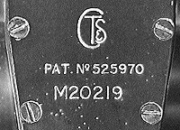
Image left shows the 'CT&S'
logo often used on their stands, eyepieces and accessories. The serial
number indicates it is based on the M2000 stand.
 General
features
General
features
I was fortunate to acquire my M2000
stand for a very modest sum when it became a cast-off at work. My model
is equipped for phase contrast and has a set of phase objectives, a bright-field
/ phase condenser with centring telescope.
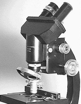
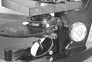
The image right shows some of the main features. It is a moving body tube design with a 160mm fixed tube length (although see notes on the fine focus below). It is finished in a satin black enamel with chromed controls and although not a large stand (at 35cm high), it is heavy (ca. 6Kg) and very stable. The body tube with inclined binocular head is on a sliding brass dovetail which can be removed and replaced with a vertical monocular head (which I also possess). Each head is held in place by a knurled screw on the left of the stand near the fine focus. The large coarse focusing and condenser knobs allow easy manipulation of the controls. This model has the M1336 mechanical stage with vertical adjustments, which is probably not as convenient as the alternative M1341 stage illustrated in the catalogue with horizontal side mounted controls.
Focusing
The feature most 'CTS' microscope
owners comment on is the fine focus, and the author is no different in
this respect .... the fine focus is superb. 'CTS' in their catalogue (ref.
7, p. 68) succinctly describe the focus as follows.
'The fine focus mechanism which is of a novel design, moves in ball bearings and is frictionless and "dead beat" in operation. A sector takes the place of the usual bell crank lever and carries an involute cam which imparts a uniform movement to the slide, a division of the scale thus has exactly the same value throughout the whole range of movement.'
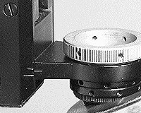 The fine focus only acts on the objective changer and nose (see image right)
thus the mechanism is not overloaded by the weight of the binocular head.
One division on the graduated focus knob corresponds to a 2µm vertical
movement. The movement is limited by stops to 1mm each way and shown on
the stand by limit lines. 'CTS' state that the slight change in the tube
length when fine focusing is not sufficient to affect optical performance.
The motion is so lightly loaded that accidental contact of the objective
and slide is not sufficient to damage the slide.
The fine focus only acts on the objective changer and nose (see image right)
thus the mechanism is not overloaded by the weight of the binocular head.
One division on the graduated focus knob corresponds to a 2µm vertical
movement. The movement is limited by stops to 1mm each way and shown on
the stand by limit lines. 'CTS' state that the slight change in the tube
length when fine focusing is not sufficient to affect optical performance.
The motion is so lightly loaded that accidental contact of the objective
and slide is not sufficient to damage the slide.
This type of fine focus was also used in the M3000, M4000 and the more advanced polarising stands of the M7000 series but not in the M1000 and simpler M7000 stands, which used a conventional fine focus with bell lever crank.
Image right above shows the objective changer and fine focus limit lines. (Body tube removed for clarity).
 |
Reproduced from ref. 7, page 68. |
 |
 Substage
The condenser substage is also rack
and pinion with a cylindrical (38.6mm diameter) simple clamp fitting for
the condenser. The phase condenser supplied (an Abbe condenser N.A. 1.0)
is non-centring although the phase rings selectable on a rotating disc
are of course centrable.
A useful feature is that the plano-convex
mirror can be clamped on two axes to avoid knocking, whereas on my Russian
Biolam microscope it is easy to knock the mirror when changing filters.
Objectives
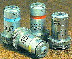 When I obtained the microscope it had 20X (N.A. 0.45) , 45X (oil, N.A.
0.95) and 95X (oil, N.A. 1.3) achromatic phase objectives. There was a
10X phase ring on the condenser and I had spent some years waiting for
a 10X 'CTS' phase objective to turn up on the second-hand market. My patience
was recently rewarded and I was able to purchase one for less than ten
pounds to complete the set. The objectives are shown right, and although
the 10X (N.A. 0.28) objective is slightly newer in style to the original
series it is close enough!
When I obtained the microscope it had 20X (N.A. 0.45) , 45X (oil, N.A.
0.95) and 95X (oil, N.A. 1.3) achromatic phase objectives. There was a
10X phase ring on the condenser and I had spent some years waiting for
a 10X 'CTS' phase objective to turn up on the second-hand market. My patience
was recently rewarded and I was able to purchase one for less than ten
pounds to complete the set. The objectives are shown right, and although
the 10X (N.A. 0.28) objective is slightly newer in style to the original
series it is close enough!
The coloured bands were not used by 'CTS' to specifically indicate phase objectives, but as they state 'the colours have been arranged in order of the spectrum, so that the colours representing the various powers may be more easily memorised'. The fluorite objective was in addition marked with a black band to aid recognition. 'CTS' also marketed a range of other objectives including apochromatics and objectives for dark ground illumination of opaque objects.
The objectives are complemented by a pair of 8X and 12X compensating eyepieces.
 I'm not in a position to comment on the quality of the optics compared
with other manufacturers of this era, but to me the 45X (oil, N.A. 0.95)
fluorite phase objective in particular is a real delight. It was a revelation
when I first looked at the classic diatom test slide Pleurosigma angulatum
with this objective using bright field. The dots were very clearly resolved,
whereas although the Russian 40x achromatic objective (dry, N.A. 0.65)
on my Biolam stand can resolve them, it requires contrast enhancement such
as oblique illumination to see the dots convincingly.
I'm not in a position to comment on the quality of the optics compared
with other manufacturers of this era, but to me the 45X (oil, N.A. 0.95)
fluorite phase objective in particular is a real delight. It was a revelation
when I first looked at the classic diatom test slide Pleurosigma angulatum
with this objective using bright field. The dots were very clearly resolved,
whereas although the Russian 40x achromatic objective (dry, N.A. 0.65)
on my Biolam stand can resolve them, it requires contrast enhancement such
as oblique illumination to see the dots convincingly.
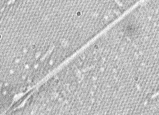
As an experiment the image left above shows the dots of this diatom using the 'CTS' 45x objective and 8x eyepiece in bright-field from an image captured direct to the PC via a video camera. This is pushing my outdated capture card and modest colour video camera to the limit, the visual image is crisp and clear! (The horizontal field of view shown is ca. 20µm with dots ca. 0.5µm apart).
The 45x fluorite objective was specifically intended for medical workers and described by 'CTS' (ref. 7, p. 105) as 'the ideal lens for examining blood films for differential counts and stained sputum for tubercle bacilli. It has a large field, long working distance, brilliant detail owing to its high numerical aperture, and great depth of focus.'
Final thoughts
I don't regard the M2000 as an elegant
stand, but more as a good example of quality optics and engineering in
an era before the rapid decline in British microscope manufacture. (As
an aside, my vote for an elegant microscope would probably go to the Wild
M20, Zeiss Universal 1 or the current Zeiss Standard KF2 student stand).
The 'CTS' microscope I have, has to my knowledge never been stripped down
and serviced, yet the mechanical and optical components are still in sound
condition although showing some wear and tear after 40+ years. Like many
microscopes of this era and earlier they still have a lot of life in them,
and more importantly give great pleasure to the amateurs like myself who
regularly use them.
Comments to the author Dave
Walker welcomed.
| Please note: Unfortunately the author cannot help with sourcing information on Cooke/Vickers instruments, or on obtaining spares or manuals. |
References and Further Reading
1) Cooke, Troughton and Simms Polarising Microscope by A. Burton, Balsam Post, 1997, no. 36, pp. 18-19. ('Balsam Post' is the quarterly magazine of the Postal Microscopical Society, UK).2) Vickers 'CTS' 400/4000 Microscope by N. N. Bradpiece, Micro Miscellanea, 1994, no. 28, pp. 8-10. ('Micro Miscellanea' is the Newsletter of the Manchester Microscopical and Natural History Society).
3) 'Notes on Modern Microscope Manufacturers' by B. Bracegirdle, Quekett Microscopical Club, 1996, p. 24.
4) 'Instrument Makers to the World; A History of Cooke, Troughton and Simms' by A. McConnell, Sessions, York, 1992. See the publisher's Web site for details.
5) 'A History of Vickers Instruments' Microscopes - Part 1' by A. J. Munro, Microscopy, 1980, 34, pp. 81-101. 'Microscopy' is the Journal of the Quekett Microscopical Club, now the 'Quekett Journal of Microscopy'.
6) 'A History of Vickers Instruments' Microscopes - Part 2' by A. J. Munro, Microscopy, 1981, 34, pp.162-183.
7) 'Cooke Microscopes', Publication No. CM1000A, Cooke, Troughton and Simms Ltd., York, ca. 1950.
On-line resources on CTS/Vickers microscopes: The author is not aware of any at the time of writing. If readers know of any I'll be pleased to link to them.
Image details
Images digitally captured direct to the PC via a Panasonic CL350 colour CCD camera (430 horizontal line resolution) fitted with Nikon SLR lenses (via adaptor) and using a Creative Labs Videospigot capture card. Image processing using Photostyler 2.0. Apart from the diagrams of the fine focus mechanism, microscope images were video stills of my microscope not scans from the catalogue.