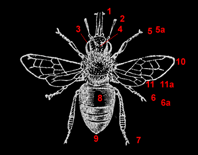
Key:
1 - mouthparts
2 - antenna
3 - compound eye cornea
4 - ocelli (simple eyes)
5 - first leg, 5a detail
6 - second leg, 6a detail
7 - third leg
8 - abdomen
9 - sting
10 - fore wing
11 - hind wing, 11a detail.
|
Interactive honey bee (Apis) worker Explore the parts of a honeybee.
The images are freely downloadable by David Walker, UK |
The honey bee worker has a number of fascinating features on the microscopic scale that enable it to fulfil its role in the bee colony. It's preferable to study these features 'hands-on' using live or preserved specimens of the worker (the latter available from biological suppliers) under a stereo microscope to appreciate the 3D structure in conjunction with prepared microscope slides of key parts which permit the smaller features to be more easily studied. A simple student compound microscope with a 5x - 10x objective with 10x eyepiece can show these features using prepared slides.
As a complement to these studies, images for viewing or printing out may be of use and some large detailed images are offered below. The images have intentionally not been annotated. The reader unfamiliar with the honey bee is invited to explore the many resources online and books to understand what is seen in these images. A good illustrated overview is in ref. 1 and probably later editions.
Each image gives a length for the longest dimension to give a sense of scale.
Note that they are images of prepared microscope slides so three dimensional parts when flattened won't represent their original shape (hence the importance of complementary studies of the whole insect).
|
|
Key:
1 - mouthparts |
Click on each red number above to see an
image of that part of the honey bee.
Close new window opened and/or save image to PC if
desired.
This
resource, including images, can be used offline if desired by downloading this
2.5 Mbyte micscape-honeybee.zip
file. Unzip and save to PC directory
of choice
and open dwapis.html in web browser.
Notes on images: They were created by scanning prepared microscope slides in a Minolta 5400 Scan Elite 35 mm film scanner. For notes on the use of film scanners for scanning microscope slides, see this article. The cornea image was taken on a microscope using a 10x objective and phase contrast. The first leg detail image was taken on a microscope using a 10x objective in brightfield.
The slides were prepared by the author using material kindly supplied by the late and sadly missed Eric Marson (Northern Biological Supplies) at one of his excellent slide making courses at Belstead House, Ipswich .
Notes on image use: The images can be freely downloaded for personal use, e.g. for part of a school project or by educators. Further distribution, commercial use or display on other websites is not permitted without the author's permission.
The worker bee drawing above is a 'negative' of the scanned Figure 181, page 204 'The Hive Bee (Worker)' from W S Furneaux's 'Butterflies, Moths, Other Insects and Creatures of the Countryside', published London, 1927.
Comments to the author David Walker are welcomed.
Suggested reading:
1) Animals Without Backbones Vol. 2 by R Buchsbaum. Pelican Books 1951 (and other editions). This classic two volume work has an excellent illustrated summary of the adaptations of a honeybee leg and how they are used to collect pollen.
Published in the July 2005 edition of Micscape.
Please report any Web problems or offer general comments to the Micscape Editor .
Micscape is the on-line monthly magazine of the Microscopy UK web site at Microscopy-UK
©
Onview.net Ltd, Microscopy-UK, and all contributors 1995 onwards. All
rights reserved.
Main site is at
www.microscopy-uk.org.uk
with full
mirror at
www.microscopy-uk.net
.