
|
A Sponge With A Tail by Richard L. Howey, Wyoming, USA |
Most of us are intrigued by unusual things around us. However, many of the most interesting are not readily visible. Some are in difficult locations and others are so small that they are accessible only with a microscope.
Now, I’m going to tell you a tale about a sponge with a tail–that’s how I’d start this off, if I were a fiction writer, but this sponge really exists. O.K., it’s not exactly a tail, but I think you’ll agree after seeing it that this is one very weird sponge. Technically, the “tail” is an anchor although I don’t think that this terminological shift helps us understand anything any better. Some monkeys have prehensile tails, others use tails for balance; lizards have tails which they sometimes sacrifice when attacked and then go off and grow a new one. All kinds of mammals have tails that serve a variety of functions. We might even say that the ciliate Urocentrum turbo has a tail; it has a bundle of posterior cilia which can secrete a kind of anchoring “glue” and then it spins like a whirling dervish. Some kinds of rotifers may be said to have tails; we talk about the tail of the flatworm Planaria, so why can’t a sponge have a tail? The point is that the notion of tail is not exactly scientifically rigorous, so we can feel free to talk about sponge tails.
On the specimen I have, the tail is almost 2 feet long, is a bundle of glass fibers, and is said to be, as a transmitter of light, a fiber optic, superior to anything we have as yet been able to produce. The “head” or main mass of the tailed sponge is a brown, amorphous mass which, when alive, housed countless numbers of tiny organisms which produced an incredible number of glass spicules in a variety of shapes and forms.
These present a real challenge photographically. 1) They are silica, that is, essentially glass, so achieving contrast becomes a major issue. 2) Many of the spicules are quite small but, nonetheless, have considerable depth–especially hexactinids or six-sided spicules. 3) They occur in intricate, interlocking networks which must be dismantled to examine individual spicules. 4) Clearly, spicules fuse to form these complex matrices and trying to figure out the relationship of individuals in a fixed matrix is an observational and logistical nightmare. The “tail” consists of a collection of thin glass fibers which have better light transmission than any optic fiber we have yet created. Why these should have the property to conduct light is unknown. In the process of collecting these specimens, the “tail” is usually cleaned of its surrounding detritus which actually forms a sort of sheath around the bundle which means that in its natural state, if there were light transmission (perhaps some sort of bioluminescence) virtually no light would be visible. To grasp the magnitude of the strangeness of these fibers, we need only remind ourselves that these are being produced by a colony of animals!
In the main “body” of these creatures, there is a considerable variety of spicules which are composed of silica, thus the designation “glass sponge”. A distinctive type that is definitive for this group is the hexactinellid or six-pronged spicule.
In the remainder of this article, I will show you some of the types of spicules which I have found thus far. For the sake of convenience, I have provided some of these with my own descriptive (some might say, whimsical) names.
The image below appears to be a cross, but it is in fact an hexactinellid. From the center there is another prong extending directly up at the observer and another on the other side extending down.

In 2009, I submitted an image to the Nikon Small World Contest and to my astonishment ended up with an Image of Distinction Award. (“I’m not bragging.” “Yes, you are!”–“No! I’m not!”–“Yes, you are!”–Well, we all know that argumentative exchanges such as this one are endless.) In any case, it was an image of a “dumbbell” sponge spicule, 3 or 4 of which I found in the detritus at the bottom of a jar of miscellaneous preserved specimens and there was no clue as to what kind of sponge it had come from.
Well, recently while browsing on the Internet, I came across a drawing which looked very similar to my spicules, so I began checking further. Additional poking around led to the discovery that even the famous German biologist and illustrator of the 19th Century, Ernst Haeckel, had encountered this extraordinary creature and had drawn these “dumbbell” spicules. Here is an image of the spicule

For a significant number of these spicules or matrix sections, a computer image stacking program becomes essential.
So, let’s begin by taking a look at the whole organism. I’ll show you 4 images; the first being the entire sponge; the second, the entire sponge with a small ruler to give you a sense of its size; and the third, a closeup of the “head, and finally, a closeup of the tail showing that indeed, it is a bundle of long fibers.


I was curious as to whether or not there was any discernible substructure in the fibers that could be revealed using optical microscopy, since neither my wife nor my budget will let me have a Scanning Electron Microscope. Maybe when Bill Gates or Warren Buffet or Mark Zuckerberg give me a 10 million dollar grant, I’ll get one. In the meantime, I’ve had to improvise and try some eccentric techniques of my own. I knew that dyes wouldn’t actually stain the glass fibers, but I wondered if they might deposit on the fibers in such a way as to reveal some surface detail. I published a short article on this using Euplectella rather than Hyalonema. Here is a link to that essay, which is titled “A Note On Technique: Staining Glass Sponge Fibers”
I tried 5 different stains and 3 of them provided results that gave me more than I had anticipated. They did indeed reveal some surface detail of the fibers and a couple of bonuses. In some places, the deposition was such that I was able by shifting the angle of illumination slightly, to achieve refraction which suggested some possibilities that there might be an optically fairly complex internal structure to the fibers. Furthermore, this was reinforced at points where I had broken the fiber and the tip was uneven strongly suggesting layering, a kind of stratification in terms of the way the fiber was formed and the suggestion that the fiber was an assemblage which had been added to over and over again. It is, after all, rather unrealistic to imagine that this 2 foot long anchor was constructed all at once; it makes much more sense to regard it as a work in progress which was constantly being added to, but it’s important always to double check, triple check, retest and rethink, because Mother Nature is so full of unimaginable and preposterous tricks. Consider the platypus. When a preserved specimen was first brought back to England, most “experts” dismissed it as a hoax perpetrated by creative taxidermy.
Let’s consider the “head” of this bizarre sponge for just a moment. If we were to find such a mass in a tidepool (without the tail), then we might well glance at its amorphous character and say, oh yes, a sponge–nothing special there. That however will not be the case since most of the glass sponges occur at considerable depths. One of the many mysteries of this extraordinary group of creatures is the great morphological variation. Some of the glass sponges are beautifully structured and could win prizes for underwater architecture. Consider Euplectella, the Venus Flower Basket


or the wonderful “cup” sponges

or the type frequently found on Xenophora shells

Now, let’s look at some particular types of spicules which are frequently found in glass sponges.
Here is a tetract and they are abundant, but there is an anomaly here in that the shape is quite irregular and one would usually expect something more geometric in the way of a cross.

There are indeed spicules which look like crosses or swords and I’ll show you 2 examples. You may remember the first spicule which we looked at above. If we zoom in a bit and look at the center of the cross, we can see the darkness of that area with one bright spot in the center; this is because a prong is projecting directly up at us.

Here is an example of the “sword” spicule and if you look closely at the intersection, you can see the blur produced again by a prong extending directly upward.

Next is a hexact which characterizes this group of sponges as hexactinellids.

Another unusual form that shows up in these sponges is the “dumbbell” spicule.
As you can see these are fairly complex for a spicule and they are somewhat difficult to isolate without damage. Here are 2 more views and in the second, you will also see some monaxons and hexactids.


This is a type which I encountered before I had any access to glass sponges. I found a few spicules at the bottom of a jar of miscellaneous specimens which didn’t include any glass sponges. At the time I was very puzzled, but I did manage to get a decent image of one by stacking 26 images. I realize that I already showed it to you, but I’ll let you see it again, since it underscores the very strange world of glass sponge spicules.

One of the things that fascinates me most about these creatures is the wild variety in the morphology of the spicules. This next one is a pentact which I call the “Christmas tree” spicule.

And here is another view along with a large spicule which may or may not be a pentact, but which certainly has a highly unusual form.

The next spicule is highly unusual and I have found only a very few of these. They rather remind me of the Japanese Shuriken or “throwing stars”.

Another anomalous type which is extremely small is a band-shaped form which is also rather scarce in the samples I examined.

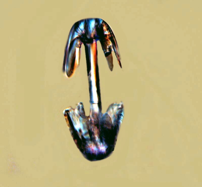
Some of you may be familiar with an “anchor-shaped” spicule which occurs in the genus Synapta of the sea cucumbers and is, therefore, calcareous rather than siliceous.

Not to be outdone, glass sponges also have some anchor spicules, albeit of a quite different sort. These are, of course, silica and have a small hooked anchor on a long clear fiber or rod.

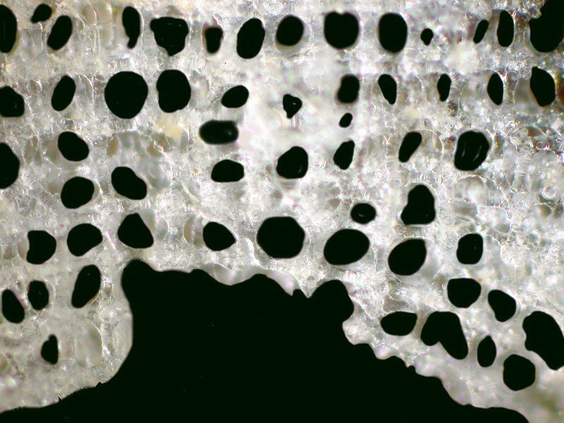
From what we have been looking at, one might get the impressions that these spicules are just randomly scattered around in some sort of organic matrix. This is, of course, rarely the case. There are apparent instances of individual spicules being isolated and separate, but that may very well be a consequence of the process of extracting and sorting in order to examine the structure of the different types, although it is possible that a few are embedded in such a way that they are not part of a lattice. However, that is the issue which now confronts us. These sponges do tend to form lattices, some of which are exceptionally ornate and complicated and they do this by having cells which are capable of fusing spicules together. So, now we need to look at some examples of the types of lattices which are constructed. Some are rather ordinary in appearance; namely, like a wide opalescent strip punched full of holes.
A more complex lattice is evident in this example from one of the cup sponges and rather resembles a dense bramble bush.

This next image shows a sample where we get a glimpse of the way in which some spicules have fused.

Now I am going to show you a close-up of the classically beautiful Venus Flower Basket (Euplectella aspergillum) and you can consider the enormous number of spicules involved in being fused in just this one section.

Euplectella does have a tail of sorts, but it is a much more modest affair than that of Hyalonema or Monorhaphis chuni, the latter having fibers over 10 feet long!! I just recently acquired a section of the tail of one of these with part of the sheath still intact and that will, I hope, soon be the subject of a follow-up article on these amazing creatures.
All comments to the author Richard Howey are welcomed.
Editor's note: Visit Richard Howey's new website at http://rhowey.googlepages.com/home where he plans to share aspects of his wide interests.
Microscopy UK Front
Page
Micscape
Magazine
Article
Library
Published in the January 2017 edition of Micscape Magazine.
Please report any Web problems or offer general comments to the Micscape Editor .
Micscape is the on-line monthly magazine of the Microscopy UK website at Microscopy-UK .
©
Onview.net Ltd, Microscopy-UK, and all contributors 1995
onwards. All rights reserved.
Main site is at
www.microscopy-uk.org.uk .