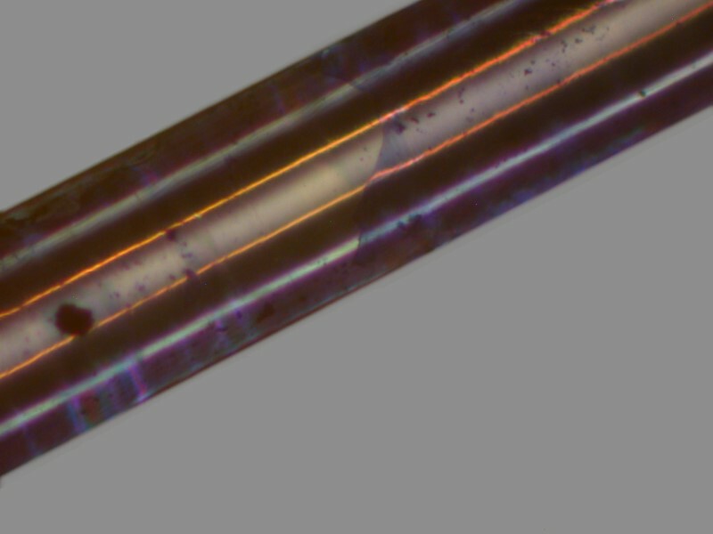
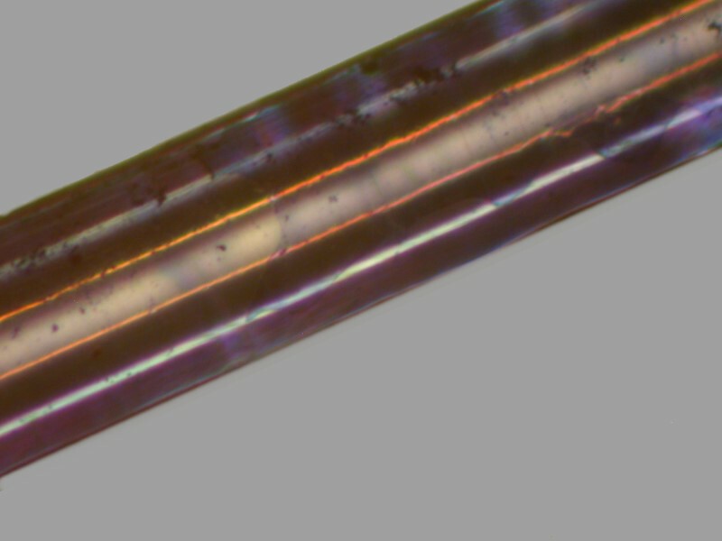
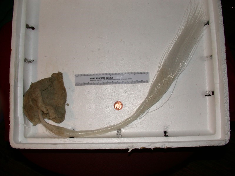
Technically, this technique doesn’t really stain the fibers, but rather coats them in the dye in such a way that detail is revealed that is otherwise almost invisible. However, I will call this process staining nonetheless now that we all understand that it really isn’t.
For some time now, I have been investigating glass sponges and particularly Euplectella aspergillum and you can find some earlier essays here:
Thus far, I have applied this technique only to the long, clear fibers which extend down from the base. However, I am anxious to try it out on some other parts of the sponge and some other species of glass sponges as well.
I carefully removed several strands from the base, coiled them into a 100 mm (about 4 inch) plastic Petri dish and covered them with distilled water. Then I used a pair of micro-forceps and a micro-chisel to break them into lengths of about 2 to 3 inches. This I do under water to avoid small chips of glass popping up out of the dish. Next I transfer a length or 2 of the fiber into a small plastic tube which I half fill with stain. I use the tubes rather than dishes in order to conserve the stains. I let these sit for about 2 months hoping to get a significant deposit on the surface. On reflection, I am not at all sure that it necessary to wait so long. This initial experiment involved the use of 4 different stains each at a concentration of 1% with the possible exception of the Trichrome stain: 1) Trichrome (I don’t know its concentration because it is a proprietary stain which was sold by Ward’s Natural Science, but is no longer listed), 2) Toluidine Blue, Methylene Blue, and Malachite Green.
I removed the fibers, rinsed them very carefully in distilled water and placed 1 or 2 of each type of stain I used into individual Petri dishes; in other word, I placed them in 4 different dishes according to the kind of stain and let them dry. On examination under the stereo microscope, I was pleased to see that there was indeed some deposition of the stain on the fibers and I could now get a clearer idea of the construction of these fibers. In Euplectella, some of the basal strands are at least a foot in length and in Hyalomena, I have some strands that are over 2 feet long which will be the target of my next such experiments. So, clearly it makes sense that these were constructed by the addition of small sections of silica over considerable time. If the fiber were a single long piece, it would not have much strength.
The Toluidine Blue and the Trichrome stains show clearly that the fiber consists of a series of sections and that there are “barbs” that distinguish the sections and are reminiscent of certain kinds of mammalian hairs. First I’ll show you the Toluidine Blue and then the Trichrome.


Both images suggest that there is a different sort of composition in the middle of the fiber than that on the outer surround area.
Wonderfully, the other 2 stains reveal something about what’s going on in the center. First, I’ll show you 3 images stained with Methylene Blue, each of slightly higher magnification.


Here the optical properties of the fiber indicate a complicated internal layering that runs parallel to the outer surface of the strand.
This becomes even clearer in the images in which Malachite Green was used. Again, I’ll present 3 images, each of slightly greater magnification.


The last of the Methylene Blue images seems to hint at the center core being hollow (or at least, having a very different refractive character than the surrounding layers). This is strongly reinforced by the 3 Malachite Green images and would be consistent with the great flexibility and strength of the strands.
I hope in the near future to be able to get some fairly thin cross sections in order to examine the arrangement of the deposition of the crystals. In looking at a few broken tips of sections, there are some intriguing hints which I’ll save for the next essay when I have more information.
Finally, I will conclude by mentioning some of my notions for future investigations along this line and if you beat me to doing some of them, please write an article for Micscape and share your results.
1) I mentioned above that I want to try this technique out with some strands taken from the very, very strange glass sponge Hyalonema and just to whet your appetite for sponge weirdness, I’ll show you an image.

2) I have a few other odd glass sponges from off the coast of British Columbia that certainly will be candidates as well.
3) There are also some very intriguing little cup sponges with lots of interlocking “feathery” spicules.
4) I want to try a series of different sorts of stains. Some years back, I developed a technique for “pyritizing” forams and other micro-fossils using Silver Nitrate and then using Ascorbic Acid to precipitate the silver and deposit it on the specimens which you can read about here.
Now, I want to try it on some spicules and sections of glass sponges.
5) Some other stains and reagents which I think may be worth trying:
a) Iodine
b) Crystal Violet
c) Nigrosin
d) Alizarin Red S
e) Orange G
f) Gentian Violet
g) Hematoxylin
h) Janus Green B
and I might also try out a few fluorescent stains.
I look forward to these experiments and I will dutifully report any interesting results.
All comments to the author Richard Howey are welcomed.
Editor's note: Visit Richard Howey's new website at http://rhowey.googlepages.com/home where he plans to share aspects of his wide interests.
Microscopy UK Front
Page
Micscape
Magazine
Article
Library
© Microscopy UK or their contributors.
Published in the August 2015 edition of Micscape Magazine.
Please report any Web problems or offer general comments to the Micscape Editor .
Micscape is the on-line monthly magazine of the Microscopy UK website at Microscopy-UK .
©
Onview.net Ltd, Microscopy-UK, and all contributors 1995
onwards. All rights reserved.
Main site is at
www.microscopy-uk.org.uk .