Many new microscope models have LED illumination and homebrew conversion of older stands from tungsten to LED is being widely embraced by many enthusiasts with commercial LED retrofits also available for some stands. The key benefits of white LEDs over tungsten are well known—near constant white balance at variable intensities, essentially no near UV / IR, relatively cool running, relatively cheap, compact size, long life, stable white, power supply benefits.
As a microscopy enthusiast I have a foot in both camps, using both LED and tungsten halogen lighting depending on the microscope. My compound microscope, a Zeiss Photomicroscope III, I prefer using as the maker's intended with the 100W halogen lamphouse (see September 2013 Micscape article 'Tungsten filament microscope lamps in the early 21st century: A case for the defence'.)
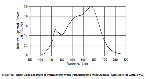
One of the widely cited advantages of tungsten and remarked on in the article above is that the typical single emitter white LED is less suited for critical work e.g. quantitative polarisation studies and critical colour assessment in stained specimens—the white LED's spectra is atypical as a consequence of the exciting blue LED's output mixed with that of the phosphor's emission. The standard white's have a marked blue spike, the warm white's less so but the latter at the penalty of a lower colour temp—often comparable to a 6V 5W tungsten lamp. I'm a great believer in being aware of theory where possible but then putting it to one side and to 'try it and see' with our own equipment.
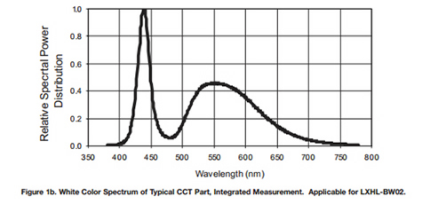
Left: Typical spectrum of a white LED equivalent to the one tested, cited as having a 'typical' colour temp. of 5500K.
Right: A comparable 'warm white' with a markedly lower blue spike, 'typical' color temp. cited as 3300K.
Source: Philips Lumileds Luxeon Emitter Technical Datasheet DS25.
Some manufacturers offer microscopes with LED lamps based on RGB LED sources to give a more balanced light output for critical work. Olympus state they are using 'mixed-matrix brightfield LED technology' in some stands e.g. the BX46 which they call 'True Color LED'.
Side by side test of a 100W halogen lamp and typical white LED: I was interested to see using my own microscope and camera whether photomicrography using the typical daylight white LED I currently had was noticeably different from the 100W halogen lamp. Subjects were chosen that may best reveal any differences i.e. stained sections and thin rock sections.
The set-up used a half-silvered mirror as shown below to allow an immediate switch between the halogen lighting lamphouse and an external LED. Ideally, exactly the same optics path should be used for both light sources but LED conversion was not trivial for the 100W lamphouse. The key details are summarised below.
|
Tungsten system |
White LED system |
|
| Lighting | Zeiss 100W halogen lamphouse, with Osram Xenophot bulb |
Philips Luxeon III LUMILED LXHL-LR3C |
| Conditions | 12V max. with Zeiss CB12 filter. Neutral density 0.5 filter set in base for comfortable visual work. | 100mA constant current. Set to give same intensity for halogen. Good even coverage of field of view with objective used. |
| Colour temp. for conditions | Stated 3400K without filter, CB12 brings to approx. daylight 5500K | Luxeon typical CCT 5500K at 700mA. CRI 70 (the warm equivalent was 90). The lower current used shouldn't shift temp. significantly. |
| Objective |
Zeiss 10x NA0.32 planapo set for Köhler no diffusers in light path. |
Zeiss 10x NA0.32 planapo set for Köhler by focussing LED in back focal plane of objective. |
|
Camera: Canon 600D, Zeiss 10X Kpl W eyepiece on short collar for projection. 'Neutral' colour tone set. |
ISO 100, 'Custom white balance'. Shutter speed adjusted for highlights without clipping on live histogram. RAW images used, exposure readjusted if necessary to reset highlights. Black level on tonal balance set if needed to lower left of tonal curve. No intermediate tonal balance levels made on any image. |
As for tungsten. Post capture adjustments required of exposure required for both tungsten and LED were minor. |
*CRI = 'Colour Rendering Index. A black body illuminator is assigned a CRI of 100. CCT = 'Correlated Colour Temperature'. Values cited are from the maker's datasheets.
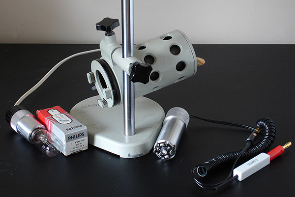
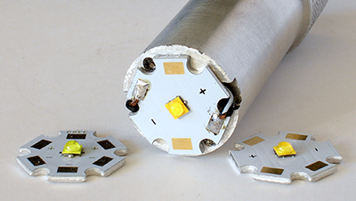
Left: Russian LOMO OI-14 lamp. The original tungsten lamp fitting is shown and the conversion to a 3W white Luxeon III LED 5500K (lamp fitting kindly made by Robert Pavlis. This design also works well on Zeiss Standards models that use the rear 6V 15W lamp.) Electrical fittings with in-line 4.7 Ohm 10W resistor by Ian Walker.
Right: A limited study was also made with a Cree XT-E 'warm white 2700K'. Despite its smaller die size, it gave excellent even coverage of the 10X objective with the LOMO lamphouse. The Cree 'cool white 6500K' and 'neutral white 4000K' are also shown. Typically <£4 each fitted on the star bases on eBay UK.
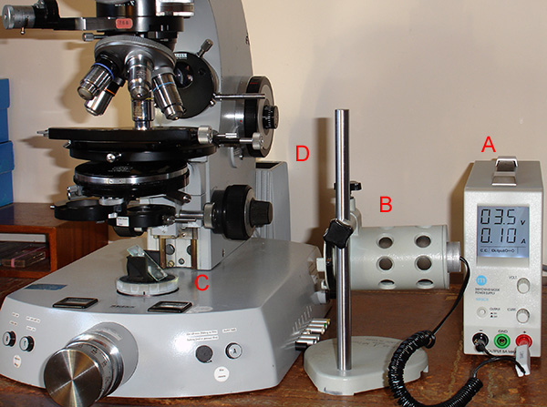
Set-up used for the lighting comparison:
(A) The LED was driven using a switched mode bench variable power supply (20V, 5A max.). (N93CX model, constant I or V, ca. <£50 used eBay, £90 new from Maplin UK.)
(B) The LOMO OI-14 lamp with two element condenser and field iris. Lamp set-up for Köhler.
(C) A half-silvered mirror 50/50 beam splitter at 45° from Knight Optical UK is on the lamp field iris housing on base.
(D) Zeiss Photomicroscope III with the 100W halogen lamphouse at rear driven from the Zeiss power supply. Blue CB12 filter below silvered mirror.
Image comparisons
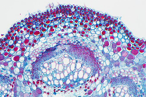
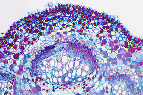
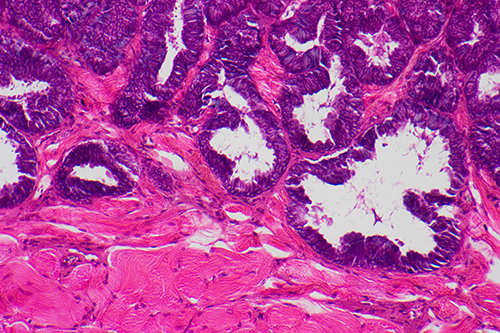
All lefthand images: 100W halogen ca. 5500K. All righthand images unless stated: 3W Luxeon white LED ca. 5500K.
Comments on colour differences were when viewing on an HP All-in One 3420 LED screen.
Any differences were much less marked on my HP TM2 laptop with lower available brightness range on the LED display.
Sea grape, Coccoloba uvifera T/S leaf, Biosil slide.
Halogen Luxeon LED
The colour differences are subtle (repeated a few times to check not random variation) although any differences are likely harder to spot on this type of subject with relatively low areas of colour. Blue a little more purple and reds darker for the LED. Other plant sections tried with other stains also only showed subtle differences.
Frog tongue T/S Numount, iron haematoxylin and eosin (IH&E).
Made by myself on an NBS course tutored by the late Eric Marson.
With repeatability check starting from new custom white balances a day later.
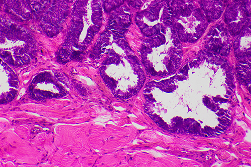
Halogen Luxeon LED
The differences between halogen and LED were more marked for this colour palette so a repeatability check is shown below.
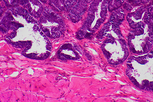
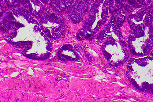
Halogen repeat Luxeon LED repeat
There are slight hue differences for the repeat of the halogen lighting but there is a more marked difference in the rendition of the eosin and iron haematoxylin for the LED. Histology is not a subject I explore so can't comment whether the colour changes are of significance, although the differentiation of cellular structures by the two stains is still marked.
Olympus used I&HE and PAP stained histology sections to demonstrate the superiority of their 'Mixed Matrix' 'True Color LED' system commented on earlier.
Rat testis T/S Numount, iron haematoxylin and eosin (IH&E).
Made by myself on an NBS course tutored by the late Eric Marson.
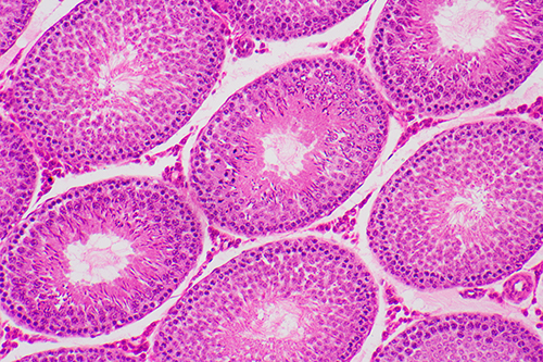
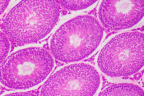
Halogen Luxeon LED
Eosin a more purple hue with LED, possibly to the extent of lowering the differentiation slightly between the two stains?
Horsetail Equisetum sp., growing tip L/S, Alcian blue and safranin.
Prepared by John Nichols 1988.
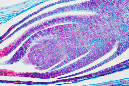
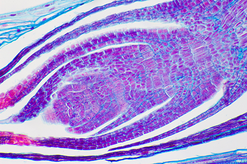
Halogen Luxeon LED
Again the more saturated purples and the reds are evident for the LED lit image.
Gabbro thin rock section, Open University, UK S260 slide K.
(These slides are finished with a slightly gritty varnish rather than a coverslip.)
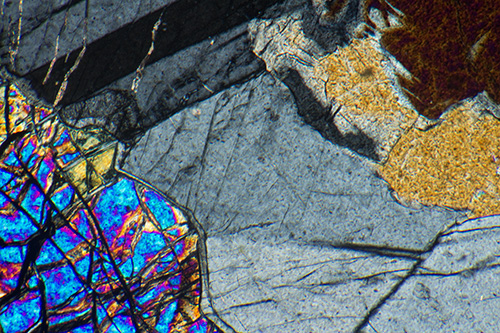
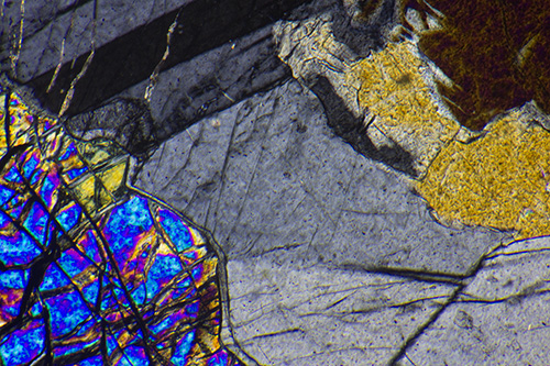
Halogen Luxeon LED
Above, crossed polars only to include a range of grey tones. The greys seem slightly warmer for the LED which again is giving more saturated blues and purples, yellow perhaps cooler for the LED. The LED also seems to be distinguishing subtle birefringence differences in lower left mineral out of camera.
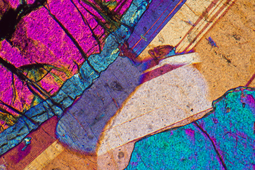
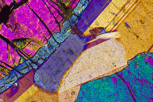
Halogen Luxeon LED
Above, same subject (different area of slide) with lambda red tint plate to give a wider birefringence colour palette. The 'cooler' look to the more expansive area of yellow hues is more apparent on this image.
Strychnine crystals, Victorian unnamed papered slide.
Crossed polars, with lambda red tint plate.
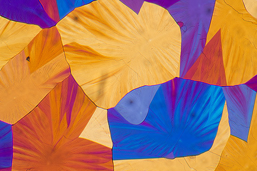
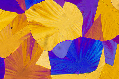
Halogen Luxeon LED
'Artistic' type polarised light studies may not require critical colour reproduction but the gamut of colours they can show does illustrate when the results from halogen lamp and a white LED can differ. The increased saturation of blue and purple is seen but yellow/orange hues also differ slightly.
White LED variants, what colour temperature to choose? Developments to improve the white balance of LEDs are continuing apace e.g. by using advanced multi-phosphors on a monochromatic diode or the multiple emitter approach. The 'colour rendering index' CRI is used as an indicator of colour fidelity cf a black body illuminator with the latter assigned a CRI of 100. The Wikipedia CRI entry notes that LEDs claiming a CRI up to 98 are being developed and they cite the products of the company Beijing Yuji Science and Technology Ltd. The older Philips Luxeon III I used had a CRI of 70 (the warm equivalent was 80, but both now discontinued)
Browsing around the latest offerings, some manufacturers now offer a range of whites. Cree in their XT-E range for example offer 'cool', 'outdoor', 'neutral', 'warm' plus four others with stated CRI number. I chose a Cree 'warm' white CRI of 80. This had a marked yellow tint for microscopy (comparable to an unfiltered ca. 6V 15W tungsten lamp) and would not use without a blue filter, but did use without a filter as an extreme for the colour tests. The Canon 600D had no problem setting a custom white balance for this colour temperature.
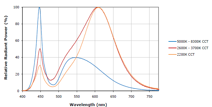
'Relative spectral power distribution - white'
Source: Cree® XLamp® XT-E LEDs (data sheet CLD-DS41 REV 10B).
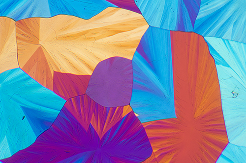
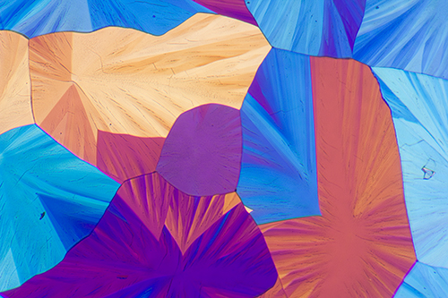
Left: Halogen. Right: Cree XTE warm white LED. Dealer cited as colour temp. 2700K.