Samworth's Snippets
Why use video?
by Mike Samworth
I am interested in Protozoa. There are none more
beautiful or more generally attractive to observers than the
Stentors, well known to many microscopists so far as their more
obvious characters are concerned, but which present considerable
difficulty when it is desired to make out their details of
structure.
It is only by the frequent examination of a great number of
individuals, and making the most of fortunate opportunities, that
the various organelles can be distinctly seen. Often there is
disappointment felt as many efforts have to be made before
success is achieved.
 Drawings in texts exhibiting
anatomical details are almost invariably the result of combining
in one view parts that cannot be simultaneously seen in the
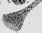
living organism. One Stentor shown left, for example,
may at one time show the mouth or gullet well, while the nucleus
is scarcely visible, and some other details cannot be seen at
all. Another time the nucleus may be prominent throughout more or
less of its length, and a complete delineation can only be made
by putting together the information contained in a long series of
observations. As microscopists if we are not fully aware of this
fact we are not only puzzled, but are discouraged by the wide
difference between what we see and what we find drawn by others.
Previously, the only remedy against this disappointment was
patience, and a knowledge of how to use the published figures. I
believe there is another solution open to the microscopist of the
nineties, namely video. Recording the organisms on tape allows
future study and the option of slow-motion or even still- frame
images. This makes it far easier to piece together detailed
structure of the organism in question. Indeed, moving images
often appear to show greater detail to the human eye.
I have borrowed a video camera for mounting on my microscope a
couple of times now and am considering purchasing one for myself
eventually. Though not able to get to see any of the very few
texts published on the subject, it is interesting that much of
that I have seen is written by amateurs. Certainly it is a
rapidly developing area of microscopy. If anyone has any
particular advice or tips from their experience with the medium,
then do not hesitate to forward this to the Editor (see contact
below) for inclusion on the site.
Image derived from a still of a video by Maurice Smith.
Read an illustrated Micscape
Article to learn more about Stentor.
Download
a Micscape Movie of Stentor.
© Microscopy UK or their
contributors.
Please report any Web problems
or offer general comments to the Micscape Editor,
via the contact on current Micscape Index.
Micscape is the on-line monthly
magazine of the Microscopy UK web
site at Microscopy-UK
WIDTH=1
© Onview.net Ltd, Microscopy-UK, and all contributors 1995 onwards. All rights
reserved. Main site is at www.microscopy-uk.org.uk with full mirror at www.microscopy-uk.net.
 Drawings in texts exhibiting
anatomical details are almost invariably the result of combining
in one view parts that cannot be simultaneously seen in the
living organism. One Stentor shown left, for example,
may at one time show the mouth or gullet well, while the nucleus
is scarcely visible, and some other details cannot be seen at
all. Another time the nucleus may be prominent throughout more or
less of its length, and a complete delineation can only be made
by putting together the information contained in a long series of
observations. As microscopists if we are not fully aware of this
fact we are not only puzzled, but are discouraged by the wide
difference between what we see and what we find drawn by others.