HOW MANY
ONION SKINS ARE THERE?
Walter
Dioni - Cancún, México
When we speak
of "onion skin" it seems that everyone knows what we mean. But in reality the onion, which is the bulb of Allium
cepa (and a species with
several varieties), has many "skins", as this simple check will
show
|
|
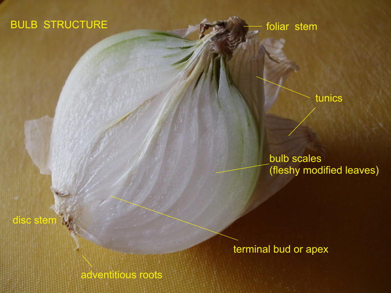
It's evident that there are many skins:
every scale has two.
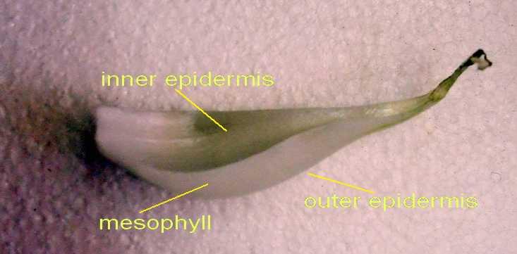
FIG. 1, 2 (Fujifilm FinePix A900 Camera)
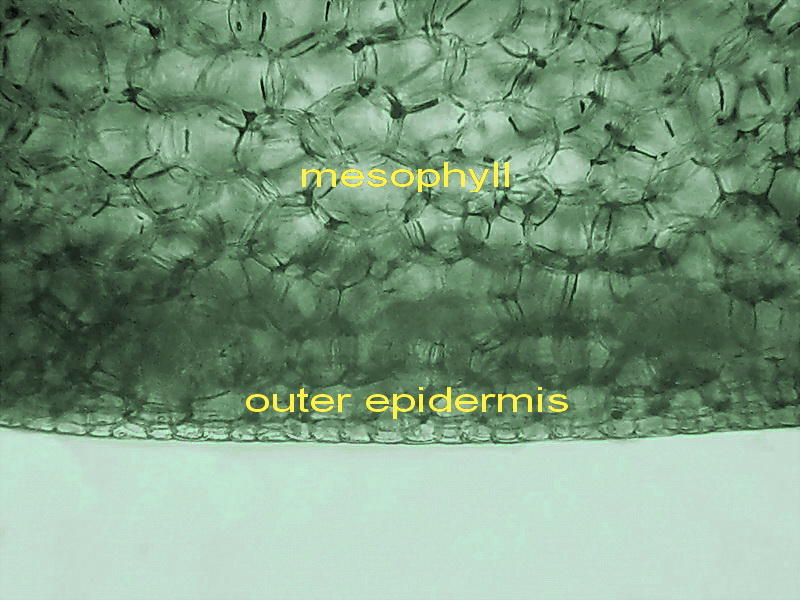
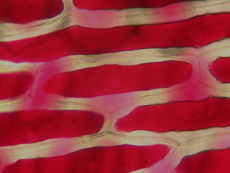
Fig. 3 the mesophyll is composed of large polyhedral
cells. As this is an underground leaf, the mesophyll lacks the chlorophyll of
the aerial leaves. In comparison, the epidermic cells, here seen in transversal
section, look tiny. Mesotome section; mounted in glycerin. 1:1
cut from the original 3 Mpx picture. Canon Powershot A75
As you can see (fig
2) each onion bulb scale has two epidermal layers, one internal and one
external. Almost all protocols of practical exercises that use
"onion skin", makes it clear "to use the skin from the inner
side of the bulb scale." Would both be different?
|
|
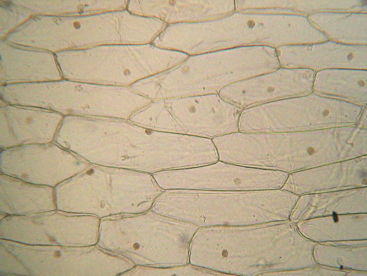
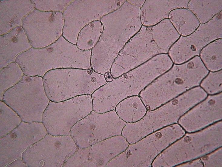
Fig 4 Inner
epidermis without stomata Fig. 5 Outer epidermis with stomata
No stain applied, mounted in glycerinated water 10x objective 1:1 crops from the 2 Mpxs originals, additionally reduced to be included in the article.
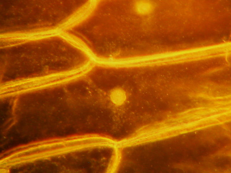
Fig 6 40x obj. Inner epidermis - Water
mount, no fixatives. Rheinberg disc (black center, orange border). The cytosol
is seen as a gel with clouds of granulations (probably mitochondria).
Canon Powershot
A75
The following
pictures (6 and 7) shows a bigger image of one stomata (x40, reduced to be included
here) and a topographic view of the outer skin (x 10, also reduced; the stained
glass effect is due to an irregular staining with 1% aqueous methylene blue).
It does not have scientific value but shows what I want.
|
|
|
|
|
Fig 7 and 8 Outer epidermis two images by the DC3 Motic camera. The methylene blue solution has not permeated the cell walls, and the nuclei are not stained.
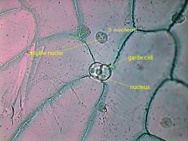
What are these
differences? I think
they are easy to see. The outer
epidermis has cells of many sizes, with an irregular design, and has stomata
(not excessively frequent in this case, but undoubtedly present). Note also that
at least four of the cells in this tiny fragment of the surface of the scale (fig 5), has 2 nuclei.
The inner one (fig. 4), on the contrary, has
the architecture we are used to assign to the onion skin. The skin that is
shown to us over and over ... over decades. Narrow elongated polygonal cells in
a well builtstructure, each cell with one nucleus, and its nucleoli (1,2 or 3).
|
|
|
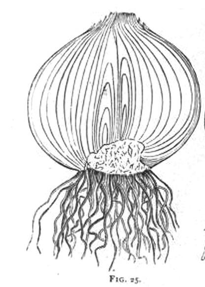
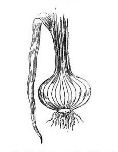
Fig.9, left, onion bulb drawing, in Bastin, 1895 Fig.10, right, onion,
with its foliar stem, and a fully developed leaf that normally we dont see ,
Bentley, 1887.
The scales
(food reserve leaves) form the bulb of the onion (the underground part of the
plant). But if you compare it with leeks (Allium ampeloprasun) although it has
no bulb, you see the same concentric structure on the stem, and you could see that the "white part" is continuous with
the "green leafy part" generally discarded by chefs, and which in
onions is cut flush to the bulb by the vendor. (Our fig. 1, and also fig. 8, from
Bastin 1895). The green part,
if it could grow freely in the plant, could become long narrow leaves that by
its own weight would bend to the outside, (fig 9, from Bentley, 1887).
In such
a position, which so far we call "the outside", would be "the
underside" of the leaf. A lightweight review in your Botany book providing
elementary notions of the structure of the leaf, will tell you that it is at the inferior face where
there are the stomata, exclusively, or more numerous, than at the "top
side of the leaf.
Leaf sheaths (scales) have shown with
their stomata that they actually are modified onion leaves, and that its
external face, which have the stomata, is equivalent to the underside of an
aerial leaf.
By using the "inner skin" the biology teacher focuses the
student to the study of the plant cell, and one plant tissue, the epidermis,
without having to divert their attention to explanations about the stomata, its
structure and its function, nor on the homology
between a bulb scale and an aerial leaf. It
is a useful trick.
And... the third
onion skin
Normally the
other "onion skins" usable for scholarly lectures (the outer papery layers
or tunics), are reserved for more advanced
courses, in which it is studied the secretion by plants of different chemicals,
especially calcium oxalate, which
form clearly visible and very interesting crystal structures.
To see them,
cut with scissors a 1.5 x 1.5 cm of the tunic (the upper portions are richer in
crystals) and hydrate it in plain water for many hours. If colored, the tunic
would diffuse some color (it's a useful pigment that people use to tinge textiles
or Easter eggs) and become pliable. The more pigment lost, the better image
you have. It only remains to make a wet mount.
Really the tunics are not an epidermis. They are
the compressed dry remnants of one, two or more exterior scales of the bulb.
Some focusing would convince you that the crystals are embedded in the dry mesophyll. This is why
also you can see, as in any leaf, the veins with the vessels that conduct water
through the scale. The skin for this picture (fig. 14) was first embedded in nail polish solvent, and mounted
in nail polish, to make it more transparent. (3 pictures stacked with CombineZ)
|
|
|
|
|
|
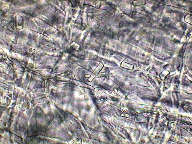
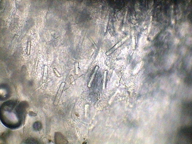
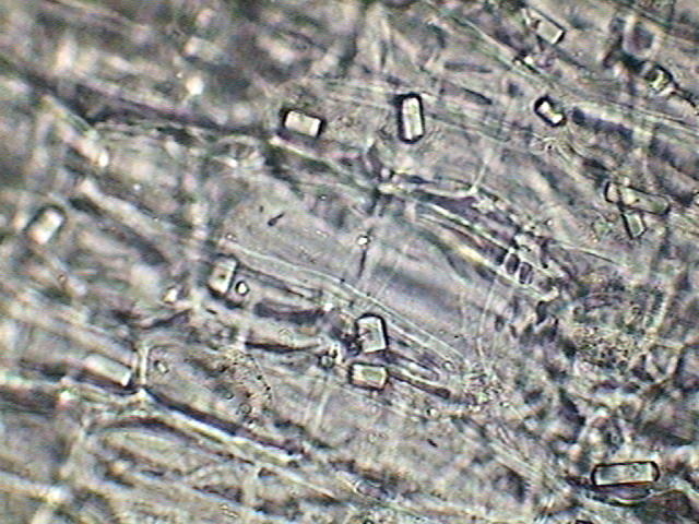
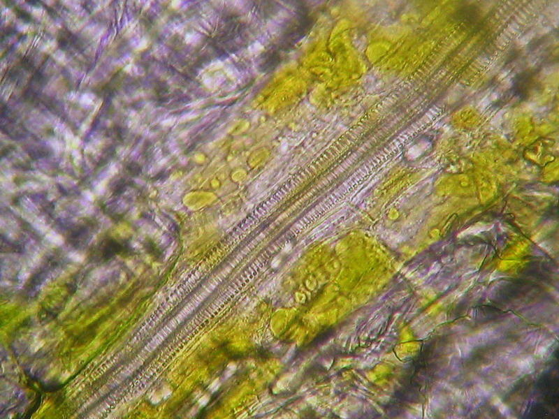
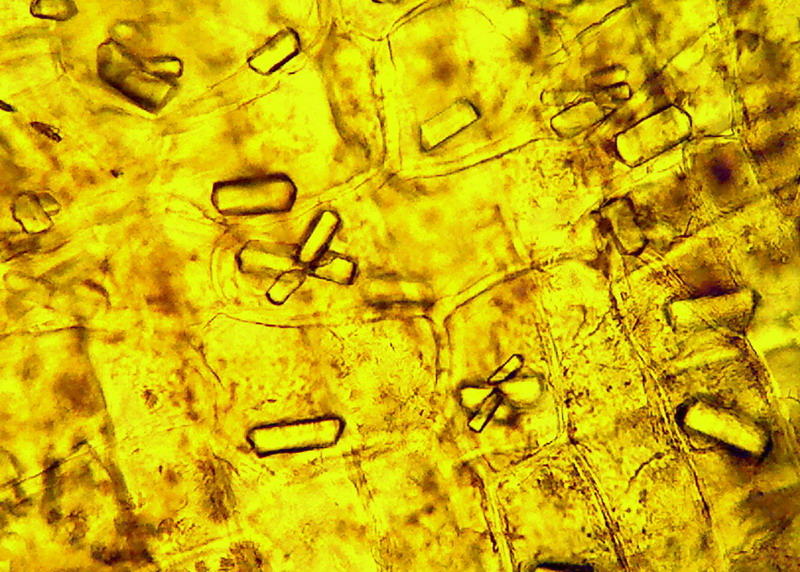
Fig 11 to 13 shows crystals of oxalate found in the tunic of a white onion. Fig 14 conductive vessels, white onion (all pictures with 40x obj.) Logitech 9000
|
|
|
|
|
|
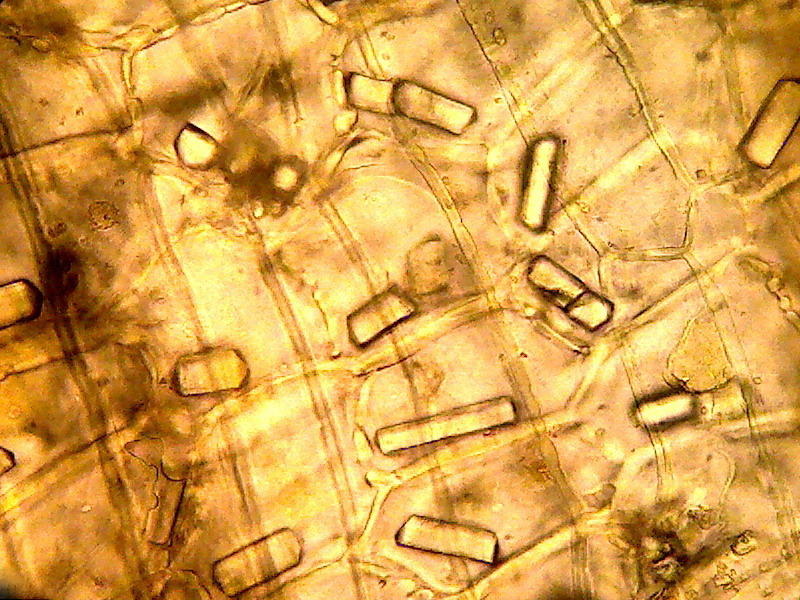
Shallots tunic, fig
15 10x; 16, 17, 18 40x
Logitech 9000 - 800x 600 cuts, reduced to publish it here.
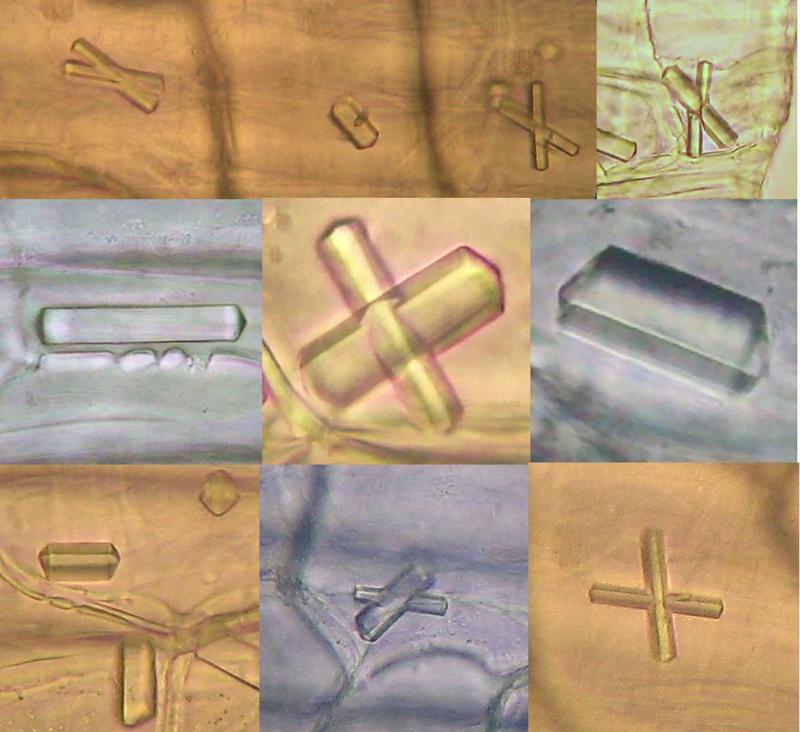
Fig 19: Some additional pictures of individual
crystals in the tunic of a white onion and a shallot. Clips from 2 Mpx pictures
taken with the Canon Powershot A75. (obj x 40) but the central row ones, which
were taken with the 100x, these 3 pictures were cropped and reduced.
In all the pictures crystals are embedded in the
mesophyll, and are seen through the superficial layer of the epidermis. As my
microscope is not a DIC with the power to make net optical sections, the images
are not very sharp.
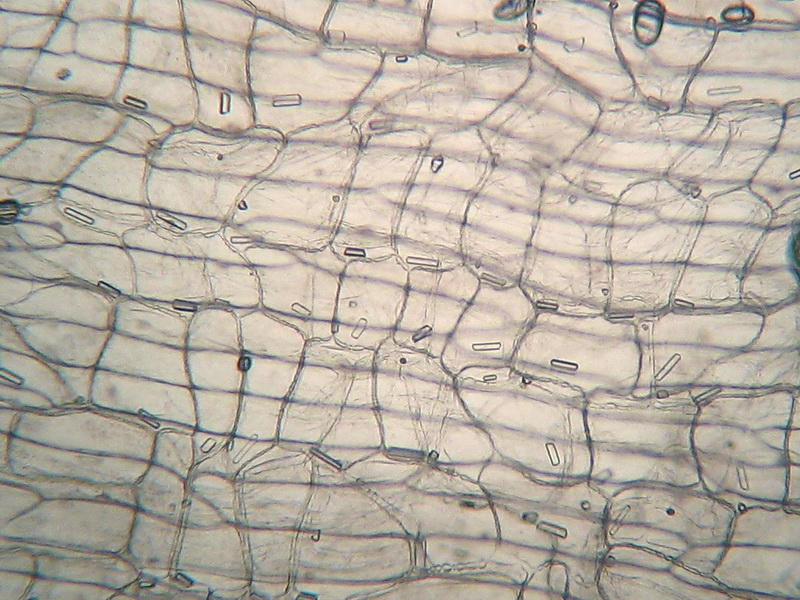
Fig. 20: 10x obj. distribution of crystals (mostly one in
a cell) in the mesophyll. Shadows of the limits of the long cells of the
epidermis are seen, out of focus. Canon Powershot A75
In other
plants are also common calcium carbonate
crystals. Why do I know that the onion ones are calcium oxalate? Because it's said (by microscopists from the 18
century to now) that is not difficult to establish a diagnosis, using two
chemical reactions, which can be controlled with the microscope:
In principle it
is interesting that, fortunately for us microscopists, these crystals are
insoluble in water, alcohol, acetic acid, or even in the hypochlorite, which is
commonly used to destroy the cytoplasm and clean plant sections to be colored
and mounted. This is
why we can see them
easily.
But...
Oxalate crystals are insoluble in acetic acid, but soluble
without effervescence in hydrochloric acid
The
crystals of carbonate by contrast
are soluble in acetic acid and hydrochloric acid, with effervescence
As in my samples I submit to, and they
resist the acetic acid, I can say they are oxalate, but ... I dont want to
risk my microscope objectives, exposing them to vapours of hydrochloric acid, so, please, believe me, as I believe all the botanists
that spoke about these crystals from the 18th century to today, that they would
dissolve readily. Amen.
What are the
calcium oxalate crystals for? Included as they are in the external wrapping (the
tunic) of the bulb, it is generally considered that they may be, by its
taste, and its sharp edged structure, an element of deterrence for those
underground invertebrates that could be tempted to attack the onion. And
possibly they are also a form to sequester, and to turn inoffensive, the oxalic
acid produced by the plant metabolism itself.
|
|
|
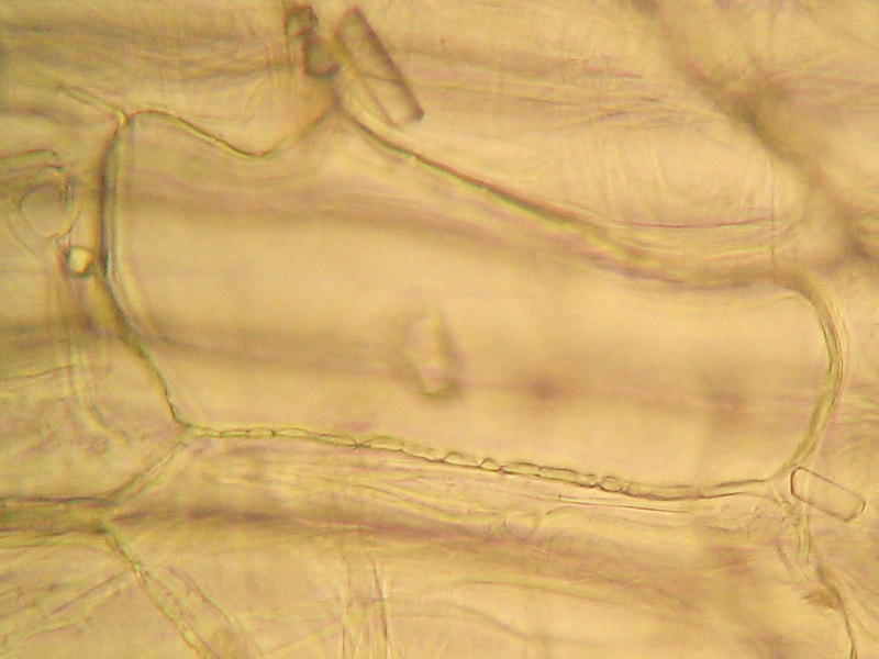
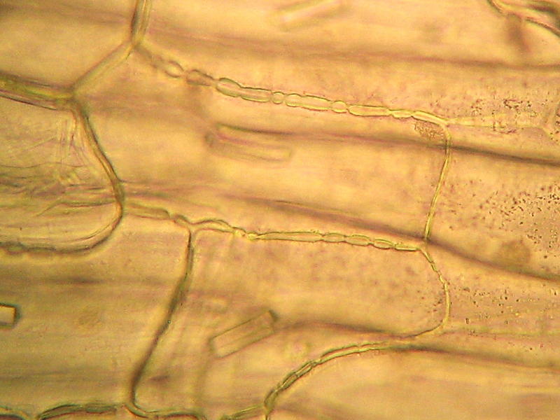
Fig. 21 and 22: plasmodesmata in mesophyll cells
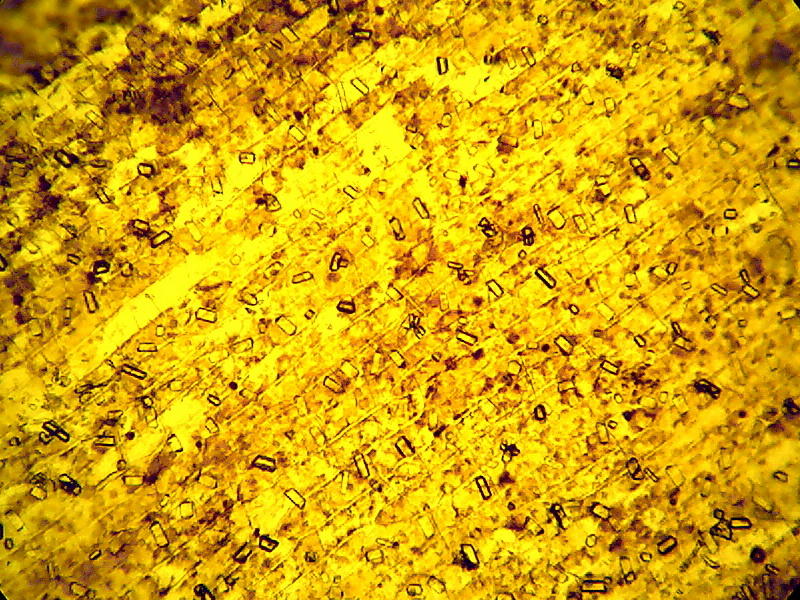
Another interesting
trait was the very long cells, that build the upper (internal) tunics epidermis
.
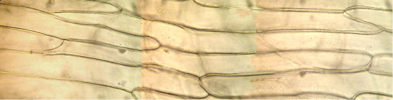
The above picture (fig.
23) is a montage of 3 individual pictures showing the special epidermis of the
tunica: cells are very long and narrow. See the position and size of the
nucleus that have to manage the long cell in the second row from the bottom. It
is out of focus because my interest was to show the cellular walls. Original
pictures taken with 40x obj. with the Logitech 9000
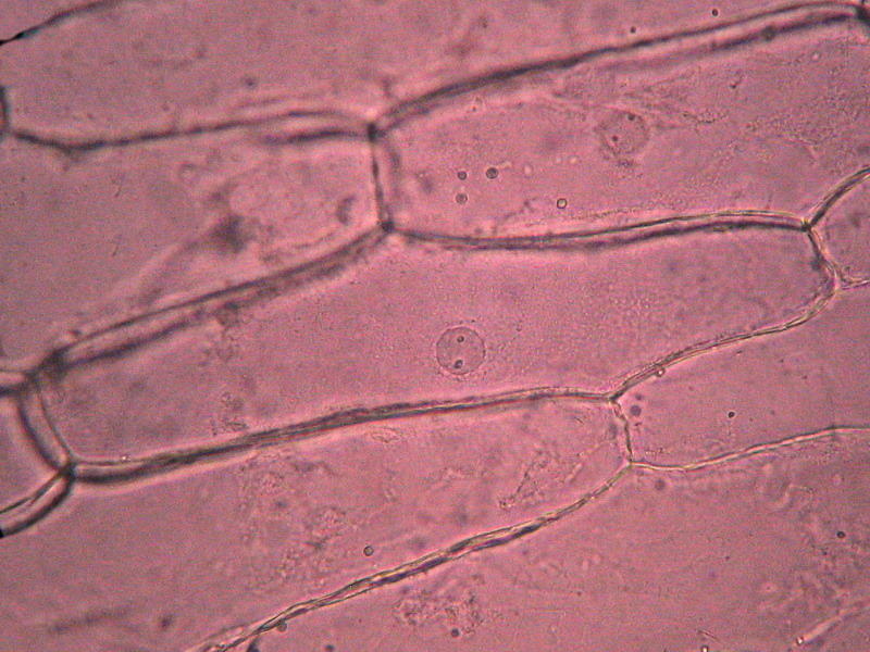
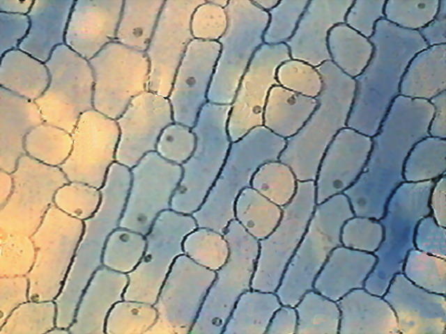
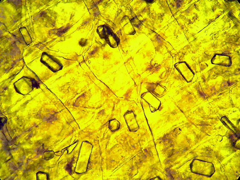
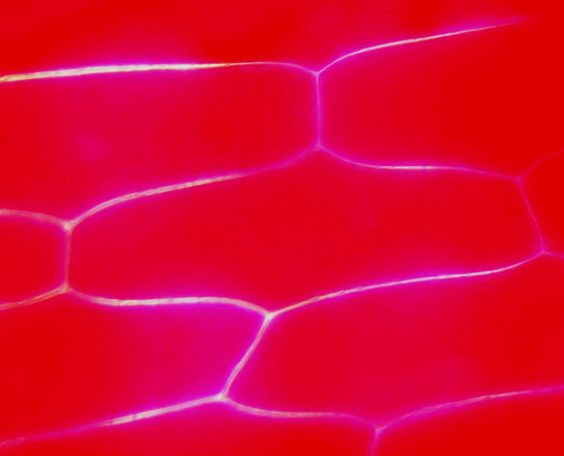
Fig 24: 40x obj. - a water mount of the outer epidermis of
a scale of red onion. The nucleus is perfectly round and with a clear limiting
border. Very faint color. Probably there is only half a cell here. Compare with
fig. 26 Canon Powershot A75.
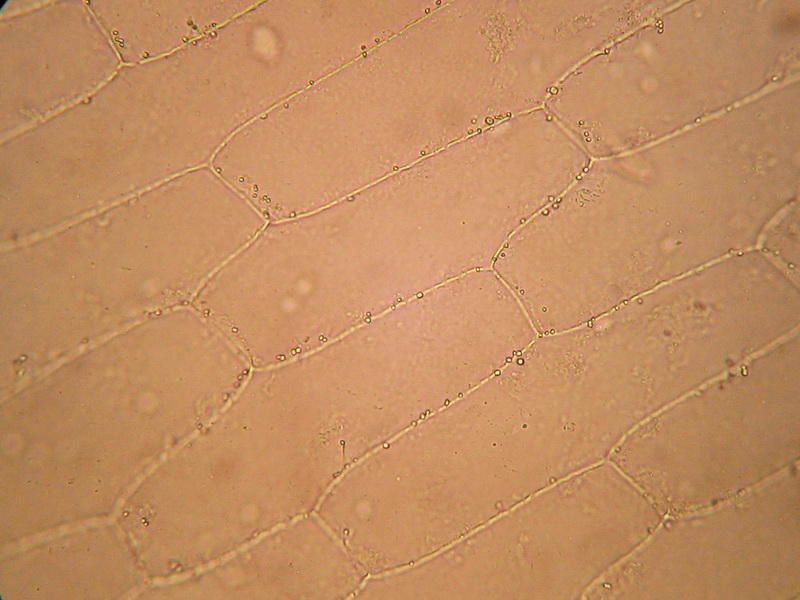
Fig 25: a peel with only the upper wall of the cells
with a very thin layer of citosol, no nucleus.
People dont tell
about this when explaining the protocol of the laboratory practices. The
following two pictures (26 and 27) were taken from a thin tangential section of
no more than 0.5 square centimeters that interest not only the epidermis, here
shown, but a thin section of the mesophyll, which, being out of focus is not
showing here. The short focus deep of the 40x allows this. Pictures were taken
with the Canon Powershot
To see the famous
plasmolysis you must prepare a concentrated sodium chloride solution (kitchen salt you
know). It is enough to make a solution with 1 teaspoon of salt (more or less 6
g) in 100 ml of clean freshwater, and replace the water under your coverslip
with the salt solution using the usual trick of putting a drop of salt solution
at one border of the coverslip and absorbing water with an absorbent paper from
the other side. In no more than 10 minutes
the effect is totally displayed. In YouTube there are some good videos, showing
not only the plasmolysis but also the deplasmolysis when returning the
epidermis to fresh or distilled water.
Can the onion skin give us more fun, for
so little money? ...yes! it can ... see the next article.