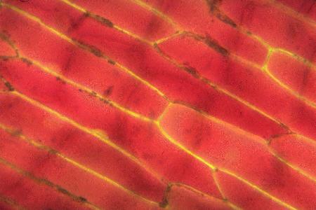
Making epidermal peels of onion skin is a common subject for the first plant cell studied at secondary school. To best see the rather translucent cells of the common onion needs staining, but if you use the red onions, and use one of the outer layers, then this is not necessary. A temporary mount can easily be made by placing a small piece of tissue on a slide with a drop of water and covering with a slip. Normal epidermal cells look like those in the photomicrograph below.
Normal cells.

These cells are what is called 'turgid' that is they are pretty full of water and hence there is enough pressure against the rigid cell wall for the cells to keep their brick-like shape. Notice that the red cytoplasm is throughout the whole cell and the only non-red part is between the cell membrane and the cell wall.
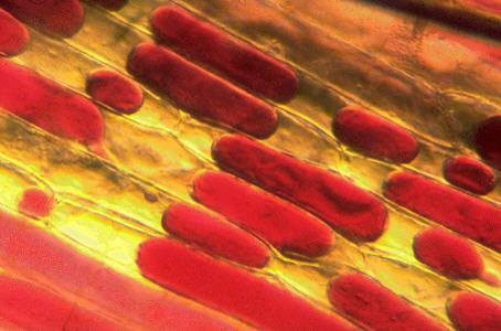
Now, if a strong salt solution is drawn under the coverslip, by means of some blotting paper; a technique known as 'irrigation', then the effect of the salt can be seen. Water is drawn out of the cells, shrinking the vacuole and the cytoplasm. This is seen in the photomicrograph below, the red areas being much smaller.
Flaccid cells.

This process is called 'plasmolysis' and the cells are now said to be 'flaccid'. Such cells lend little support to the structure of the pant, indeed this is the very reason why plants wilt if put into salty water. Unless they are saltwater plants, but that is another story!
Both photomicrographs by Mike Samworth.
Please report any Web problems
or offer general comments to the Micscape Editor,
via the contact on current Micscape Index.
Micscape is the on-line monthly
magazine of the Microscopy UK web
site at Microscopy-UK