Notes on near IR microscopy
with a tungsten lamp and visible light blocking filters
by David Walker, UK.
As for many hobbyists,
the advent of cheap LEDs used with suitable consumer digicams has prompted my interest in digital photomicroscopy
just outside the visible light spectrum. Both at shorter wavelengths (near UV)
for enhanced resolution and longer wavelengths (near IR) for superior light
transmittance with near opaque subjects like insect exoskeletons.
To date, I've used near
infra red (NIR) on my LOMO microscope using its external tungsten lamp and an LED soldered
onto
a blown bulb stub with a suitable low voltage power supply (not mains!). Near IR, near
UV and violet LEDs on blown bulbs allow quick interchange for multi-wavelength studies.
More recently I've been
trying a Zeiss Photomicroscope for near IR and UV studies.
I've also tried LEDs on a blown bulb stub for the 12V 60W lamphouse, but unlike the external LOMO lamp, readily interchanging between LED and
tungsten lighting was inconvenient. So as an alternative I tried filters to block visible
light from the tungsten lamp but pass the NIR component. A reader kindly remarked that this technique
can work well from their own experiences but hadn't tried it to
date. Experiences using two types of blocking filters are summarised below
as they may be of interest to others wishing to try NIR with tungsten lamps.
Hoya R72 filter: This
filter or equivalent is commonly used in conventional near IR photography.
With a bright microscope lamp it transmits a visible
deep crimson red and near IR (50% transmittance at 720 nm). The full transmittance
curve can be viewed on the Hoya website. Purchasing tip: I already had a
Hoya R72 52 mm lens grade filter for normal NIR landscape photography, but these are expensive. A cheaper optical
grade and smaller area can be used for microscopy as the filter sits on
the field iris below the microscope objectives.
Total visible light
blocking filters: I'd used a 940 nm NIR LED to date so was interested in
a filter that gave deeper NIR than the R72.
I bought an 850 nm 'long pass filter' from www.knightoptical.co.uk
(a 5 cm² acrylic filter was less than £10). From specs supplied by Knight Optical, this filter
had a sharp cut off, passing no light
at all < 800 nm and 50% at 850 nm (i.e. a similar transmittance
to that reported for a Hoya
IR85 filter). A tungsten lamp at a typical filament temp has
excellent emission below 700 nm into the near IR wavelengths (see ref. 1).
The author used both
an OpticStar 1.3 Mpixel 'C' mount monochrome
camera (sold for astronomy) and a Nikon D50 DSLR. There's many resources on the web discussing which
unmodified digicam models are most suitable for NIR. From browsing web camera forums,
my relatively old D50 seems to be regarded as 'weakly filtered' for NIR compared
with newer models like the D300. The live video feed from the OpticStar and
similar cameras
could be used instead of stills for demonstration purposes.
Compound microscope
(Zeiss photomicroscope III).
|
A typical monochrome CMOS sensor camera
has good sensitivity into the
near infra red as no blocking filter is usually on the sensor
and one must be used for visible work.
Right: The dense brown insect skeleton is
in many parts opaque to visible light and where fairly transparent
can give too high a contrast with areas like wings
to image well.
Left below, R72 filter: Detail now seen in exoskeleton
apart from the thickest areas.
Right below, 850 nm filter: Detail is seen even in
the thickest areas.
The images are of the full sensor capture
field. A suitable relay lens would need to be found to give
a good coverage of e.g. an insect mount.
|
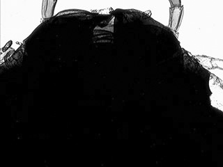
Brightfield.
Visible light with near IR blocking filter.
Leaving
off the filter gives a mix of visible light and near IR
but
gives washed out soft images as the focus positions are mixed.
|
|
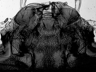
Hoya
R72 filter on field iris.
|
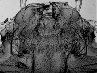
850
nm long pass fillter.
|
Nikon D50 DSLR.
Out of camera resized images. ISO 200. Zeiss 12V / 60W lamp at 12 Volts.
Diving
beetle, Zeiss 1x NA0.04 planachro objective. Prepared by the author on the late
Eric Marson's course.
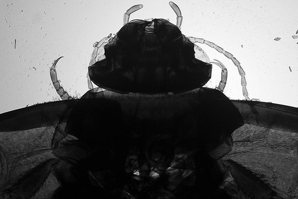
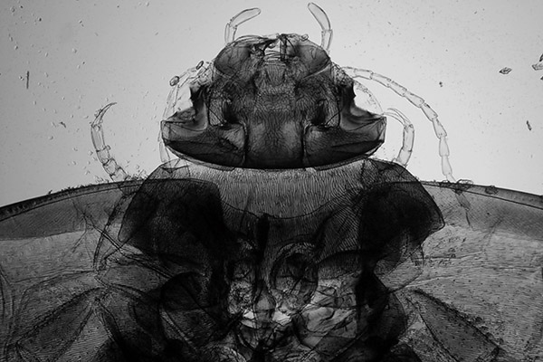
Hoya R72
filter on field iris. Converted to monochrome, the master is a deep
red. Same lamp setting, ND1.0 light reduction, 1/13th sec. Detail now
seen in exoskeleton apart from the thickest areas. The filter gives
a deep crimson image which can be focussed visually.
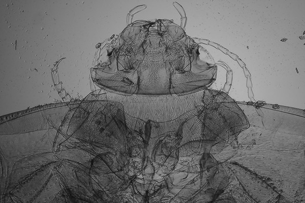
850 nm long
pass filter on field iris. 4 sec. Although 6 - 7 stops greater
exposure was required cf. the R72, sensitivity of the D50 was still
good and detail is seen even in the thickest areas. No visual image
is seen but possible to calibrate each objective's new focus position
with a camera that gives live image. (For example, with the Zeiss 2.5x
objective on the photomicroscope, 3.5 revolutions up on the fine focus position
for white light was the near IR focus.)
The limited tonal range
of a NIR image at this and longer wavelengths seems fairly typical from the author's experiences
with the D50 or other digicams tried,
so the images are more informative than artistically pleasing. Depending
on subject and degree of internal detail that can be revealed, the more
contrasty R72 filter may be preferred.
Not surprisingly, the D50 is much
more sensitive to wavelengths of 720 or 850 nm compared with the 940 nm
LED used previously, where very high
ISO and long exposures were needed. The author briefly assessed the recent
Nikon D300 model using the same filters and found that depending on wavelength
it was 3 - 7 stops less sensitive for transmitted NIR than the D50. Discussions
in online camera forums seems to suggest that modern DSLRs are often
using more extensive NIR blocking.
Stereo microscope
The
large insect slides are perhaps better suited for wider field imaging on a stereo although
my Meiji EMZ1 lacks a trino' port and, like many stereo light-bases, has
poor uneven lighting for camera work. But was interested to see how
it performed for NIR and the setup is shown below. The OpticStar camera was
used because of its live view and sensitivity. The under driven quartz halogen
lamp was apparently emitting more NIR than
visible as a neutral density filter had to be added for NIR work to stop overloading
the camera.
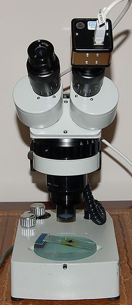
Left above: Meiji
EMZ1 stereo with the 6V 7W quartz halogen lamp base. The OpticStar camera
sat in an eyepiece tube with 'C' mount adaptor or for wider field a
Moticam relay lens on the Meiji W10X eyepiece. The Meiji supplementary
0.5x lens was added to the 1-3x zoom head. To improve the lighting, strong diffusers
were added below the ground glass base plate.
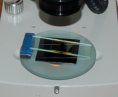
Right above: Set-up used for NIR
imaging with the 850 nm filter under slide (it's totally opaque to the
eye). One slide edge sits on a strip of 6 mm thick mouse mat to present
the slide at approx. the correct angle to one train of the Greenough stereo optics
to give a flatter imaging plane.
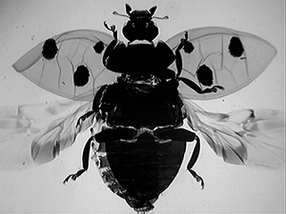
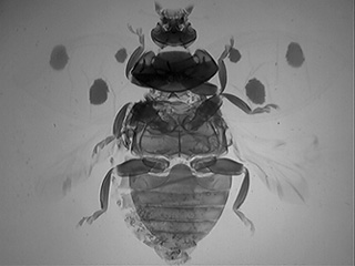
Seven spotted
ladybird, unnamed Victorian whole mount. The exoskeleton on
this subject was very dense.
Left above: Brightfield with NIR blocking
filter. Little internal detail was seen.
Right above: 850 nm long pass filter.
Good internal detail was seen.
Images below:
as above, optical zoom in to show abdomen detail. Left - visible light,
right - NIR.
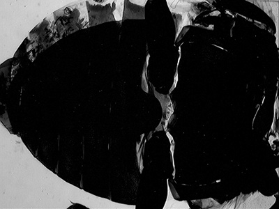
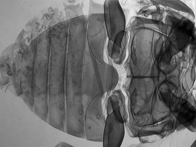
There is of
course a loss of resolution using NIR light but for the gross insect detail
it was typically useful for it wasn't noticeable, except for whole insect imaging
on the low NA stereo objective with 0.5x supplementary lens where the images
did lack bite.
Comments to
the
author David
Walker
are welcomed.
Acknowledgements:
Thank you to Marien van Westen who offers
his versatile Micam camera
control, imaging and measurement software for
free download and use. All image capture with the OpticStar above used Micam. I
much prefer Marien's software to control the camera than the camera maker's
supplied software.
The
author's own casual interest in NIR microscopy was inspired by the notes of Tony Dutton in the Quekett Microscopical Club Bulletin (ref.
2). Hopefully my modest trials may also
encourage others to have a go.
Thank you to the reader who suggested I tried
blocking filters with a tungsten lamp, prompted by my earlier article using
NIR LEDs. Unfortunately it was some years ago and can't remember their name,
but will be pleased to acknowledge them if they'd kindly contact me.
References
1)
R. P. Loveland, 'Photomicrography. A comprehensive treatise', Volume
I, 1970. Figure
7-11, p.338, 'Spectral distribution of radiation from tungsten lamps'.
From this graph, a bulb run at 3000K has its peak wavelength in energy emission
at ca. 800 nm i.e. in the near IR region. The Zeiss 12V 60W lamp when set at
ca. 11.5V is at 3000K from a graph in Zeiss' Photomicroscope III manual
'G41 170 1-e', p.13.
Volume
2, chapter 14 ' Use of the whole Photographic Spectrum' of Loveland's book
presents a valuable discussion of the application of infra red and
ultraviolet microscopy in photomicrography with examples. The author notes the
value of NIR for insect photography.
2) 'Constructing and adapting sub-miniature CCD
TV cameras to the microscope' by J A Dutton. Bulletin of the Quekett
Microscopical Club, 1997, 30, p. 21-22. Also the record of a demonstration by J
A Dutton at the QMC Annual Exhibition (1997) on p.41-42 of the same
issue.
Microscopy UK Front Page
Micscape Magazine
Article Library
©
Microscopy UK or their contributors.
Published
in the February 2008 edition of Micscape.
Please
report any Web problems or offer general comments to
the
Micscape
Editor
.
Micscape
is the on-line monthly magazine of the Microscopy UK web
site at
Microscopy-UK
©
Onview.net Ltd, Microscopy-UK, and all contributors 1995
onwards. All rights reserved.
Main site is at
www.microscopy-uk.org.uk
with full mirror
at
www.microscopy-uk.net
.


