All comments to the author
Ted
Clarke are welcomed.
Related Micscape articles:
Topical tips
8 - a simple way of measuring the eyepoint of eyepieces. Designs with
higher eyepoints are recommended for digicam use.
Footnotes:
1) The MegaPlus 1.6i/AB is a monochrome
1.6 megapixel scientific grade camera. Its performance can't be directly
compared with a consumer colour digital camera of similar 'pixel count'.
Consumer digicams use interpolated pixels from using a Bayer color mosaic
filter over the sensor. The consumer camera requires twice the pixel count
to achieve the same spatial resolution.
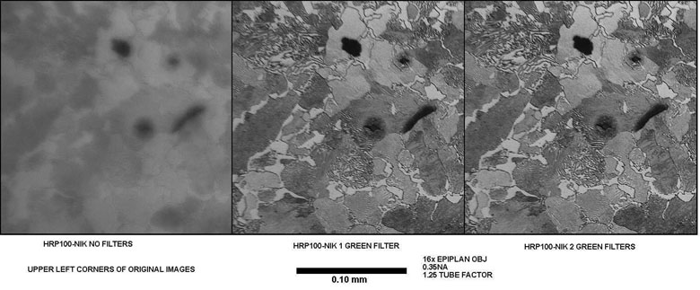
2) The specimen in Figure 2 is a
metallographic specimen of pearlitic gray iron. Clifton Sorby was
the first person to see such a microstructure. The pearlite makes
a fine resolution test subject in reflected brightfield illumination because
it is composed of alternating thin layers of nearly pure alpha iron and
cementite (iron carbide). This microstructure in a properly polished
section is made evident after etching with a 2% solution of nitric acid
in alcohol (nital etch) which selectively dissolves the alpha iron leaving
the cementite plate edges standing in relief.
Sorby discovered the two-phase structure
associated with pearlite in about 1880. He is probably the one who
named it pearlite because his early metallography in the mid 1800's was
done at too low a resolution to resolve this two phase structure and it
looked "pearly" to him. His early work was with low power objectives
and the vertical illumination introduced by a reflector between the specimen
and the objective. His later work was with the light coming from
above a high powered objective with the objective acting as its own condenser.
References
1) Clarke T, M., 'High-Res Digital
Camera Provides Fast, Flexible Defect Imaging'. R&D
Magazine, June 1998, 49-50.
An article describing the author's
use of the Kodak Megaplus 1.6i/AB digital camera for photomicrography when
metallurgist at Case Corp., Racine, Wisconsin.
2) Clarke T, M., 'Digital Imaging
in the Materials Engineering Laboratory', The Microscope, 1998,
46(2), 85-100.

