|
Winter and Microscopy Part 2
Richard L. Howey, Wyoming, USA |
In Part 1, we looked at a variety of subjects for examining during wintry weather. However, as it turned out, I was left with a considerable number of other items which I felt it wisest to postpone so that I didn’t end up producing a piece that would cause both you and me to doze off and have to finish reading later.
This time I would like to discuss radiolaria, Thecamoebae, hair, and some crystals. I touched on these in Part 1, but here, we’ll take a bit of a closer look. Let’s start with radiolaria. As I mentioned these “shells” are composed primarily of silica which makes them a challenge to photograph, because we are using glass lenses to try to image specimens which are essentially glass themselves. I also mentioned that whereas one used to be able to buy small vials of washed and cleaned material containing thousands of shells, now, from my most recent Internet search, the only items which are available are microscope slides containing strews of the shells. These are selling for about $6.50 U.S. The images I’m presenting you here are from slides which I made up from specimens in vials from Barbados or the Andaman Sea. However, I also have some slide specimens from a specialist which are from the Pacific and I will indicate those for you so that you can compare and contrast.
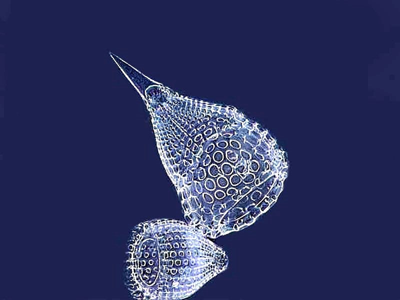
First, a couple of marvelous space capsules from Barabados and I used the “Invert” function on the graphics program to increase contrast. (PhotoImpact Pro 13)
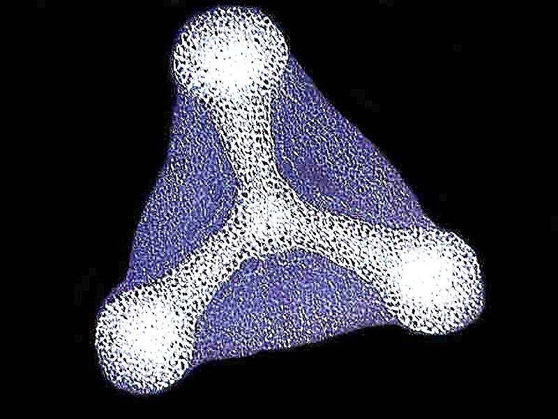
And here’s another which is still out in space making its approach to a new planetary system. Don’t you wish that we could devise such elegant space vehicles; instead we have to rely on amoebae for inspiration.
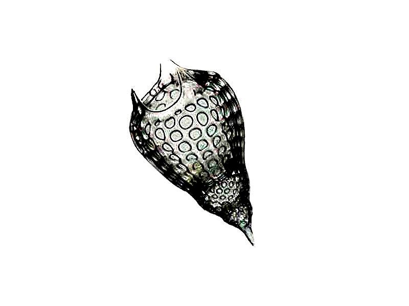
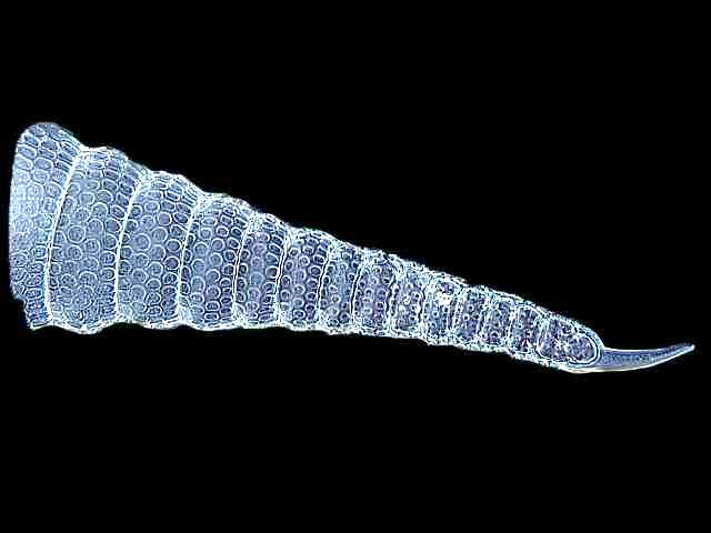
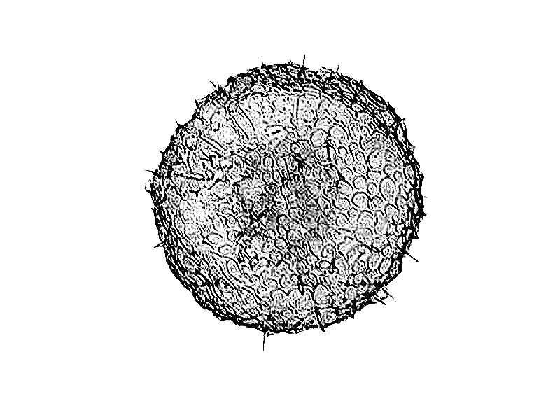
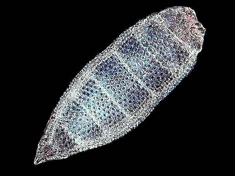
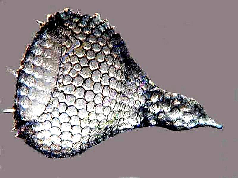
Next, we have a cornucopia or, at least, the horn without the “plenty”.
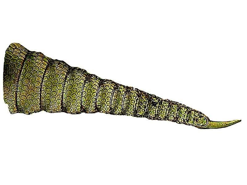
If we use the “Invert” function, we can see more detail in the surface of the shell, especially all the tiny circular “plates”.
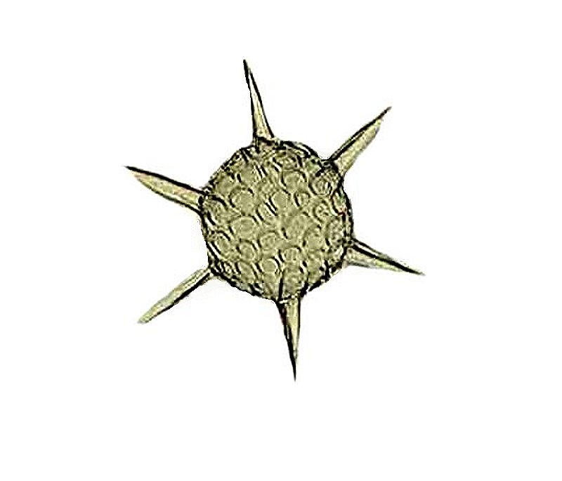
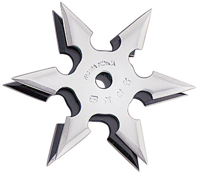
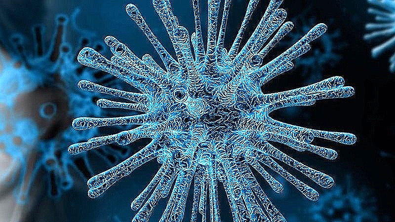
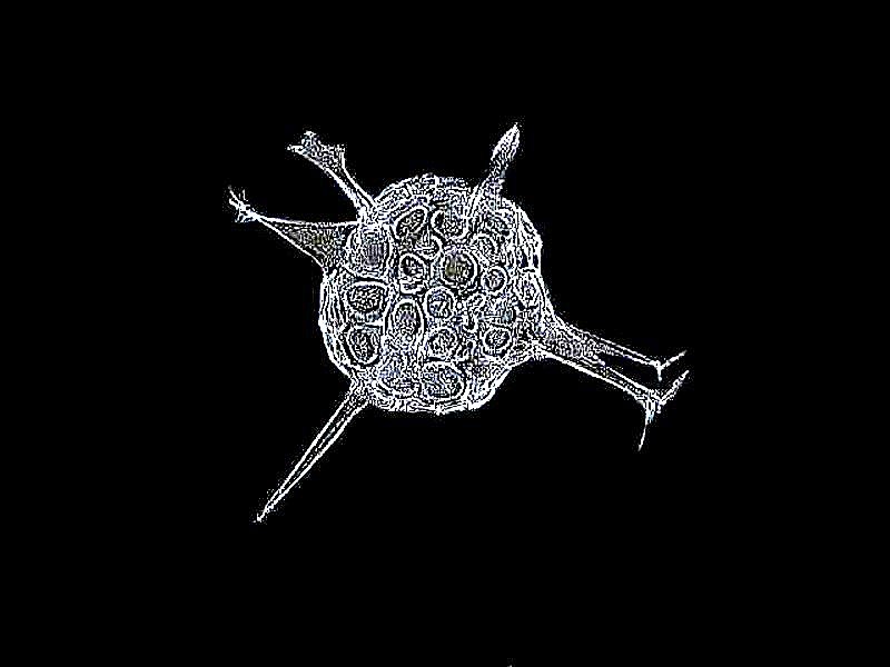
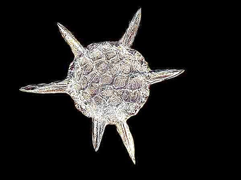
Here’s a spiky one which could be a primitive virus or perhaps an Oriental throwing star. I’ll show you the radiolarian and then a throwing star; at least they both have 6 projections. Also a representation of a virus particle which is much more spiky and looks like some of the more complex radiolaria or certain sea urchins. Mother Nature repeats patterns from the ultramicroscopic level up to the cosmic level.
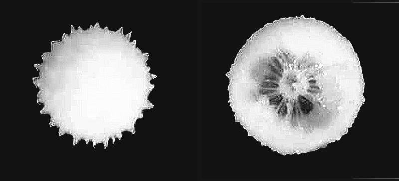
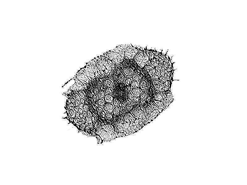
In the material from the Andaman Sea, I found a number of small spherical forms and using a micro-needle managed to chip a few in such a fashion as to reveal that inner structure and was surprised to discover a series of internal struts. I have stitched 2 images together here to show you the complete spherical form and then the “broken” form to reveal the struts.
The next 4 forms are from the Pacific ocean and I obtained these from a researcher whose assistant had already mounted them on slides.
I just can’t resist radiolaria (that would make a good song title), so here are 3 more forms from a Barbados deposit.
For those of you, who find radiolaria as fascinating as I do, be sure to investigate the extraordinary drawings of Ernst Haeckel on a wide variety of life forms, both botanical and zoological, with a generous helping of radiolaria.
Next, I would like to move on to an amoeba that doesn’t have a shell, but does have a pellicle which can get quite wrinkly and resistant. The image will be of Thecamoeba, which I discovered in a bucket which I had placed under a large hanging basket of flower on our patio. This, of course, took place during the summer. I noticed a delicious looking green scum on the surface of the bucket after a couple of months. I watered the flowers heavily with a bit of plant fertilizer mixed in and the excess water would flow down into the bucket. A great place for algae, I deduced and so I took some samples and not only did I find algae, lots of small amoebae, rotifers, hypotrichs, but also lovely specimens of Thecamoebae as well. As it turned out with an occasional bit of care, adding some water very lightly laced with plant fertilizer; and an adequate amount of indirect light, these cultures with a resplendent variety of micro-beasties survived well into the winter for detailed study. I became engrossed with the Thecamoebae in a fashion similar to my absorption with radiolaria. So, I am going to show you a variety of images using several illumination techniques.
My first encounter was not particularly impressive even though I was using DIC (Differential Interference Contrast); it looked like some green glob into which someone had dropped a half-smoked cigarette which, of course, was a bit of an algal filament.
I was curious about whether or not such filaments were a staple in the Thecamoeba’s diet so, I isolated some filaments, which were present in abundance, and stained them with Neutral Red. I conjectured that this dye in a very low concentration would be essentially non-toxic. I isolated some Thecamoebae and added the stained algal filaments to the culture dish. A couple of days later, I extracted a specimen and photographed it. You can see the result below with a very small piece of a stained filament just above the large vacuole.
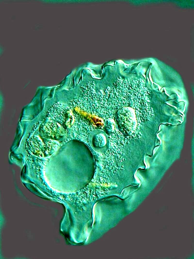
I found it curious that the large vacuole seemed to curve down inward into the protoplasm whereas the 3 small structures just above it and slightly to the right seemed to project upward, as did the algal fragment. I decided to alter the color for contrast and then to invert the image and the result was, as you can see, that all of those items were reversed.
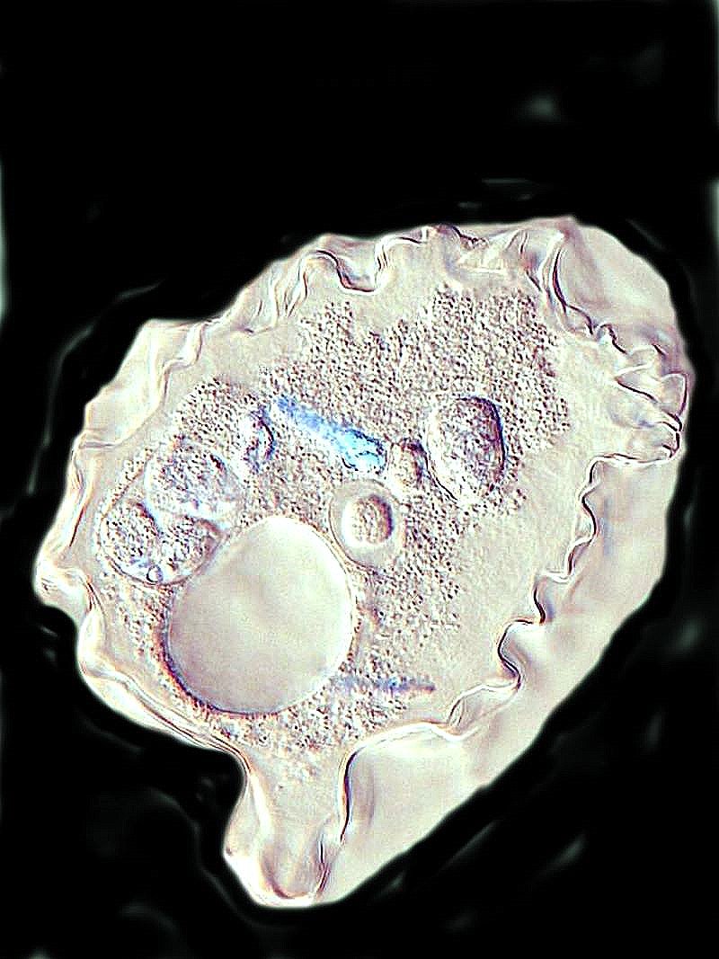
So, do the vacuoles project up from the pellicle or are they embedded such that they are like a bowl? The former, I think.
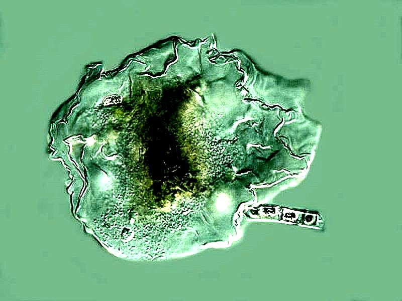
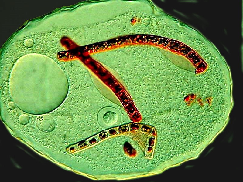
A few days later, I extracted another Thecamoeba after they had had a chance to indulge their appetites further and the results were much more interesting. First of all, there were 3 relatively long algal filaments and 2 remnants of what also appear to have been filaments packed into little pear-shaped structures. (small vacuoles?). Again, there is a very large spherical vacuole which appears to be empty, this time on the left side of the organism. Just below center is a small, clear vacuole which has content, but it is not stained, so it may well be a tiny protist that the Thecamoeba ingested. If you look carefully, you can see partial vacuoles which have formed along portions of the long filaments.
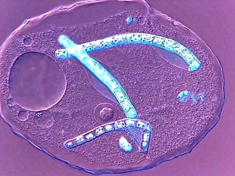
Once again, I decided to apply the “Invert” function and the result is interesting, because the filaments now stand out in a sort of neon or electric blue.
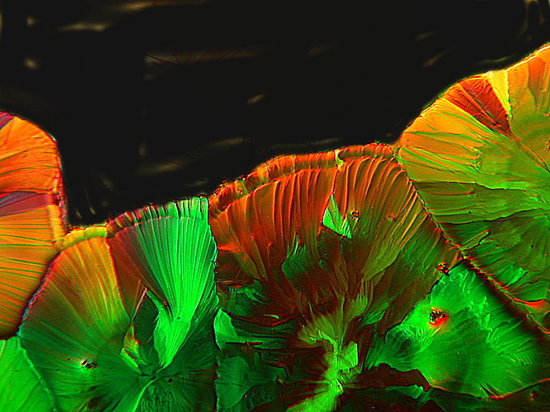
Next up, I’d like to show you some of my more recent images of crystals formed with a variety of chemicals photographed with polarized light. Some examples are ones where the slide has been allowed to dry and then is subjected to low heat to remelt the crystals. I will indicate such images.
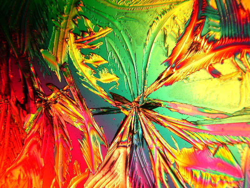
The first image is of crystals of Sodium citrate which was dissolved in hot water when initially prepared. To me, it looks like a cluster of alien carnivorous plants.
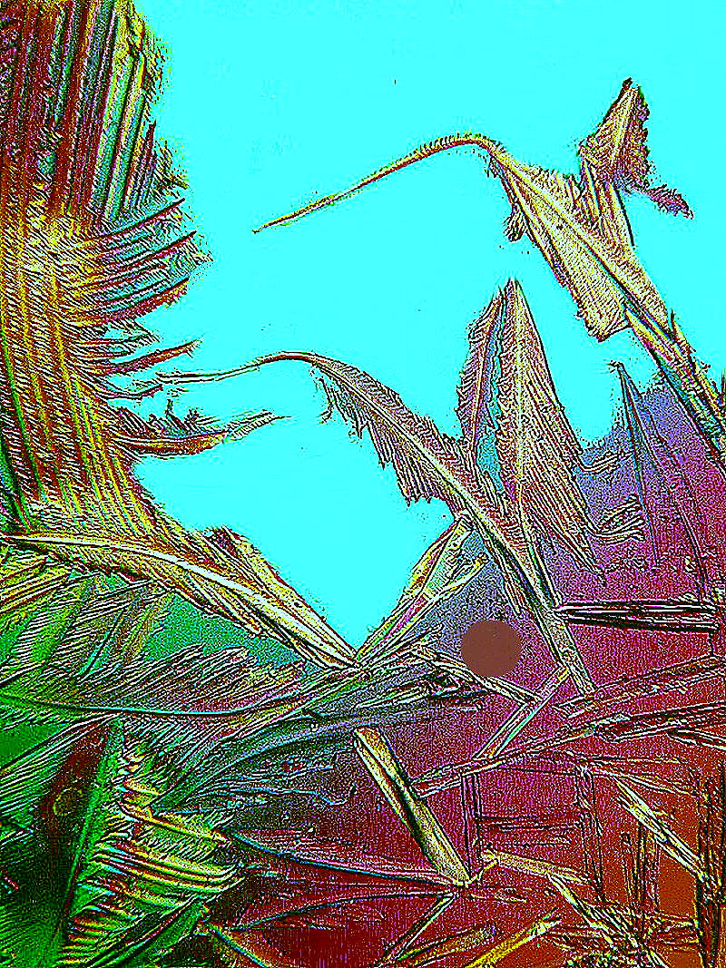
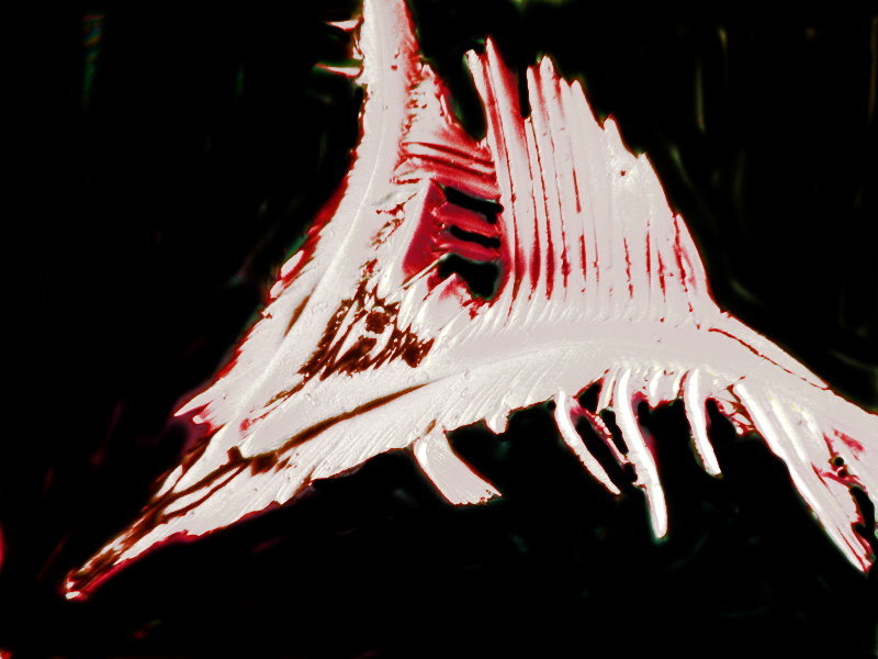
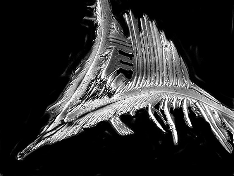
The next image is also Sodium citrate prepared hot. It looks like an alien warrior whose face shield has been blasted by a laser. I know, I just can help it; I’ve been isolated too long so my imagination runs rampant. It’s a severe case of pareidolia and the doctors tell me that it won’t end before this article is finished. So, consider yourself warned.
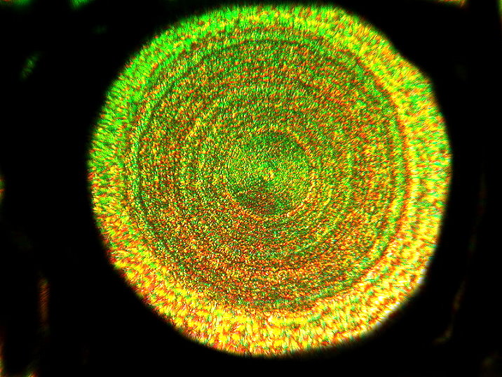
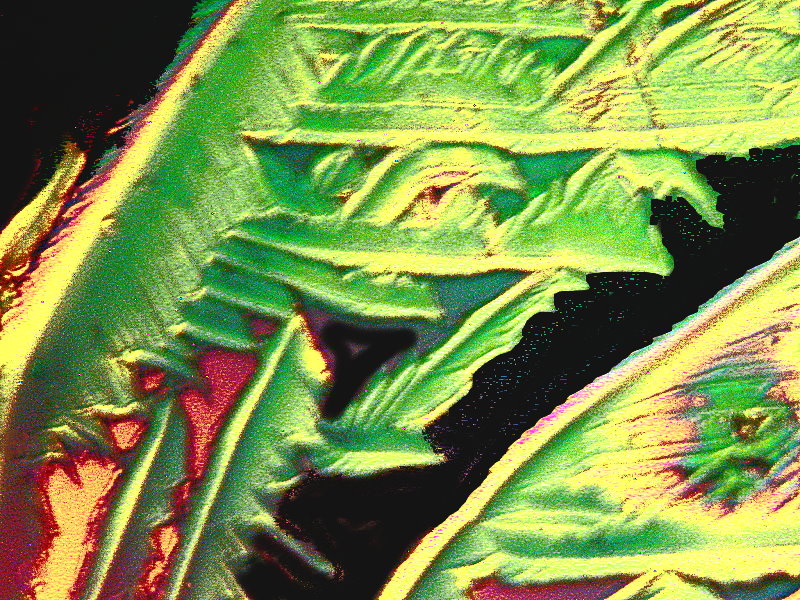
When I was growing up, we had a number of “rag” rugs in our house, some large and rectangular, and others small and round like the one below. This is a mixture of Cream of Tartar from our kitchen and Ascorbic acid and the preparation has been remelted.
The image below was taken from the same slide.
Back to Sodium citrate; this one reminds me of the leaves of some exotic plant.
And clearly, another aspect of alien carnivorous plants.
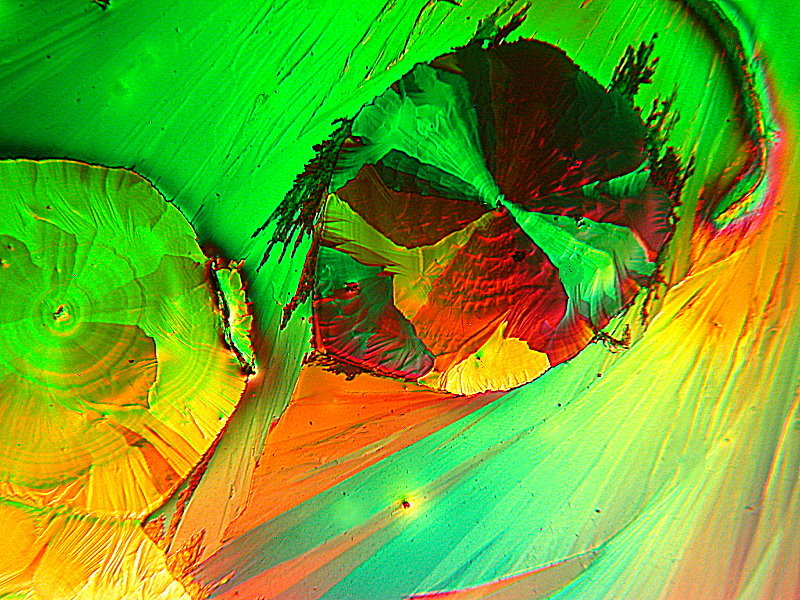
Sodium citrate has, in terms of crystal formation, some of the characteristics of Ascorbic acid. Here is an example where we get a disk rather like those of Ascorbic and, in this case, other flattened disks radiating out from the center.
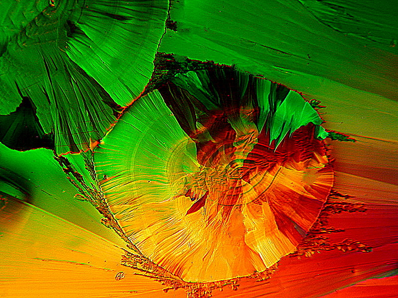
The next Sodium citrate image clearly represents a cosmic event of magnificent proportion.
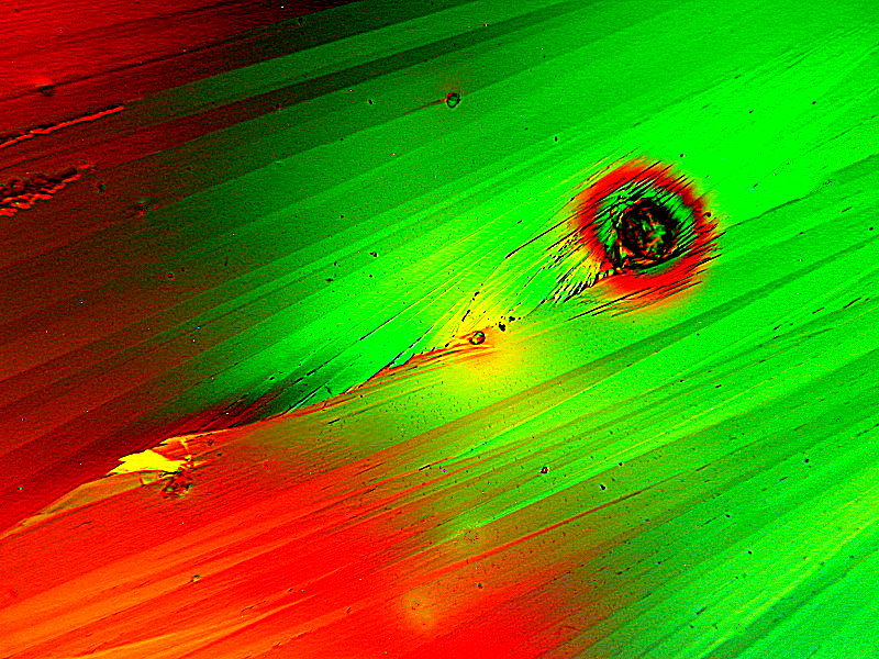
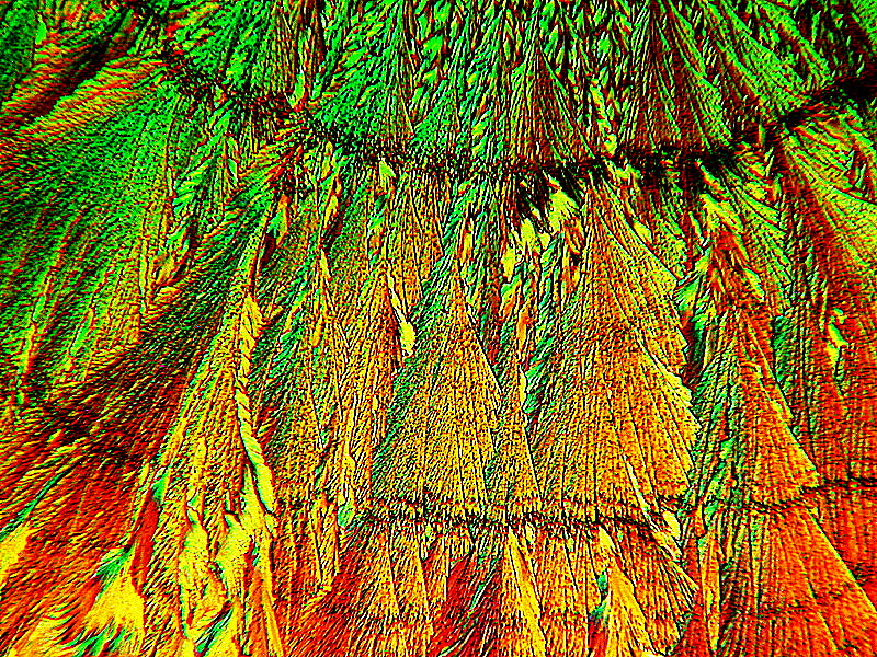
And finally, just to show you how diverse crystals on the same slide can be, I’ll show you 3 images. The first one is a whirl of color; the next 2 I have modified the color, but not the forms and I named this “Skeleton of Dimetrodon”–an ancient fossil reptile. Dimetrodon was actually a type of prehistoric reptile known as a pelycosaur, and it lived during the Permian period, 50 million years or so before the first dinosaurs had even evolved. Both the “Whirl” and the “Dimetrodon” images were on a slide which was mixture of Urea and the biological stain Nigrosin and then the slide was a subjected to remelting.
In my introduction, I mentioned hair as another source of objects for wonder micro-recreation, but this article has already gotten quite long and I remembered that I had done a couple of articles on hair and one of them is a fairly good survey, so I’ll provide a link to it here.
It’s in 3 parts, so if you get interested, you can look up the other 2 parts. I hope you enjoyed this winter excursion and can find many other specimens to help get through the inclement weather.
All comments to the author Richard Howey
are
welcomed.
If email software is not linked to a browser, right click above link and use the copy email address feature to manually transfer.
Editor's note: Visit Richard Howey's new website at http://rhowey.googlepages.com/home where he plans to share aspects of his wide interests.
Microscopy UK Front Page
Micscape
Magazine
Article
Library
Published in the August 2022 edition of Micscape Magazine.
Please report any Web problems or offer general comments to the Micscape Editor .
Micscape is the on-line monthly magazine of the Microscopy UK website at Microscopy-UK .
© Onview.net Ltd, Microscopy-UK, and all contributors 1995
onwards. All rights reserved.
Main site is at
www.microscopy-uk.org.uk .