|
Winter and Microscopy
Richard L. Howey, Wyoming, USA |
Every self-respecting microscopist, who lives in an area where winters allow very limited, if any, possibilities for field trips and collecting, should make plans for having lots of specimens to carry one through the inclement periods. Where I live, it’s a matter of snow, ice, subzero temperatures, and high winds. That is not a combination which is conducive to outside ventures. As a consequence, most of my comments will be directed to areas with cold climates.
So, here are some things you might try to keep yourself in a position to indulge in your passion for microscopy and natural history.
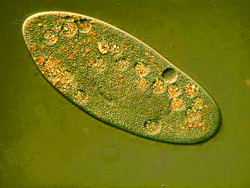
1) While you still have access to live samples, start some cultures in your lab. I always like to have Paramecia cultures going. They are usually quite easy to keep going and produce abundant specimens. They are also amazingly complex and challenging and just because they are relatively common, one shouldn’t dismiss them as uninteresting.

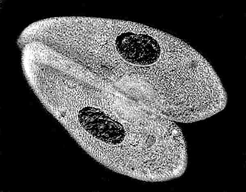

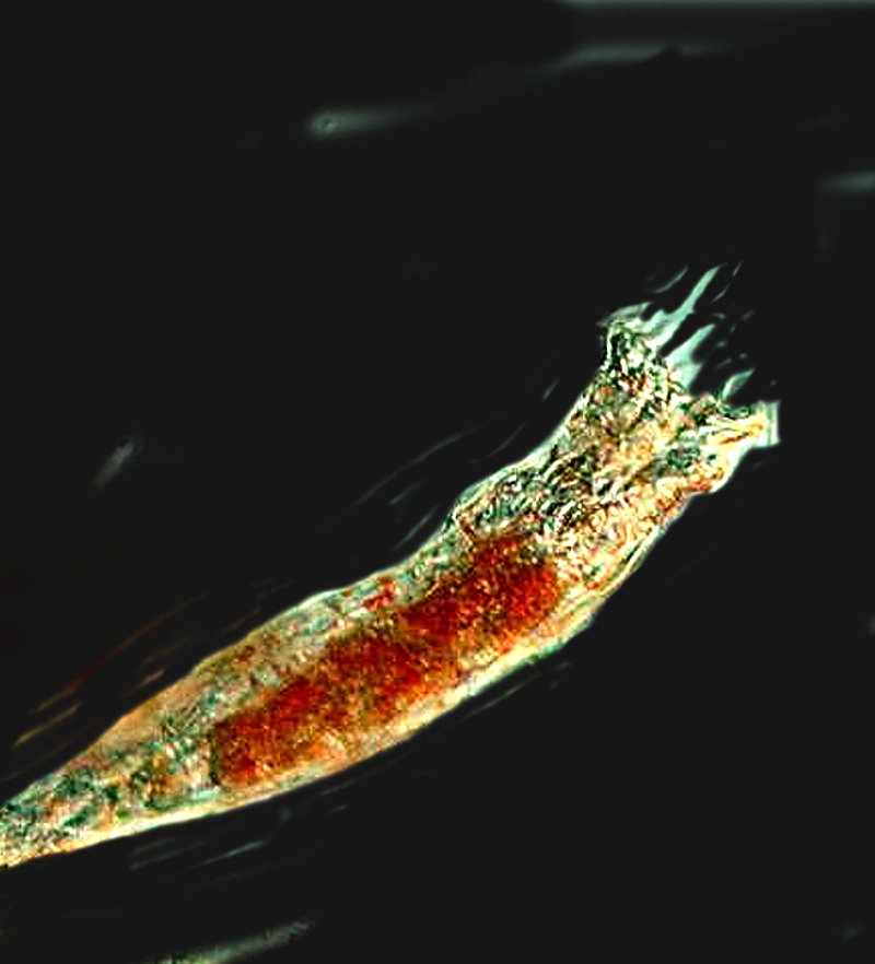
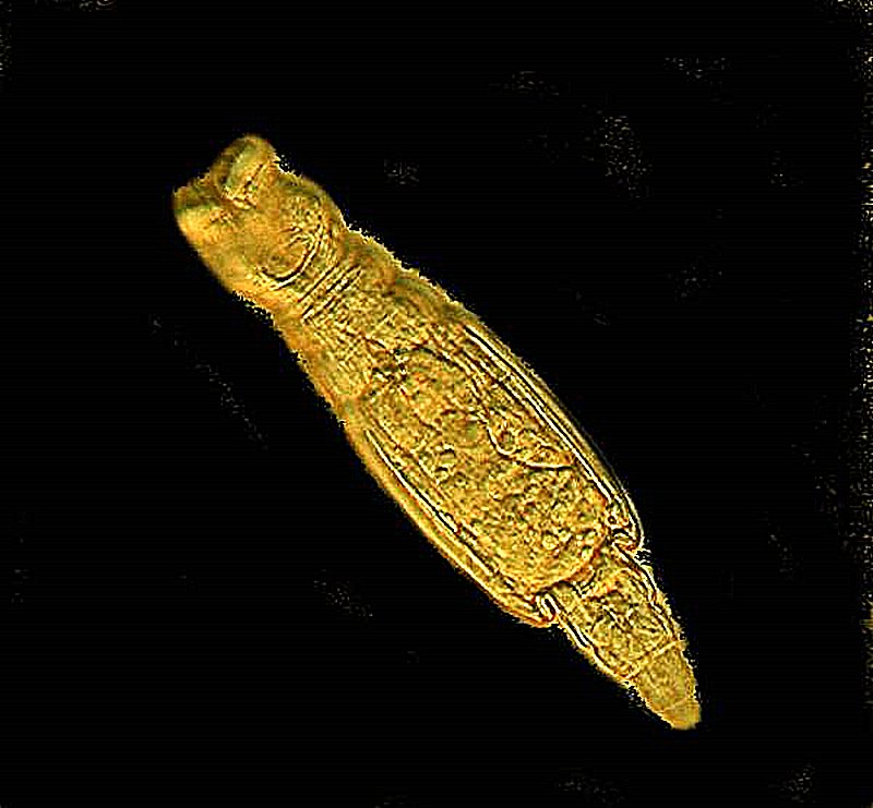
They offer many avenues of exploration. Rotifers, particularly, bdelloid forms are readily cultured and can multiply in large numbers producing eggs which are interesting to examine. Here is an example of a bdelloid form.
Nematodes are also readily available in soil samples and occur in a surprising variety.
If you have some dried plant matter or animal dung, these can separately or together produce some interesting cultures. Cow dung can provide Spirostomum in enormous numbers. First, an image of a single specimen. The largest species S. ambiguum and S. teres can reach lengths of over 2000 microns and they have a chain or beaded macronucleus. They are highly contractile and claims have been made that they set the speed record in the biological world for contractility.

Here is an image of a cluster of Spirostomum and this is only a small portion which I separated out from a larger mass.

Soil and dung samples are also a source of a variety of Colpoda which will encyst and divide while encysted. These samples will also provide a variety of tiny amoebae, some of which are highly unusual and some are potential pathogens, so should be handled with some care.
The first image is of naked amoebae found in soil and the second shows some of the types of testate amoebae.

Naked amoebae isolated from Sourhope soil (selection). a Saccamoeba sp., b Mayorella vespertilioides; c Korotnevella stella; d Vexillifera bacillipedes; e Platyamoeba placida; f Acanthamoeba sp. Scale bar: 10 μm.
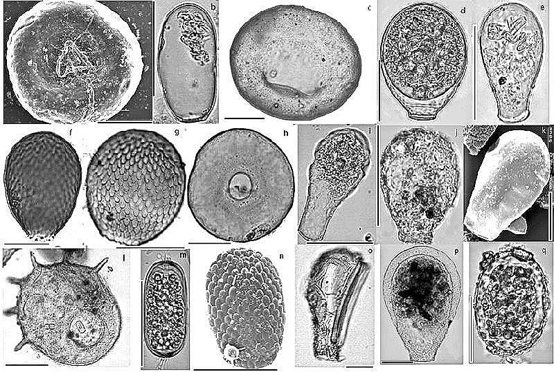
Examples of the diversity of testate amoebae from soils and mosses: a) Trigonopyxis arcula, b) Hyalosphenia subflava, c) Bullinularia indica, d) Nebela tincta, e) Nebela militaris, f) Assulina muscorum, g) Assulina seminulum, h) Arcella arenaria, I) Hyalosphenia elegans, j) Physochila (Nebela) griseola, k) Hyalosphenia papilio, l) Centropyxis aculaeta, m) Archerella (Amphitrema) flavum, n) Placocista spinosa, o) Difflugia bacillifera, p) Nebela carinata, q) Amphitrema wrightianum. Scale bars indicate approximately 50 µm except for A. muscorum: 20 µm.
Some years ago, I had, during the summer, 20 or so hanging plants out on our patio. Under 2 of them that required a lot of flow through watering, I placed plastic buckets. Over time, these developed a ripe, green color on the surface of the water in them. I got curious and took some samples and was delighted to discover a nice variety of filamentous algae and amoebae, including some Thecamoeba which I had not seen before. I was especially pleased by the fact that in the lab, given proper attention in terms of light and a bit of plant fertilizer, they grew in abundance and I was able to study them in some detail during the winter.
First a very ambitious little amoeba feasting on an algal filament which is longer than it is.

However, I have 2 examples of yet even more ambitious little amoebae. The first one has wrapped itself around a rotifer that is either dead or dormant (encysted). You can see it has managed to go all around the rotifer to engulf it and then, I gather, to have a very, very substantial meal.

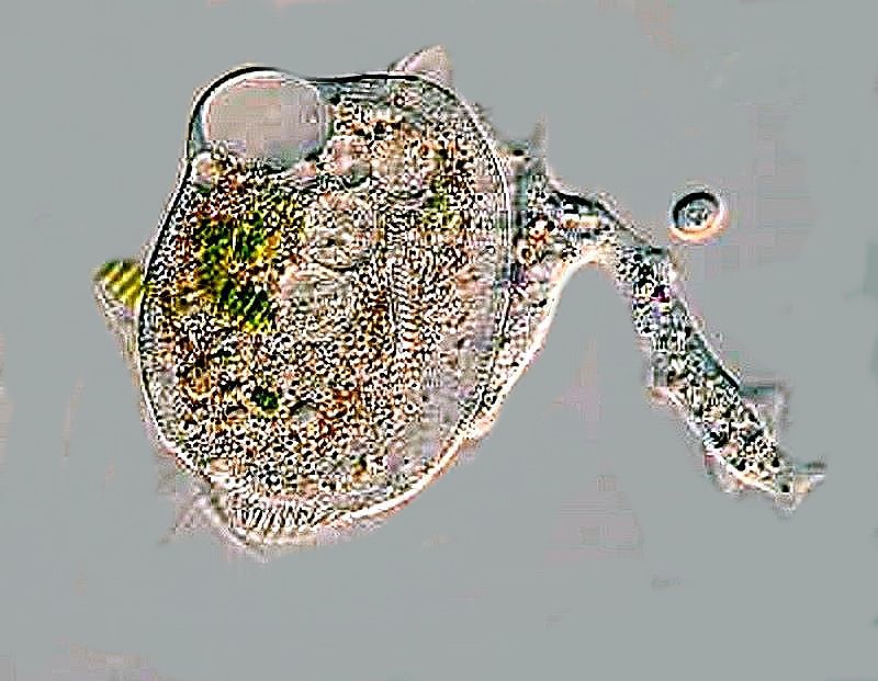
The classic textbook amoeba is Amoeba proteus which, curiously, is often difficult to find in nature. The good news is that it cultures easily and biological supply houses always have it in stock if you can’t find any in your collecting expeditions.
I’ll show you 2 images of the same specimen and even though the illumination is different displaying different coloration, it is clear why this organism has the species name of “proteus” since between the two photos, it has significantly altered shape.

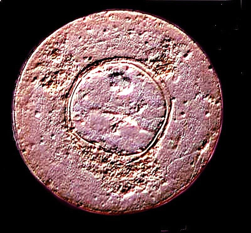
A common testate amoeba is Arcella and this is a view showing the aperture through which the protoplasmic pseudopodia flow to feed and move the “shell”.
A fascinating, very strange critter related to the foraminifera is Gromia, and there are at least a couple of freshwater species, although the vast majority are marine. For a bit over 2 years, I maintained a small marine aquarium and I found a few specimens and my friend, Mike Shapell and I, studied them in amazement. We looked at them using a Wild Heerbrugg inverted microscope and observed that these specimens of Gromia could extend their pseudopodia out to 8 or 10 times the length of their shell. At higher magnifications using phase contrast, we noticed that protoplasm was flowing in both directions along the pseudopodia in the manner of a 2-lane highway. We watched in astonishment as small diatoms would be transported back toward the “oral” aperture while protoplasm on the other side was flowing in the opposite direction. So, keeping a small marine aquarium to grow algae and protists without worrying about fish food and predators is enormously rewarding and relatively easy. It can be a source of many, many wonders for the microscopist.
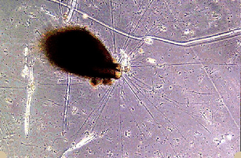
Another delight are the freshwater Helizoa. This example looks like a micro-pincushion with its spherical form and spines.

I hope that you made a wise investment a few years back and bought a vial or two of radiolarian ooze. Each vial, which cost between 10 and 20 U.S. dollars, contained thousands of shells and was a source of hours and hours of wonder and delight. Today, I did an Internet search and was unable to find any radiolaria for sale, except for prepared slide strews and each slide is about 5.50 + shipping. So, if you have a friend, acquaintance, or fellow member in a microscopical society, who has a vial or two, see if you can arrange to buy a small portion. A very small amount contains enough material to keep you engrossed for a long time. I’ll show you a few examples of radiolaria shells. The living organisms are even more amazing and many look like something out of an alien world.
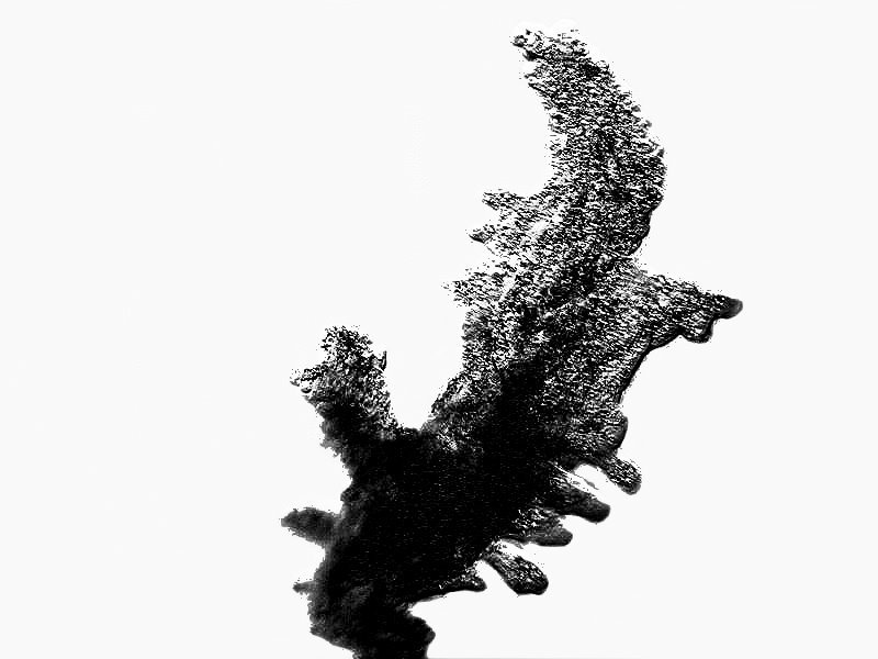
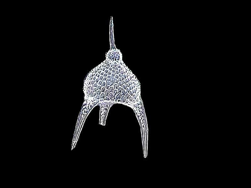
I was inclined to go into a minor discourse on radiolaria and foraminifera at this point, but have decided to postpone that for a Part 2 article on winter microscopy. However, to whet your appetite, I’ll show you an image of a radiolarian that, to my mind, looks like an alien moon lander. That will be followed by a DIC image of a cross section of a fossil foraminifer which looks like it could be part of a stained glass window.

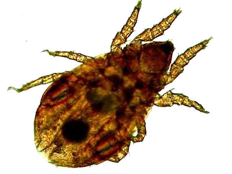
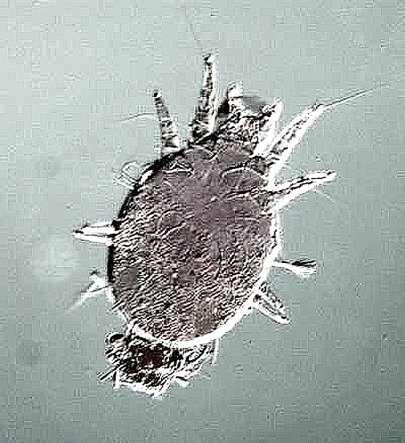
Soil samples can also frequently provide a considerable variety of specimens of tiny mites. If you can’t get soil samples from your frozen tundra, try some soil from around a houseplant.
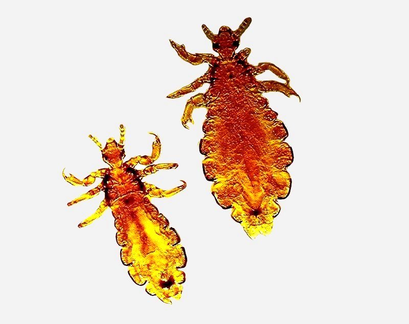
Perhaps you are fascinated by mighty mites which, by the way, are tiny arachnids, so you might then wish to forage through the gatherings of your vacuum cleaner seeking dust mites or simply scrape a bedsheet that hasn’t been washed for a month or two. Dust mites are extremely common, but somewhat elusive and some people have had difficulty finding them, whereas others find them in disconcerting numbers.
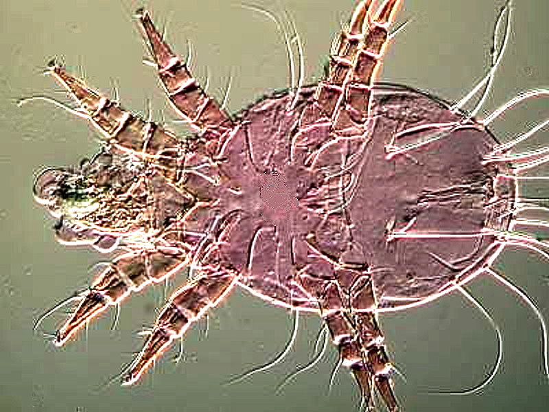
One afternoon, I was looking at a dried specimen of a large ground bee and noticed something down in the hairs which when extracted turned out to be a mite.
2) During the winter months, people often have flowers growing in pots in the house or acquire the occasional bouquet. These can provide a wealth of material; pollen, sections of pistils and stamens, leaves, and, of course, flower petals. These latter often exhibit an interesting cellular structure which can be enhanced by the colors. Other times, it’s necessary to bleach them to get a good glimpse of the cytology and then, you might want to follow up with some stains to differentiate detail.
3) Many people are fascinated by the intricate geometric forms in snowflakes. Observing these, let alone photographing them, is a major challenge. If you’re still youthful and hardy, you may want to set up a lab out in your freezing garage with a microscope (preferably one not incredibly valuable or expensive), let the stage get nice and chilly, but not frosty, then put some chilled slides out in the snowfall, rush them to the stage and photograph as fast as you can. If the snow’s not falling, then you’ll have to scoop some up, sprinkle it on a cold slide and again do the winter hustle. At my ancient age, I’d rather stay inside and imbibe some Ancient Age. (For those of you not familiar with American whiskeys, this is a bourbon of 80 proof distilled from corn, rye, and barley malt.) However, if you, like me, are averse to exposing yourself to the vicissitudes of winter extremes and yet still have a passion to photograph snowflakes, you might try one or more of the following.
A.) Use an older microscope that has plenty of room between the tip of the objective and the stage. Now comes the challenge; you need to devise a small, low box, either out of wood, metal, or plastic, slightly larger than a standard microscope slide. Ideally, you will fashion something with 3 sides and the fourth side open so that you can slide in small pieces or chips of ice or dry ice. Then cover the top of your device with a thin strip of metal that will conduct the chill from the ice. Place a chilled slide on the strip containing some snowflakes that you have harvested from outside (and kept surrounded by ice, of course) and place them on your slide and quickly focus and start taking pictures. If you use dry ice, be sure not to handle this except with tongs or cheap plastic tweezers to avoid nasty burns to your skin.
The idea of this apparatus is purely fantasy as I have not tried it and, if you do, I’m sure you could find many ways to improve it. So, write an article for Micscape and share your design and photos of both the device and some snowflakes with all of us.
As for me, I am content with the marvelous photos of Bentley which have been reprinted in Dover edition and the more recent work of Libbrecht, who is a professor of physics at Cal Tech. Both of these works are available at modest cost and save you going out to buy thermal underwear.
Snowflakes in Photographs (Dover Pictorial Archive)
by W. A. Bentley | Sep 18, 2000
Capturing Snowflakes: Winter's Frozen Artistry
by Kenneth Libbrecht and Rachel Wing | Oct 5, 2021
Another source of microscopic pleasure is hair, believe it or not, and even your own and that of your pets. I’ll give you just a few examples and then refer you to some more extensive articles which I devoted to examining various types of hair. You can also find hair samples at craft shops, barber shops, and sports shops in the section with fly tying supplies, or as I like to say to annoy my friends who are fishing aficionados, tie flying.
My hair used to be a sort of dull blond which has gotten much grayer, but here’s what it looked like a few years ago with the help of a dash of polarized light.

Quite colorful, don’t you think? Even my gray beard is brightly differentiated under the microscope, but I don’t think I would care too much for it if the whole thing looked like that in ordinary light and I had to go out in public with such a display.
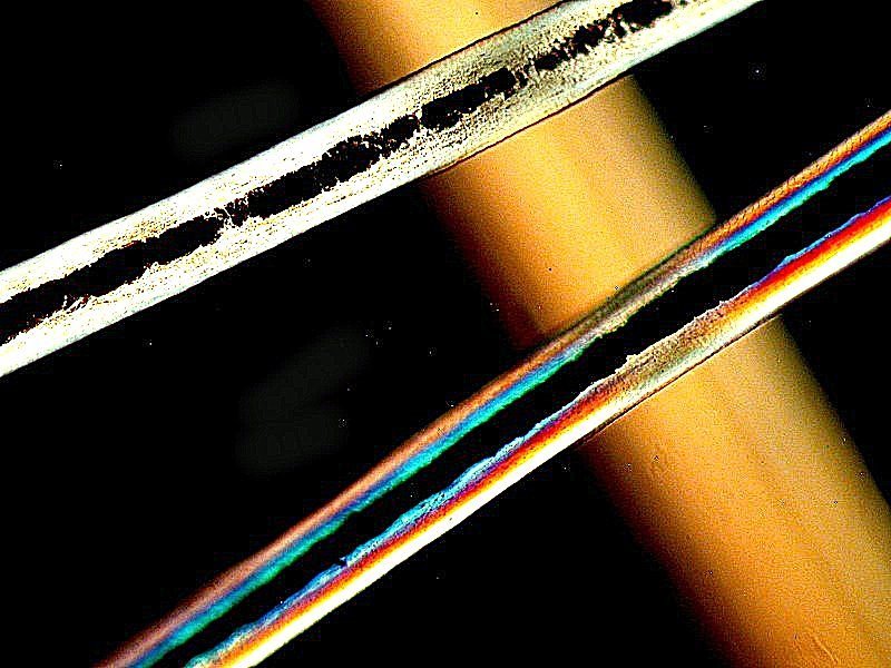
The next example is quite spectacular and is from our pet tiger. Well, O.K., it is tiger hair, but it’s on a slide which I bought some years ago on Ebay.

As I mentioned, I wrote several articles for MICSCAPE which have to do with hair, but the most extensive one can be found at this link.
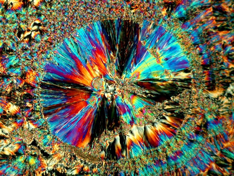
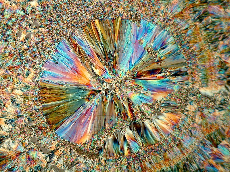
Finally, you should consider items which are readily found around the house in kitchen cabinets and medicine cabinets. I’ll give you just one example here, some crystals of salicylic acid produced from a small bottle of wart remover. This particular preparation also contained Ascorbic acid (Vitamin C) and you can see the affect of it in the “disks” in the last 2 images. Salicylic acid has a wide variety of dermatological uses and is readily available in your local pharmacy.
I’ll show you 4 images here and as you can see, they are quite vibrant when photographed with polarized light.

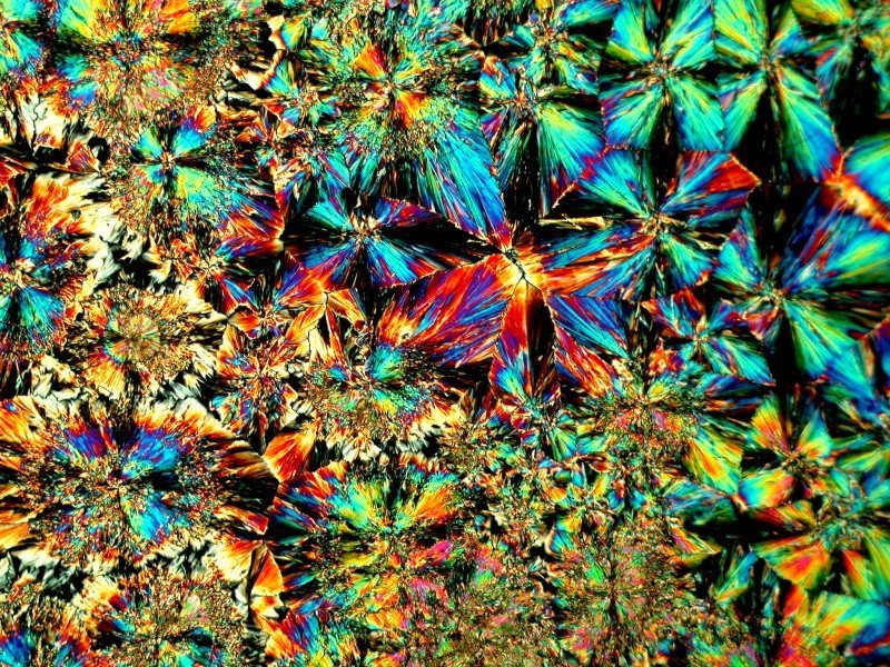
There are an enormous number of household items that can supply hours and hours of pleasure and discovery. Experiment by always using common sense and know in advance any potential risks of mixing different substances. With a bit of common sense and ingenuity, you can have a winter wonderland in your own home laboratory.
All comments to the author Richard Howey
are
welcomed.
If email software is not linked to a browser, right click above link and use the copy email address feature to manually transfer.
Editor's note: Visit Richard Howey's new website at http://rhowey.googlepages.com/home where he plans to share aspects of his wide interests.
Microscopy UK Front
Page
Micscape
Magazine
Article
Library
Published in the July 2022 edition of Micscape Magazine.
Please report any Web problems or offer general comments to the Micscape Editor .
Micscape is the on-line monthly magazine of the Microscopy UK website at Microscopy-UK .
© Onview.net Ltd, Microscopy-UK, and all contributors 1995
onwards. All rights reserved.
Main site is at
www.microscopy-uk.org.uk .