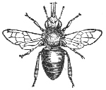


A brief look at some of the features and fascinating adaptations of insects
which can be easily seen with a low power microscope. Suppliers of prepared slides of the subjects discussed are
provided.
This month - insect eyes, and the amazing legs of the honeybee.
Note added July 2005: Also see the 'Interactive Honey Bee worker' page on this website where larger
images of the honeybee worker parts can be freely downloaded for personal and/or educational use.
How many eyes does an insect have?
 Two?
Yes some do, but in fact many insects have five. In addition to the multi-faceted compound eyes that are an obvious
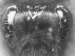
feature of larger insects, many also have an additional three simple eyes in the centre of the head, called ocelli.
If you find a dead bee in your garden have a good look at the head with a 10X hand lens or low power stereo microscope. The black beads between the compound eyes are the ocelli and can just be seen on the head of a bumblebee shown right (highlighted by the white lines).
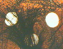 The ocelli are simple structures and are believed they only detect light levels and movement. In contrast,
the compound eyes are made up of a number of hexagonal facets which resembles a honeycomb and are capable of seeing
much more. The number of facets effects the visual acuity and varies amongst different insect species. The honeybee's
eye is estimated to have one per cent of the visual acuity of a human eye.
On the left is an image of a prepared slide of the ocelli of a honeybee (3.5X objective used). During the slide preparation the ocelli themselves are destroyed to leave the three holes in the epidermis.
The hairy eyed honeybee
 Hairs
on the eyes don't seem a particularly useful feature for an insect ..... won't the hairs obscure the field of view?
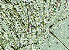
This thought crossed my mind when I noticed from a prepared slide that the cornea of a honeybee (worker) compound
eye is very hairy. An image of the cornea is shown right (using a 9X objective). The hexagonal facets of the eye
can just be seen but they are not very distinct using transmitted light. (See
reference 2).
 Hairs on the
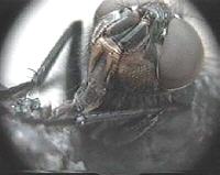
eye are not unique to the honeybee, but most insects such as the fly shown left have hairless compound eyes. If
you click on the image you get the extra treat courtesy
of Mol Smith of an avi video (128kB) showing the fly using it's proboscis to suck up nutrients.
When the honeybee visits a flower, pollen collects on the body, legs and head including the eyes and antennae. The way the honeybee transfers this pollen to the honey baskets on its hind legs is fascinating and worth a closer look ....
The amazing legs of the honeybee (worker)
The legs of many insects are specially adapted to the insect's way of life, one classic example being the honeybee
worker (Apis mellifera) shown at the head of this article.
One way to appreciate these adaptations is to purchase a set of prepared slides of a honeybee (worker) and to study
them at 40X-100X magnification under a microscope. (Click here to see suppliers of prepared slides). Also have a look at a complete bee, if you find a dead one (or
can be obtained preserved from biological suppliers), with a 10X hand lens or stereo microscope to become familiar
with the whole insect. Images of selected detail of the three legs are shown below (annotated with the aid of ref.
1), but the reader may wish to read some of the many resources on the honeybee to find out how these features play
a role.
The first leg selected detail 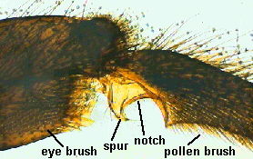
The middle leg selected detail 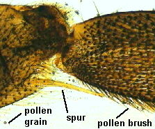
To appreciate size of a pollen grain, one that is caught in the hairs is highlighted above.
The hind leg selected detail 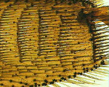
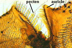
The inside of the leg is not shown, which is where the 'pollen basket' is found.
Sources of prepared slides and courses to make your own
In the US, Carolina Biological Supply Co. can offer prepared slides which may include the honeybee. Edmund Scientific offer the slide set 'Life and Structure of the Honeybee' (Catalogue No. A39,182) including slides showing all the features described above.
Microscopy can be a useful diagnostic tool for beekeepers. The Field Studies Council in the UK often offer a 'Microscopy for Beekeepers' course each year. Contact them for their Course Guide. Field Studies Council, Preston Montford, Shrewsbury, Shropshire, SY4 1HW, UK.
References and further reading
If you've ever wondered what the world looks like to a honeybee then visit the fascinating 'B-Eye' Web site. A computer program is used to mimic the way a honeybee views
the world.
There are a large number of Web sites discussing all aspects of bees, honeybees, beekeeping and bee diseases. Try a few keywords on a search engine.
1) Animals Without Backbones Vol. 2 by R Buchsbaum. Pelican Books 1951 (and other editions). This classic two volume work has an excellent illustrated summary of the adaptations of a honeybee leg and how they are used to collect pollen. The images above of prepared slides were annotated with the aid of this book.
2) Microcosmos by J Burgess, M Marten and R Taylor, Cambridge University Press, 1987. A scanning electron micrograph of a honeybee's hairy compound eye is shown on page 60 and compared with that of a hover fly which does not have hairs.
3) Encyclopaedia Britannica, 1993, 15th Edition. All the larger encyclopaedias should have a thorough introduction to insects. The ninety two page section on insects in Volume 21 of this encyclopaedia is excellent, and a book in itself.
4) Encarta 94 Multimedia Encyclopaedia has a good overview of the honeybee although it does not illustrate or discuss the adaptations of the legs.
5) My colleague Andrew Syred who is a beekeeper also recommends 'Anatomy and dissection of the Honeybee' by H.A.Dade - ' 'cos it's brilliant'.
Comments to the author (who is not a bee or insect expert) and would therefore particularly welcome comments or criticisms. Thanks to Andrew Syred for providing useful comments on the article, but any mistakes are all mine!
The worker bee drawing at the head is a scan of Figure 181, page 204 'The Hive Bee (Worker)' from W S Furneaux's 'Butterflies, Moths, Other Insects and Creatures of the Countryside', published London, 1927.
The slides were prepared by the author using material kindly supplied by the late and sadly missed Eric Marson (Northern Biological Supplies) at one of his excellent slide making courses at Belstead House, Ipswich .
Please report any Web problems or offer general comments to the Micscape Editor,
via the contact on current Micscape Index.
Micscape is the on-line monthly magazine of the Microscopy UK web
site at Microscopy-UK
NAME="emailer" ARCHIVE="email.jar" CODE="email.class" WIDTH="1" HEIGHT="1" ALIGN="BOTTOM">