Of all the attempts to produce variations on the McArthur plan, none succeeded so brilliantly as the Nikon Model H ("H" for hand held). This high-precision instrument was manufactured for only a few years in the late 1960s at reasonable cost, then quickly priced itself out of the market and disappeared. It is one of the most remarkable microscopes ever built, the ultimate in a compact instrument of unquestioned professional quality. Two models, one bright field with accessory polarizing filters and dark field stops, the other phase contrast (Model HP), performed on a par with the best bench models, all in a body 11cm x 14cm x 5cm (4.33" x 5.51" x 1.97"), weighing about 800 grams ( 29 oz). Length and breadth were similar to the McArthur, but height and weight were slightly greater due to refinements and design alterations.
Models H and HP were available with a choice of ten achromatic objectives, six of which were for bright (and dark) field, the other four for phase contrast. The high eyepoint wide field compensating eyepiece had a rubber protective ring for spectacle wearers.
 The
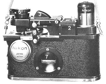
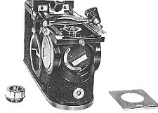
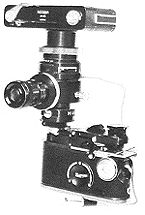
Nikon Model H (right side) with supplied metal strap, shown in closed position.
The
Nikon Model H (right side) with supplied metal strap, shown in closed position.
1) Eyepiece
2) Battery compartment
(for 2 x AA batteries)
3) Lamp housing
4) Condenser setting knob
L=low power, H=high power
5) Omni-directional mirror (closed)
6) Slide 'clips'
(spring loaded rubber rollers)
7) Focusing wheel
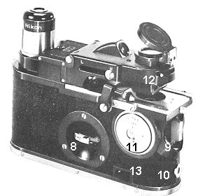
Nikon Model H (left side) in
operating position
8) Focusing wheel with lock
9) Objective changer knob (choice
of three)
10) Mini-socket for transformer
cord and house current
11) Port for immersion oil
12) Diaphragm control with filter
slot beneath
13) Lamp on-off switch
I was fortunate in obtaining a Model H in 1969, late in its manufacturing run. It has been my constant companion under every conceivable circumstance, going on expeditions from the Lesser Antilles to Newfoundland, from the high Andes to Philippine rain forests, from still-hot lava to meadows at home, and a hundred other places my studies have taken me. Despite frequent use and wide travels, this irreplaceable instrument is in nearly mint condition due to the care I have lavished on it and the protection afforded by its own fitted leather case.
 Model
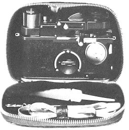
H in reinforced leather case with accessories compartment. Protective holder
for an extra objective is partly hidden at top under zipper. Immersion
oil dispenser and carrying strap also shown.
Model
H in reinforced leather case with accessories compartment. Protective holder
for an extra objective is partly hidden at top under zipper. Immersion
oil dispenser and carrying strap also shown.
The Model H with its sleek design and rounded ends resembles a classic Leica body. It is finished in chrome, black enamel, and leather like a fine camera, can be carried by a detachable metal neck strap, and is hand-held comfortably and steadily. Often I mount it on a small tripod (it has a threaded base), either for bench use, or to allow one person after the next to inspect living specimens. Biologist colleagues are frequently more interested in the Model H itself than in organisms it reveals, and invariably ask where they can get one. Unfortunately, they can't.
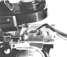
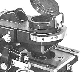 Close
view of phase contrast model with condenser annular stops selector. Phase
objectives are not interchangeable but factory aligned.
Close
view of phase contrast model with condenser annular stops selector. Phase
objectives are not interchangeable but factory aligned.
Optics are first-rate and of typical Nikon quality. The 10x ocular is threaded for security, but can be replaced by any standard eyepiece as long as the instrument is held upright. A binocular head (not Nikon) with a tubular ocular insert can also be used. The ten available objectives are special, however, with a thread much smaller than the standard Society thread. An internal rotating turret allows three to be mounted at one time, and the leather carrying case has a protective fitting for a fourth. I keep a 4x, 10x, and 20x mounted with a 40x in reserve to replace the 4x scanning lens when needed, but seldom take the 100x oil immersion into the field. (The sixth objective is a special 40X with a slightly shorter working distance for hanging drop slides.)
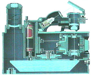 Internal
view of Nikon H. See text for description of light path. Vertical red objects
are two AA batteries.
Internal
view of Nikon H. See text for description of light path. Vertical red objects
are two AA batteries.
The accompanying illustrations are selections taken from an original 1960s Nikon brochure and the microscope's instruction manual. Note first the overall appearance of the Model H; next examine the cut-away illustration of its internal design. The long base prism, typical of the McArthur plan, reveals that the instrument is actually an inverted microscope. The optical path starts with the lamp bulb, goes obliquely to the universally adjustable mirror, is then directed downward through a diffusing disk to the substage (iris diaphragm, filter, and condenser), passes through the specimen and the objective, then to the base prism, travels horizontally through the prism, finally rises vertically into the ocular.
Cross section of rotating objective
turret. 4X in vertical position, 10X on left, 40X on right.
Objective specification (phase
objectives have same NA):
4X NA 0.10
10X NA 0.25
20X NA 0.40
40X NA 0.65 (both versions)
100X NA 1.25
 End
view showing magnification changer and external power receptacle, extra
objective, protective slide removed for exchanging objectives.
End
view showing magnification changer and external power receptacle, extra
objective, protective slide removed for exchanging objectives.
I'll take you through operating procedure step-by-step.
1. Holding the microscope in both hands, with one finger open the lock covering the indented centrally located focusing wheel. This frees the focusing mechanism which is secured during transport.
2. From beneath the stage slide out the flat plate which protects the objectives when the microscope is not in use. (An additional large aperture plate is removed when objectives are exchanged.)
3. Twist the objective changer knob at the end of the microscope to the 4x scanning lens (or 10x if the 4x has been replaced by the 40x).
4. Slide back the movable front element of the Abbe condenser to the "L" position for low power work.
5. Elevate the omni-directional mirror (folded flat for transport) and direct northern skylight into the field of view, or turn on the integral lamp (the switch is on the side of the case below the immersion oil hole). The electric bulb is powered either by two AA batteries or a 3.0 or 3.5 volt transformer and house current (a miniature receptacle lies below the objective changer).
 Condenser
assembly slightly raised to allow access to special well slide with optically
flat bottom. Note how slide is held under rubber roller clip.
Condenser
assembly slightly raised to allow access to special well slide with optically
flat bottom. Note how slide is held under rubber roller clip.
6. When a wet sample is examined, it is best to use the special well slides provided with the Model H. Each has a thin optically flat glass cemented to the bottom of the well. To eliminate jiggling aquatic specimens, an ordinary cover glass may be added to the top of the well. If a flat slide is used for an inverted wet mount with a cover glass added, the focusing range must be adjusted, after which all magnifications remain parfocal.
7. When prepared slides are used, they should be placed upside down on the stage and the focusing range must be adjusted for the additional thickness, after which parfocality is maintained.
8. To insert a slide, slightly lift the hinged condenser assembly for added convenience, although this is not necessary once you are thoroughly familiar with the instrument. (The hinged condenser assembly is lifted all the way up to change lamp bulbs.)
9. Lift the spring-loaded rubber rollers on top of the stage and insert the slide. Smooth back and forth (longitudinal) movement is provided by finger pressure on small chrome knobs on either side of the one-directional sliding stage. Lateral movement is accomplished with a single finger turning a small knurled adjustment knob below the stage. By means of gears this rotates the rubber rollers and moves the slide from side to side. In other words, the two movements provide a smoothly operating semi-mechanical stage that allows active plankton organisms to be followed with ease. (The lateral geared roller control is not shown in most of these brochure illustrations; my Model H was late in the abbreviated production run and only a few had this final modification.)
10. With one finger, or thumb and finger on either side of the instrument's body, turn the indented knurled focusing wheel until the image is sharp. Only very slight adjustment is necessary. Because all objectives are precisely parfocal, magnifications may be changed with the objective knob without removing your eye from the eyepiece. Coarse focusing is neither provided nor necessary.
11. Adjust the iris diaphragm for optimum contrast. If desired, insert a filter or dark field stop in the slot beneath the iris.
12. When changing to the highest magnifications (40x or 100x objectives), reach to the side for either of the two chrome slider knobs that return the condenser front element to the optical axis (position "H"). This can be done without looking away from the eyepiece.
13. When the 100x immersion objective is used, oil is introduced to its top element through a special port on one side of the microscope. This port has a sliding dust cover. A long-necked oil dispenser is provided.
 Model
H with Microflex and special film box. Author uses Microflex and Nikon
F3 with automatic flash instead of film box.
Model
H with Microflex and special film box. Author uses Microflex and Nikon
F3 with automatic flash instead of film box.
14. Two means of recording what you see in the field are possible. For drawing, I often use a very compact camera lucida (Leitz) that fits on top of the eyepiece. This model has no arm or mirror, but projects the image obliquely through a tiny prism. It is also possible to take excellent photomicrographs with the Model H. While any 35mm single lens reflex with suitable adapter is easily fitted to the microscope's ocular tube, I use a Nikon Microflex adapter with a focusing telescope. The brochure illustration shows this with a special, simple camera body, but I depend upon a Nikon F3 body with its automated features. I seldom take pictures with daylight, preferring to use a dedicated SB17 flash to provide high speed automatic exposure that effectively stops even the most active organisms. This combination results in a very top-heavy arrangement, so a tripod and sturdy head are essential. Once the camera is mounted and the flash is fixed in place with an adjustable arm, final adjustments are made.
15. Position the flash to shine into the mirror on the top deck of the microscope. Sometimes ambient light is sufficient for focusing, but if not:
16. The mirror should be rotated a few degrees (but not elevated or depressed) to pick up more daylight, then quickly turned back into position for exposure by flash.
17. Under dim light conditions, switch on the Model H power supply and rotate the mirror sufficiently to illuminate the field (a slight click indicates the detent position). A quick return to the flash axis allows capture of swimming specimens.
18. When specimens are moving very rapidly and the mirror cannot be moved quickly enough, I have devised a means for tracking, focusing, and instantaneously exposing the film. A small beam splitter (prism or clear plate) is fitted over the microscope's built-in lamp port, thereby allowing the flash head's optical axis to join that of the focusing light. The focusing light has no effect upon the exposure, which is determined by the flash.
19. If this system is used where electric current is available and a rheostat is placed between the source and the microscope's 3.5-volt transformer, the focusing light intensity can be decreased or slightly increased.
When a tripod is used, it must have a levelling head to compensate for uneven terrain. Sometimes I work close to the ground and use different adjustable supports - special low stands, clamps, or ground screws. Although a support is essential for photomicrography and for use by a group, it is not necessary for ordinary visual use of the Model H by a single person. (Cautionary note: I never pass the hand-held instrument from one viewer to the next for two reasons: sunlight could be directed into an observer's eye, and the microscope is so small it might be dropped!)
I tried out one of Nikon's more expensive phase models, found it excellent, but the phase objectives are factory centered, fixed in place, and not interchangeable. The standard bright field model with its several interchangeable objectives, polarizing filters, and dark field stops suited my needs (and budget) better.
Two accessories that are important when gathering aquatic samples are a compact plankton net and a homemade miniature hand-held centrifuge. (These will be described in a subsequent article for Micscape.) Both fit in a small camera case that also holds the Model H in its leather case. The whole thing can be worn around the waist as a fanny pack or slung over the shoulder. It weighs very little and is not in the way when scrambling over rough terrain.
Sensitive micro-organisms living in a temporary pool on a mountain top or in a remote forest bog may not survive a trip to a laboratory several hours away. To be able to examine specimens immediately after a strenuous climb is both a luxury and important for biological assessment of little-known life. I have been especially thankful for this microscope when on ships with severe engine vibration or in rough seas. The Nikon Model H makes such studies almost effortless.
Model H microscopes are very rare today and now extremely costly on the used market. If one is found in good condition, it most likely will go into the hands of a collector and may never again be used extensively in the field. It is a great pity that in the 1960s rising costs and a lack of demand curtailed production of this extraordinary instrument. Other variations of the McArthur design allow field observations, but none with the ease and versatility of the jewel-like Nikon Model H. Those fortunate enough to have used one in out-of-the-way places appreciate its ability to provide unsurpassed opportunities for microscopy.
It would be interesting to hear from other owners and users of the Nikon Model H. If readers have questions or comments, please e-mail me:
Biographical note: As a research and teaching biologist, Bill Amos has travelled widely throughout Asia, the Pacific and Atlantic Oceans, and the Americas in pursuit of subjects to study, photograph, and write about. One of his major interests is the biology of islands, especially those of the Caribbean, South Pacific, and the Galapagos and Hawaiian archipelagos. Originally trained as a marine biologist, today he is equally at home in fresh water ecosystems and tropical rain forests. Although his recent work has required much time in far away places studying pioneering life on active volcanoes, he always comes home to his local New England ponds that offer as many challenges and rewards as any place on earth.
Editor's note: The Micscape
Editors would like to thank Bill Amos for submitting this article on a
fascinating but little known microscope.