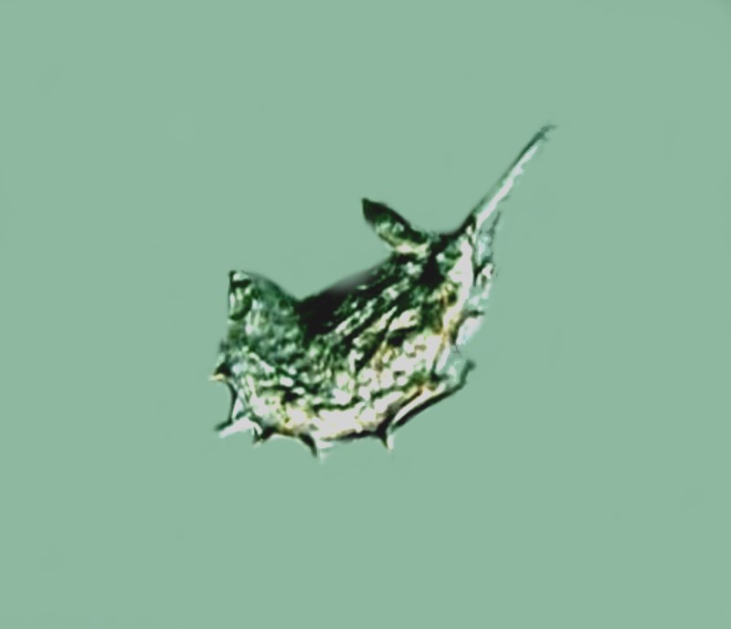
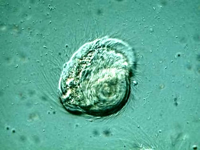
Having been caught up on the process of moving for the last several months to a smaller house to get rid of having to climb stairs, I have found myself in the situation of having not having much access to my microscopes and lab equipment and although I am gradually working on getting everything organized, it is a slow and lengthy process. One side benefit, however, has been that it has led me to go back and review the thousands of images which I have taken over the years. Many of these have never seen the light of day and some of them are, I think, rather interesting. So, I came up with the idea of creating a series of albums highlighting some of the better images. I decided to start with an album on the symbionts of termites because they are such bizarre and interesting organisms.
We’ll begin with some views of Trichonympha campanula because it is abundant and one of the most curious of these organisms. It is an active swimmer and is so flexible that it might be called a “shape shifter” which makes it sometimes rather hard to identify in particular poses. Let’s begin with a classical image that shows the basic morphology quite well.

Here you can readily see the abundant flagella, the macronucleus in the center and at the upper right a protruding structure called the rostrum. One is easily tempted to assume that here is where the “mouth” or cytostome is located. However, that is not the case and this organism ingests the wood particles through the extensive membrane along the lower part of its “body”. The wood particles are concentrated in the lower half and have a yellowish-orange appearance. This image was taken using Nomarski Differential Interference Contrast (DIC). It was once thought that such termite symbionts could not themselves break down the wood particles and were dependent upon bacterial symbionts to transform that material into a different form that the Trichonympha could digest. Now, however, it is known that they can break down cellulose on their own.
Imaging active micro-organisms is always a challenge and these symbionts present some special difficulties. As a result, I have experimented a bit with shifting contrast and altering color balance to try in some instances to reveal a bit more detail. The next image is the same as the one above, except that I have modestly shifted the brightness and color balance, such that, to my eye, certain features are more readily recognized.

The next two images are of the upper half of the organism and I will show you 2 images in the same fashion as above. Again, I think the second image provides a slightly different presentation of the morphology.


The following 2 images are again of Trichonympha but from a different point of view. I am also giving you the 2 views. I have tried to “clean up” the images in terms of eliminating distracting detritus as much as possible, but in the areas with the bundles of flagella this is not possible.


The differences are not quite as evident in some pairs of images as in others. These 2 for example are, to my eye, not as effective in showing differences as the ones above.
Another pair of examples which I think is quite effective is a view of the upper part with clear delineation of the rostrum.


Yet one more pair of views, again of Trichonympha, but once again from a different angle. This pair, does, I think, make my point rather nicely. I have altered the color balance even more in the second image than in the previous examples above.


I mentioned the problem of trying to clear the image of distracting detritus to achieve a relatively neutral background. With some images this is virtually an impossible task. Consider what kind of environment these symbionts live in. First of all, there are the fluids and random cells floating around in the intestines from the termite itself. Secondly, there are all of the bits of wood particles which are being ingested and those can form small clumps. Thirdly, these wood particles are often birefringent which means when using Nomarski DIC they can show up as bright spots in the image. Fourthly, these organisms are very active and so things are churning around and there is constant motion of all kinds of detritus, some of which will cling to the symbionts themselves. A further complication is that some of these organisms are highly flexible and can distort themselves in ways that makes them hard to recognize. This is especially true of Trichonympha and I want to show you some images that show both the problem of detritus and the problem of morphological gymnastics that makes identification difficult.
The image below shows the clumping of wood particle detritus and also a “side” perspective of Trichonympha that makes it difficult to be sure that it is indeed that organism. I, of course, had the advantage of being able to watch it as it moved and so was able to determine that it was indeed Trichonympha.

This next image is even more problematic because, while there are familiar features there seem to be others that have not been seen before. Again, because I could watch it for a bit, I was able to ascertain once again that it is indeed Trichonympha, which I would not have been comfortable saying if I had only this single image and no direct observation. I did try to “clean up” this image and also changed the color balance. However, as you can see, especially in the posterior area where there is a concentration of flagella, the effort was only partially successful and much too time-consuming.

Next is a rather remarkable, lucky shot which shows 3 specimens of Trichonympha in different positions and nicely exemplifies the morphological flexibility of this organism.

Now, let’s take a look at a different organism. This one is Streblomastix strix (a good name for a rock band). Judging from the chubby character of Trichonympha, one might be surprised to find a “twiggy” symbiont hanging out in the same gut. In this beastie, the flagella do seem to all emanate from the tip where again one would suspect the cytostome to be, however, once again, it is located at the posterior end and the tiny tube-like structure attaches to the intestine of the termite. Once again, in this first image the problem of background debris is very evident, however, the flagella are quite clearly discernible. In this case, I have altered the background color significantly in the second image and the detail of the flagella is largely lost.


Here is yet another image which nicely shows the flagella. Unfortunately, sometimes the Nikon CoolPix and the Nomarski DIC don’t like to cooperate and at certain magnifications some specimens will produce the background “rings”. I have consulted several people regarding this problem, but no one has a solution except for the advice of “cleaning” the background but, as we have seen, that is not always feasible.

Now, we come to a third kind of symbiont with another wonderful name worthy of a modern rapper, if he could get some lessons to learn how to pronounce it: Trichomitopsis termopsidis.
This one is not only difficult to photograph, but difficult to observe as it is highly active and constantly undulating. Along the “dorsal surface”, there is a jagged membrane which gives the organism the appearance of a mini-chainsaw when it is moving and twisting.

Here is a view from a different angle.

Finally, I was able to capture images of 3 other symbionts, but unable to identify them. However, they are sufficiently interesting to merit a viewing. The first one looks like a tiny seed covered with rootlets, which are, of course, flagella and are abundant over the entire surface of the organism.

By changing the color of the background and altering the color balance in the organism itself, we are able to see wood particles quite distinctly, which were not really much evident in the first image.

The next unknown has two caudal process extended and a heavy anterior “beard” of flagella. The second image with the shift in color balance and background allows the flagella to be seen a bit better.


The final unknown is a view from the top and clearly this organism has an abundance of flagella extending down from what appears to be a rostrum, which suggests that this could even be a top down view of Trichonympha, but I would be no means guarantee that.
You can see from the upper right edge of the beastie that it was caught in the process of whirling. Both image nicely suggest the organism is in motion.


As I commented in a previous article, I suspect that there are many insects which harbor an unusual fauna in their intestines and relatively few have been studied with this in mind. However, the woodroaches, termites, and even some cockroaches present a group of micro-environments of extraordinary complexity, both in their own right as environments and in terms of the diversity and intricacy of the organisms which inhabit them. Eric Gravé has a section devoted to these organisms in woodroaches and termites in his wonderful little book Discover the Invisible: A Naturalist’s Guide to Using the Microscope. He has some very interesting photomicrographs illustrating that many of these organisms are still unidentified. So, get out your plastic jars for taking rotten wood samples under old logs, your micro-dissecting tools and go symbiont hunting. You may be quite surprised at what you find.
All comments to the author Richard Howey are welcomed.
Editor's note: Visit Richard Howey's new website at http://rhowey.googlepages.com/home where he plans to share aspects of his wide interests.
Microscopy UK Front
Page
Micscape
Magazine
Article
Library
© Microscopy UK or their contributors.
Published in the September 2019 edition of Micscape Magazine.
Please report any Web problems or offer general comments to the Micscape Editor .
Micscape is the on-line monthly magazine of the Microscopy UK website at Microscopy-UK .
©
Onview.net Ltd, Microscopy-UK, and all contributors 1995
onwards. All rights reserved.
Main site is at
www.microscopy-uk.org.uk .