Minute vitreous spherules that may or may not have something to do with equilibrium, tiny globose groups of spheres with poison glands to repel unwanted larvae trying to take up residence, hundreds of Medusa-like “arms” composed of huge numbers of calcareous plates possessing sharp, curved hooks, small palmate gills, tubes that look like the accordion hose on clothes driers and which can be extended and withdrawn and often possessing a calcareous circular disk at their terminus, tube feet that aid in locomotion, tube feet that have nothing to with locomotion but are chemoreceptors that aid in the gathering of food–can’t you just image Julie Andrews singing this to the tune of “These Are A Few of My Favorite things”.
The longer I investigate echinoderms, the more truly and deeply astonished I become at the morphology of these mind-boggling creatures. I have 2 major regrets: 1) I have never had the opportunity to study live specimens and 2) my personal researches on dried and preserved specimens have been limited to a few dozen of the several thousand species. Nonetheless, what I have seen and learned has been wondrous. The ancient Greeks used the marvelous word “thauma” for designating such experience and it has a splendid multiplicity of interwoven meanings: thauma = wonder, astonishment, amazement, rapture, intensity, and a sense of being overwhelmed by the very existence of things. I suppose one could get so carried away as to have traumatic thaumatic attacks.
Whenever I am examining a specimen for the first time, even though it may be a species of which I have examined 10 other specimens, I begin by just looking at it carefully without magnification, moving and shifting it to different angles and changing the illumination. The first rule of thumb is to look for things you didn’t notice previously and the second rule is to look for anomalies–things that don’t fit what you saw in examining other specimens of the same species.
The next step is to use a hand magnifier which allows you to look for interesting areas to examine next under your stereo microscope. It is at this stage that you can spend an enormous amount of time investigating the details of your specimen, especially if you have an instrument with multiple magnifications or zoom capability and it is at this level that all of the oddball bits and pieces start to present themselves. It is also at this level where one finds all kinds of hangers-on: the spines of cidaroids in particular. These spines are different from those of other echinoids in 2 significant respects: 1) they have an extra layer, a hard thin covering called a cortex and 2) this layer bears a network of fine hair-like projections. In some cases, spines look as though they had been sprayed with a light coating of cotton fibers. It is these characteristics which permit the colonization of the spines by a large number of other organisms. At this point, we should remind ourselves that almost all of the external features of echinoids, the test, the podia, the pedicellariae, the gills, the sphaeridia, and the spines are covered by a membrane which is in part protective but, in addition, houses a range of chemo-receptors.
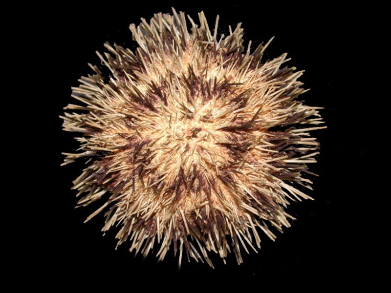
First let us remind ourselves of some of the variety of form in regular sea urchins.
We’ll begin with what we tend to think of as a typical echinoid, namely, one that looks like a pin cushion.
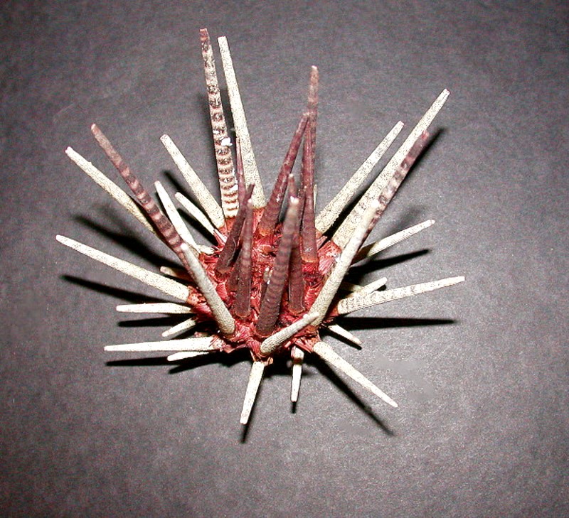
And then a spiky one with impressive spines.
And another spiky ones with thorny spines.
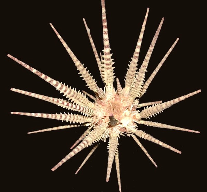
Also one with bizarre spines that look like miniature cacti and is aptly named Psychocidaris.
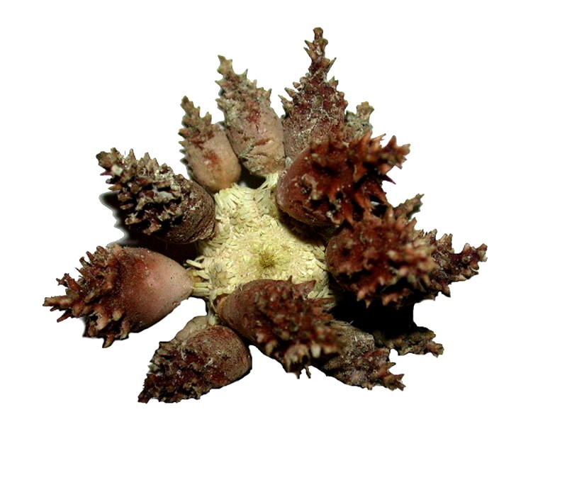
Another very strange creature with little cup spines that look rather like inverted umbrellas.
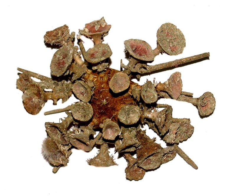
And finally, a specimen which we’ll begin with, a cidaroid echinoid which is veritably infested.
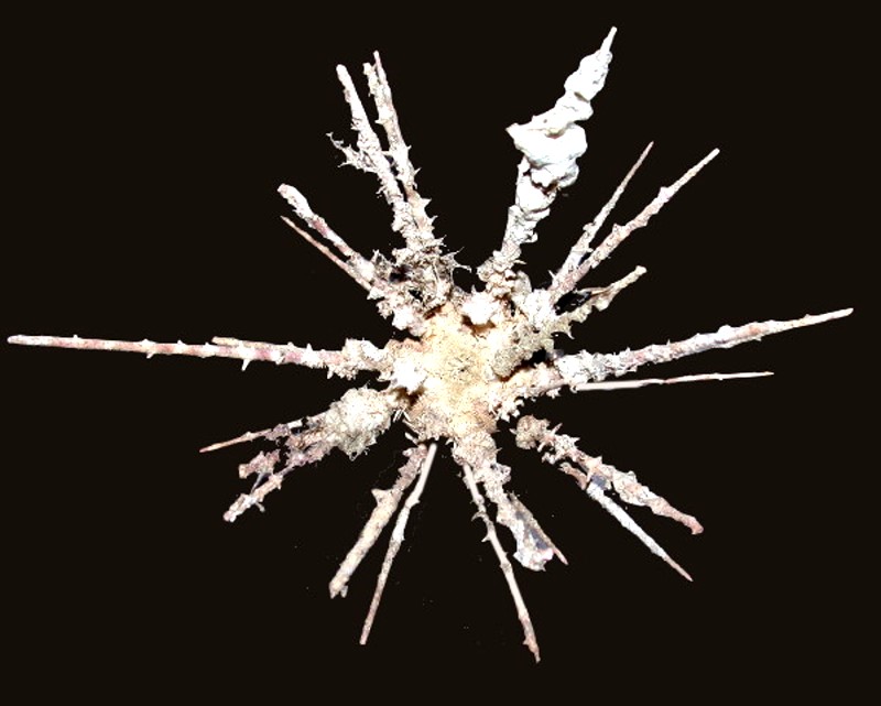
Let’s start with a look at some of the invaders of cidaroid spines; I promise you’ll be impressed. I won’t bore you with identifying the species of each spine; rather, we’ll concentrate on the organisms which have taken up residence. Small barnacles occur frequently sometimes singly, but often in small clusters.
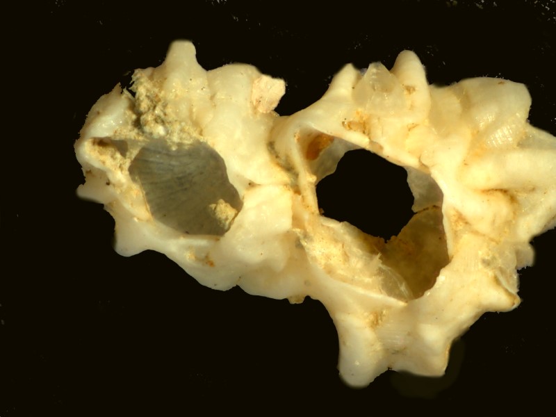
These are small specimens of a common rock barnacle Balanus. One can get some idea of how long they have been residents by their relative size. These are relatively easy to pick out and distinguish from other invaders due to their overlapping geometric plates. Here is another species of barnacle from the same urchin.
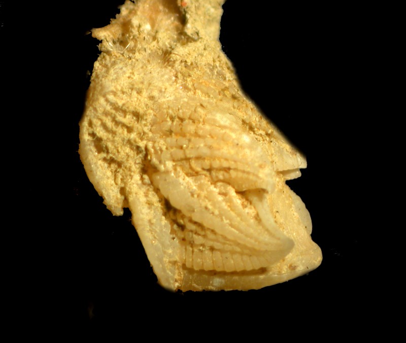
Tubeworm shells, in this case, a coiled one of the genus Spirorbis. These are also relatively common.
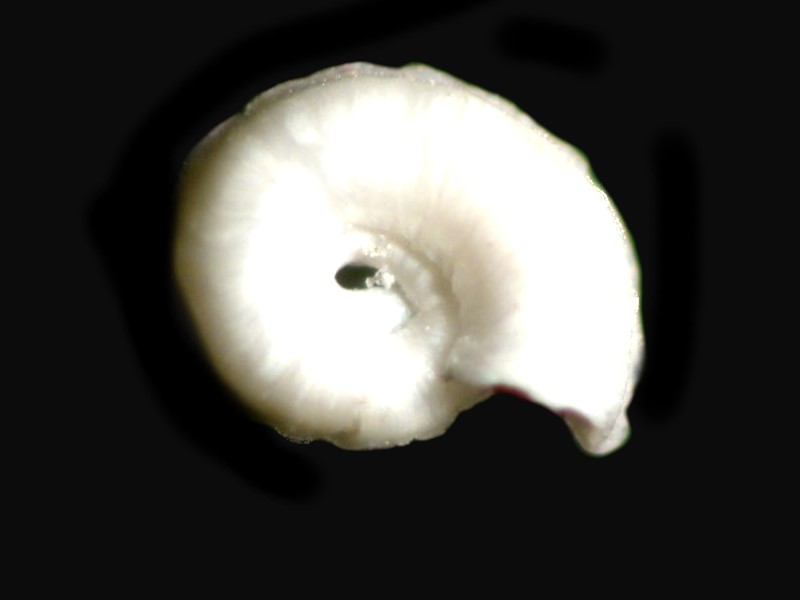
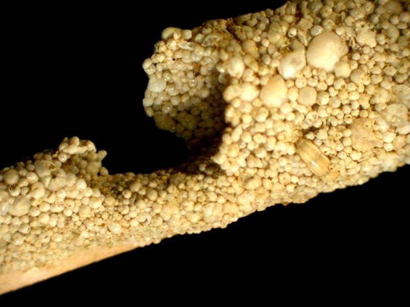
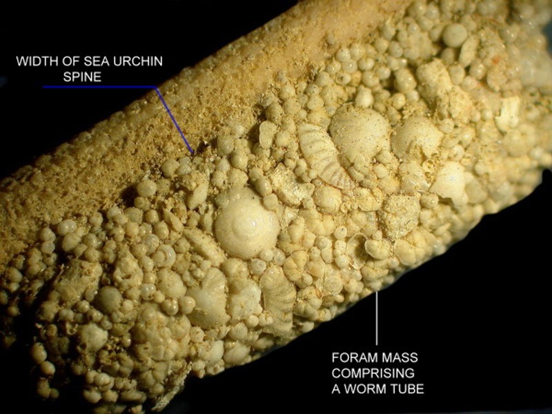
Another type which is not coiled, but hugs a small portion of a spine presents us with an amazing surprise; its entire surface is camouflaged with foram shells of various species.
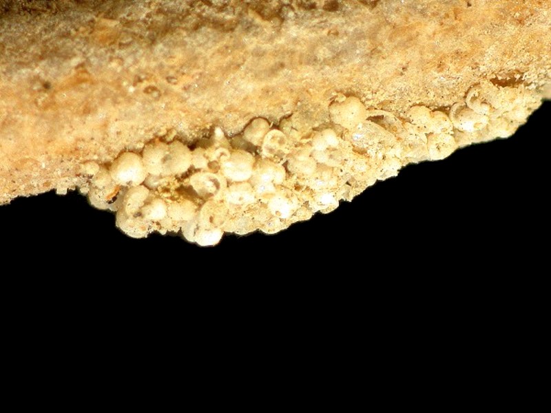
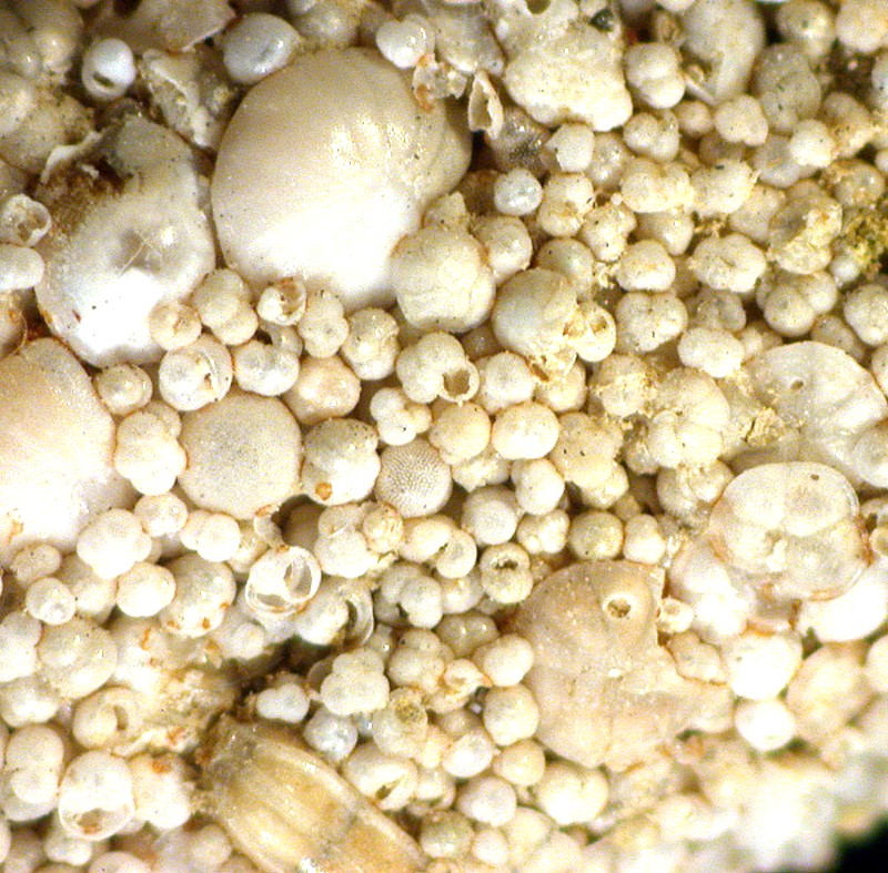
The question which jumps out at us is: How in the world did they get there? Tubeworms secrete a thin, sticky membrane which is the foundation for the tube. In addition, these incredible creatures have tentacles which are also sticky and used for food gathering. So, is it possible that they can also use these tentacles to grasp and attach forams to their membrane thus providing a protective coating? Forams are, after all, marine amoebae of considerable complexity and are capable of locomotion even though it would not be rapid. When I first encountered such a tube, my initial impression was that it was simply a peculiar aggregation of forams and, only later did I realize it was a worm tube. I was immediately struck both by the number and the variety of these tiny shells and I’ll show you some examples here.
Another frequent inhabitant of cidaroid spines are sponges of assorted types. One sort forms a delicate network along the surface of the spine and is often difficult to distinguish from the layer of fine hairs that characterizes the cidaroid spines.
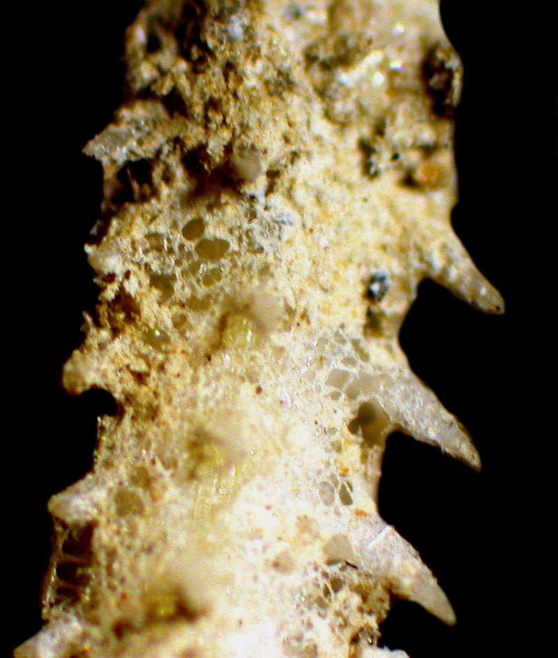
Other sponges retain a compact form and can easily be recognized as such.
Bits of bryozoan colonies can also be found.
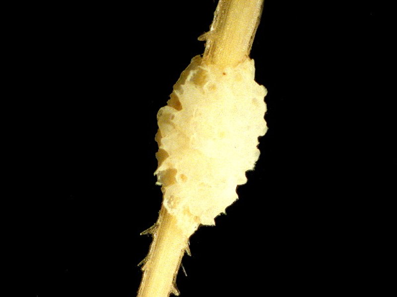
Another opportunistic denizen is the small brittle stars (ophiuroids) which are most likely just temporary visitors since ophiuroids tend to be very active organisms.
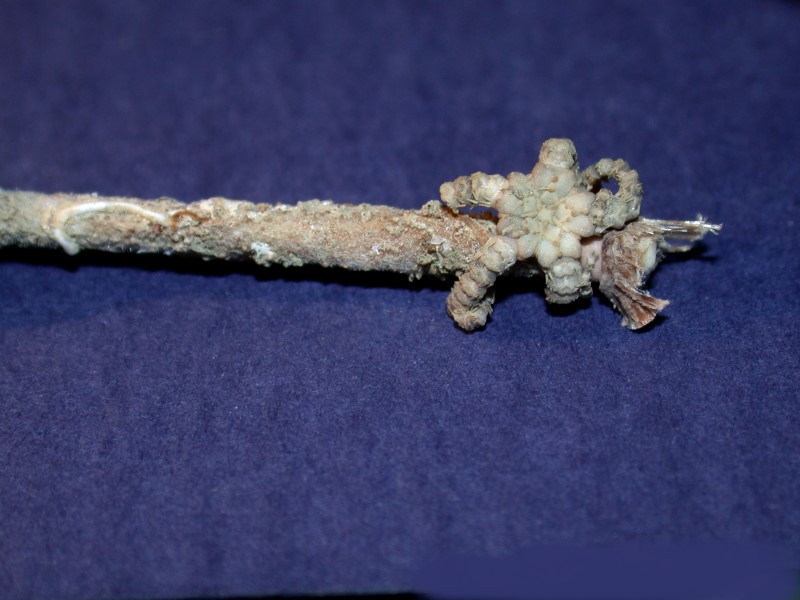
Naturally, one also expects diatoms since they occur in enormous numbers in such environments.
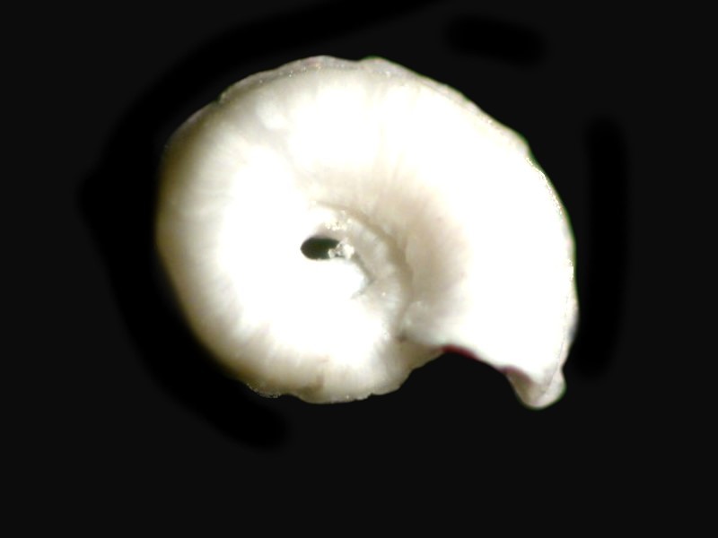
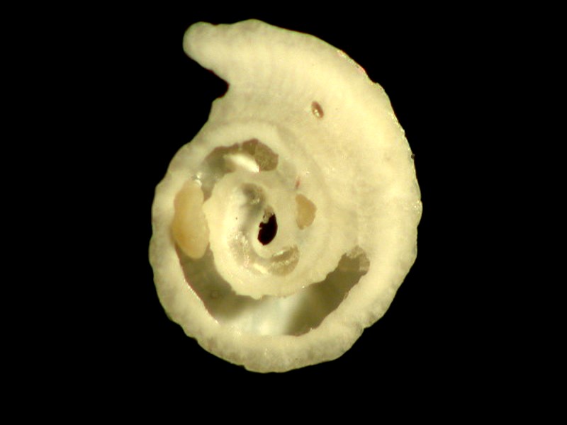
Small bivalves also put in an appearance and the tiny mussels of the genus Mytilus attach themselves by amazingly strong byssal threads which they secrete insuring that they will not be easily dislodged. This strategy of attachment can be discovered in 2 other organisms we have to look at: 1) barnacles which produce an extraordinarily strong adhesive “cement” (try prying some off of a rock in a tidepool by breaking them apart) and 2) tube worms which also “glue” themselves to spines and if we take another look at Spirorbis, we find an intriguing adaptation. From the top, it appears to be, which in fact it really is, a miniature tunnel in which the worm resides, extending its tentacles out through the anterior opening to feed. However, if we look at the underside (second image below), we observe that its surface is flattened and slightly bowed to make it fit more tightly against the spine.
Hydroids are present but are harder to spot since the “house” of the zooids and the stalk are not calcareous, but rather are composed of fairly tough organic proteins.
Micro-algae are present, but they are even harder to pick out especially on dried specimens. If you have a specimen preserved in fluid, then you have a considerably better chance of extracting some identifiable forms.
You can spend many pleasurable hours discovering, isolating, identifying, and mounting “invaders” from cidaroid spines.
Now, let us move on to those weird structures called pedicellariae. They are mini-“guard towers” found extending up from the surface of the test. They do not, however, so far as I know, occur on spines which helps to explain why there can be such accumulations of exploitative commensal organisms. So, find a nice- sized specimen and start looking at the test in the areas between the spines and soon you’ll come across a “head” usually composed of 3 jaws which are on a stalk and here is where your micro-tools become invaluable. Again, if you have a preserved specimen in fluid, your search will be easier. I recommend moving the specimen out of preserving fluid into distilled water for a day or two with a couple of changes of the water. This will make your investigations much more pleasant. In fact, you might even want to consider placing a dried specimen in distilled water with the hope that the rehydration process will reveal some pedicellariae which you can remove intact.
On dry specimens, if the pedicellariae are abundant, you can find intact ones and here is where micro-forceps, micro-scalpels and micro-spatulas become very handy. Since you are dealing with structures which are covered with a thin membrane which extends from the test, they tend to get “glued” rather firmly to the test’s surface which means that extracting them will only be possible, in many cases, in bits and pieces. However, this can be an advantage, for it will allow you to see how the various components fit together to constitute the whole entity. These structures are truly remarkable for, in one sense, they seem to have a kind of autonomy from the echinoid as a whole since there is certainly no central control mechanism directing their behavior. It is almost as though they are tiny, stalked robots with jaws which are programmed to snap at and crush anything that intrudes into their territory. Their territory varies with type and size; some stalks are quite long and flexible thus expanding their range and the head with its jaws can not only open and close the jaws, but swivel the whole apparatus. All of this suggests an impressive array of sensors and muscle fibers. It is likely that both photo-receptors and chemo-receptors are involved. There may even be thigmotactic sensors as well.
While 3 jaws are by far the most common, there are pedicellariae with 2, 4, or 5 jaws and, in addition, there are 4 basic kinds of pedicellariae. The Danish researcher, Theodor Mortensen, who produced an impressive and still highly informative set of monographs on Echinoids, argues convincingly that pedicellariae have great taxonomic importance in determining species.
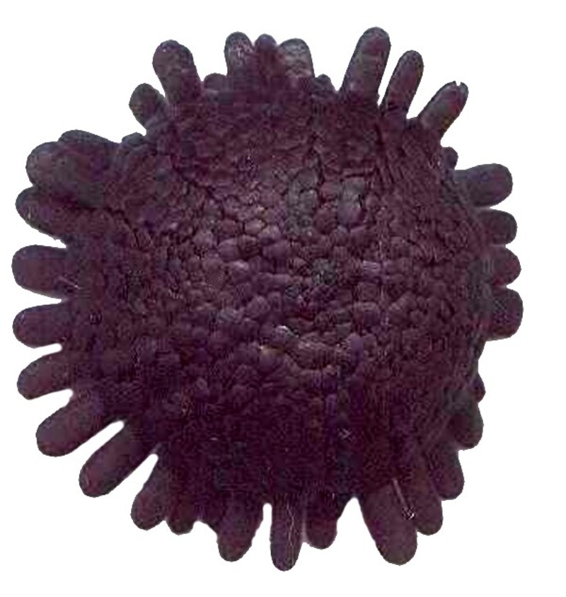
Of the 4 kinds, the most intriguing is the globiferous which has poison glands. Typically, each jaw terminates in a sharp tooth and the jaws appear somewhat bulbous due to the poison inside each jaw. A duct connects from the glands to the area of the tooth which can “inject” its toxin into whatever has triggered the response. According to Libby Hyman in her superb reference The Invertebrates, Vol. IV: Echinoderms, p. 431 and Fig. 182 E, there are also some species which have a ring of poison glands around the stalk and this can lead to a degeneration of the jaws and the development of spherical structures with a duct open in the area of the stalk glands. Colobocentrotus atrata is an example of a species in which this occurs. This is the flattened, domed species which has adapted to living in areas with heavy surf.
Next, let’s take a brief look at sphaeridia; there are tiny, glassy spheres that sit in minute pits on the surface of the test. Any discussion of sphaeridia is necessarily brief, because no one is quite sure what their function is and their morphology is not particularly helpful on this point. The prevailing theory is that they are sensors which “inform” the organisms with regard to equilibrium.
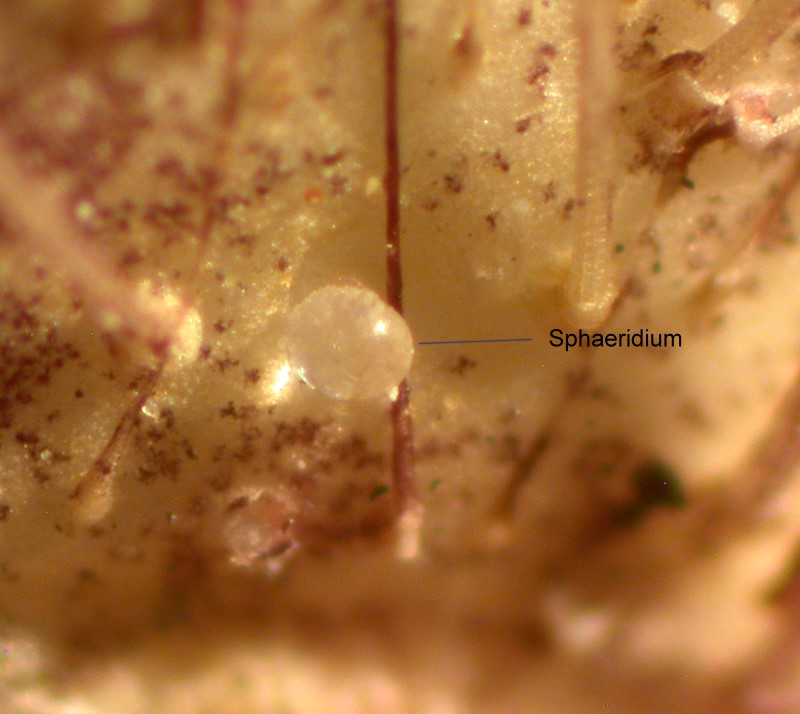
In other words, if an urchin is tipped over so that it’s upside down, presumably the sphaeridia send ”signals” to that effect and the urchin rights itself using its spines and tube feet. This scenario strikes me as highly tenuous. Recent EM studies show that sphaeridia are shaped like miniature amphora and they appear to have ridged indentations which raises a question as to whether or not they could hold fluid in which a statolith or equilibrium indicator could be suspended and function. Sphaeridia are another one of the many puzzles surrounding echinoderms.
Finally, lets take a look at the podia or “tube feet”. When we take a close look at some ambulacral plates, we notice pores which run from the exterior side of the plate through to the interior side.
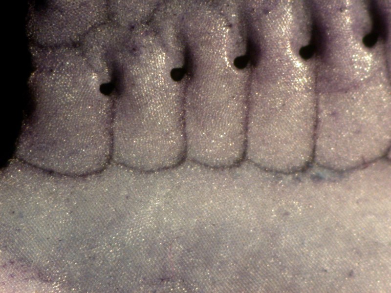
It is through these pores that the tube feet extend. Inside the test, each tube foot is connected to the water vascular ring and each has a “bulb”, rather like a pipet bulb which controls the extension and retraction of the foot. The degree of extension can be quite impressive and in many species, these feet can be extended out beyond the length of the spines. There are, of course, given Mother Nature’s compulsion to experiment, exceptions; in some of the Diadema, the spine can be over a foot long and the tube feet in these species generally cannot equal that.
On the aboral side of the test, there is a filter called a madreporite, which in many instances, looks like a small bit of coral.
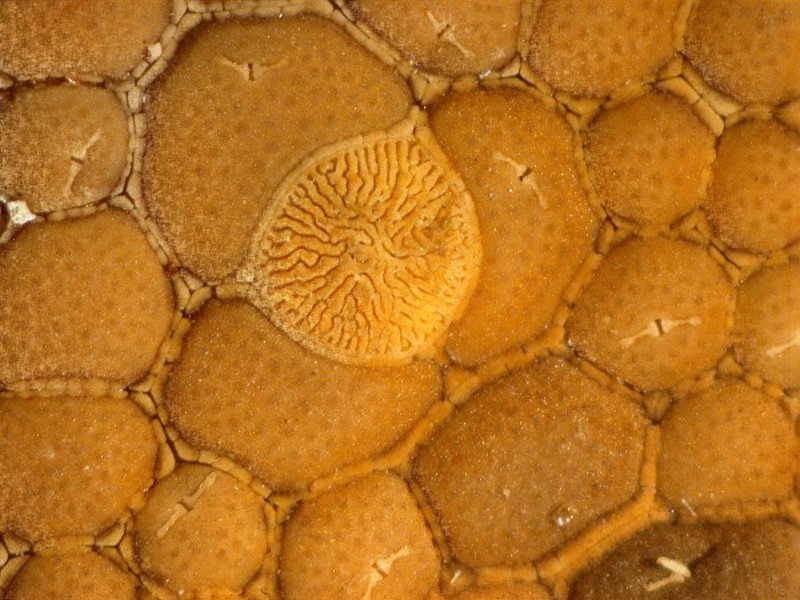
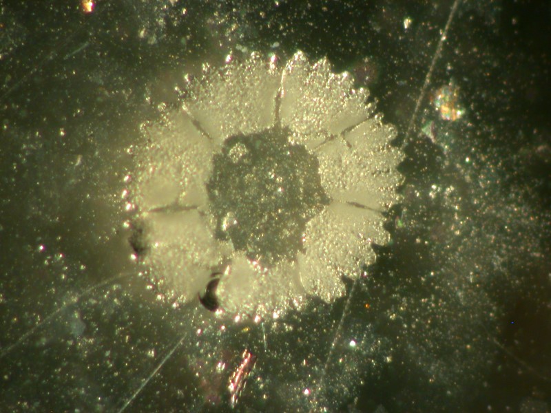
All of these folds and convolutions greatly increase its surface and thereby its efficiency as a filter. It prevents potentially clogging particles from entering the water vascular system. A tube from the madreporite connects to the radial ring of the vascular system. Running beneath the ambulacral plates with the pores for the tube feet are 2 long tubes for each of the 5 sets of plates for a total of 10. For each tube foot, there is an ampulla or bulb which regulates water pressure thereby determining the degrees of extension or retraction of each foot. As I mentioned in an earlier essay, tube feet often have terminal plates just under the tip of the skin. Some of them are quite lovely.
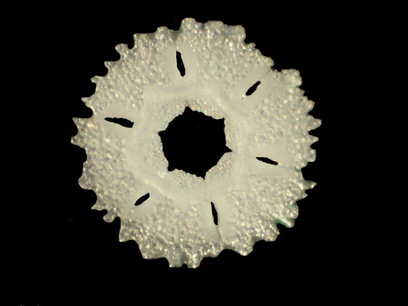
If you look closely, you will notice that this disk is composed of a series of plates which are joined together by tissue and the plates themselves are composed of hundreds of crystals of calcium carbonate. The first image shows a plate (unusually 1 of 6) which comprise the disk, followed by an image showing the disk slowly breaking apart into enormous numbers of tiny crystals.
I once described the tubes of the tube feet as looking like pieces of brown clothes dryer exhaust hose with all of their circular wrinkled banding.
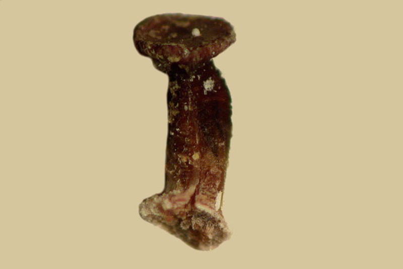
However, one has to remember that these tube feet, as well as many other structures in echinoderms, are usually not simply tissue, but are home to a variety of calcareous structures as well.
As usual, this essay has gotten a bit long, so I will conclude with showing you a few glimpses of some other structures that might well show up in another essay.
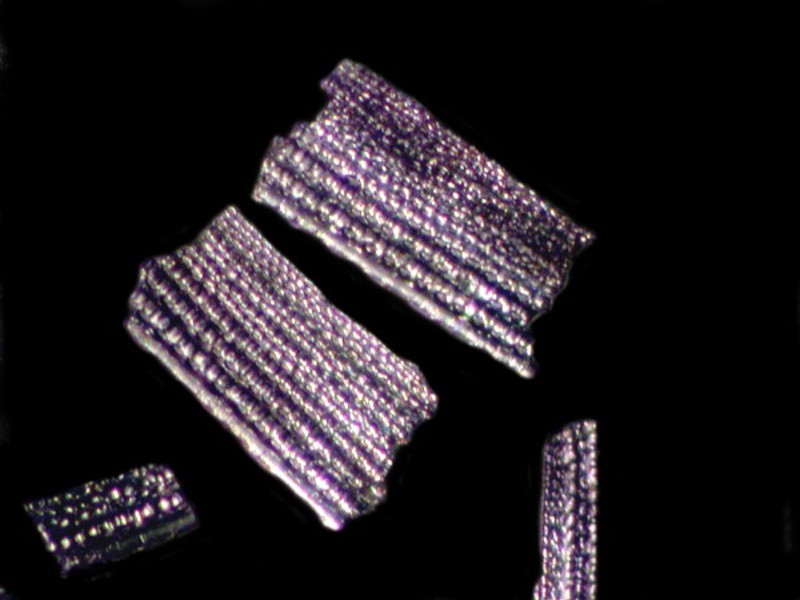
Even the spines are composed of crystals and if you crush one and look at it closely, this becomes evident.
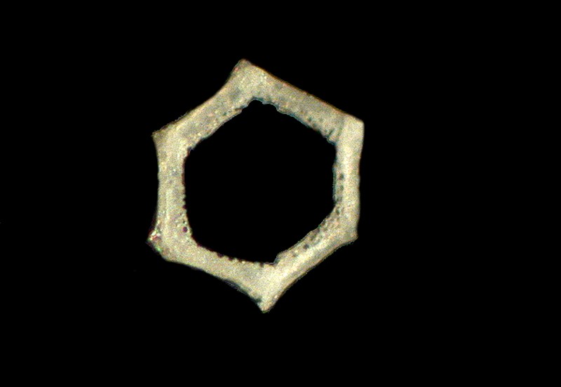
Above we looked at the circular disk in a tube foot; well, underneath the surface there is a support ring which you can see below.
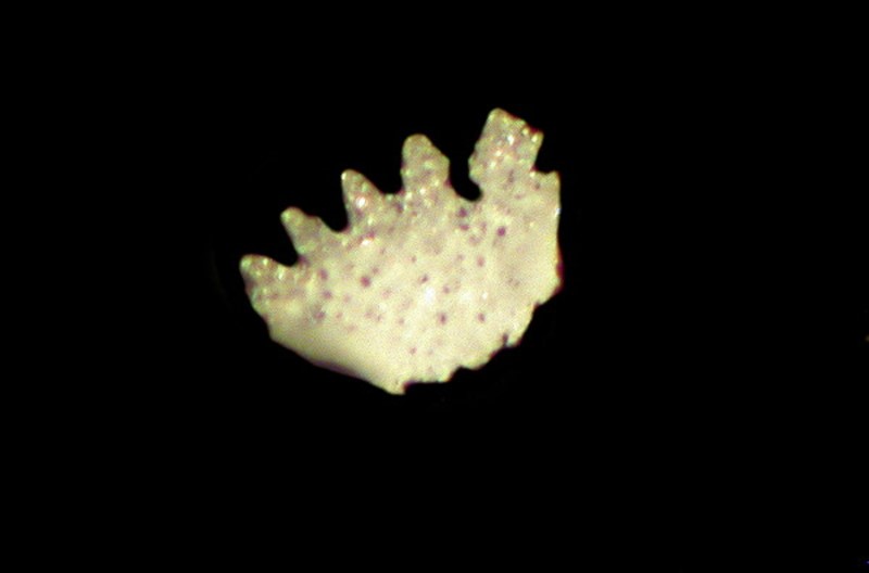
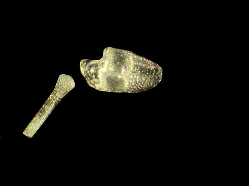
I mentioned pedicellariae and here is a fragment of one with a portion of the support rod lying nearby.
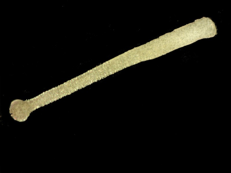
And here is a close up of a rod.
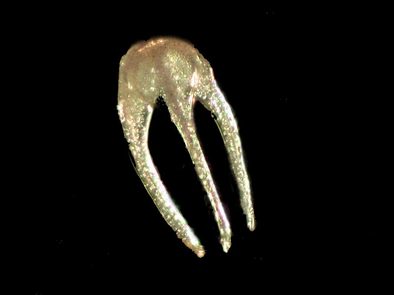
And a close up of the “jaws” of the pedicellariae.
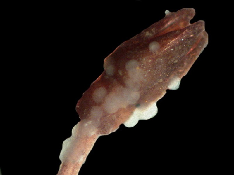
And finally, a pedicellariae that looks as though it has been infested with some kind of grotesque acne. Even the weaponry of creatures is not safe from attack.
I hope you found some ideas for further investigations of these remarkable creatures.
All comments to the author Richard Howey are welcomed.
Editor's note: Visit Richard Howey's new website at http://rhowey.googlepages.com/home where he plans to share aspects of his wide interests.
Microscopy UK Front
Page
Micscape
Magazine
Article
Library
© Microscopy UK or their contributors.
Published in the September 2015 edition of Micscape Magazine.
Please report any Web problems or offer general comments to the Micscape Editor .
Micscape is the on-line monthly magazine of the Microscopy UK website at Microscopy-UK .
©
Onview.net Ltd, Microscopy-UK, and all contributors 1995
onwards. All rights reserved.
Main site is at
www.microscopy-uk.org.uk .