A Close-up View of the

Balloon Flower


Platycodon grandiflorus
 
'Sentimental Blue'


by Brian Johnston   (Canada)


Earth laughs in flowers.

Ralph Waldo Emerson, "Hamatreya"

The unusual perennial studied in this article belongs to the family Campanulaceae, which also includes the bluebell, bellflower, and lobelia.  Native to Northeastern Asia, (China, Eastern Siberia, Korea, and Japan), it has several alternative names: Chinese Bellflower, Japanese Bellflower, and Common Balloon Flower.

The word balloon refers to the buds, which prior to blooming, swell to form (mostly) hollow, pillow-shaped structures.  Although the family name translates to ‘bell’, when fully opened, the flower more closely resembles a star.

Platycodon grandiflorus ‘Sentimental Blue’ is a dwarf hybrid which grows to about 15 centimetres in height, and possesses 4 centimetre diameter purple flowers.  These flowers can be seen in the image above, and in the one that follows.  Notice the deep purple radial and subsidiary veins that decorate the fused petals. 


Prior to blooming (anthesis), the petals-to-be are greenish-white in colour, and are joined at their margins.  At this early stage, the ‘balloon’ shows little signs of the inflation that is imminent.  Notice the pointed green sepals, (modified leaves), that ring the unopened flower’s base.


Soon however, the bud begins to swell, and five, pointed protuberances appear at the end-points of the lines where the petals are temporarily joined.


As time passes, further inflation occurs, and a hint of the final purple colouration begins to appear.


The colour continues to deepen as the bud matures.


Strangely, in the occasional bud, the distinctive protuberances that ring the bud, point inward, rather than outward.  This bud is about to open, as revealed by its deep purple colour, and the partial detachment of one of the petal tips from the rest (left image).


Viewed from above, the petal tip that has begun to separate is more obvious.


Perhaps it would be interesting to take a look inside an unopened bud.  In the images that follow, a couple of petals have been removed to reveal a bud’s interior.  The buds of most flowers are densely packed structures.  Not here however!  There is a great deal of air inside this “balloon”.  Both views below show the five, inward facing, yellow anthers, (male pollen producing structures), and their blue supporting filaments.  In the scientific terminology of botany, the anthers are referred to as being ‘introrse and weakly connivent around the style’.  Now that certainly tells us a lot!  If you look up the word introrse, it means ‘dehiscing towards the centre of the flower’.  Clearer and clearer!  If you look up dehiscing, it means ‘opening’.  As I said earlier in the paragraph, the anthers are inward facing.  Isn’t terminology wonderful!  Finally, the term connivent refers to the fact that the anthers come into contact with the style that they surround, but are not attached to it.


A view from above shows the immature pistil of the flower, which at this early stage, consists only of the style.  The pollen accepting stigma has yet to reveal itself.


In the two views that follow, one of the anthers (and its supporting filament) has been removed to show the style.  Notice how densely hairy it is.  Also note that there are furrows at its tip that will later open, (after the flower has bloomed), to allow the egress of the stigma.


The image below shows the blue, flattened base of one of the filaments.


Over a period of several hours, a bud blooms, revealing the temporary bell-shape of the flower.


Finally, the actinomorphic, 5-merous nature of the balloon flower appears.  Sorry, but I couldn’t resist the didactic urge!  Actinomorphic means ‘star-shaped’.  Or, if you wish, a shape that can be divided into symmetrical halves along any diameter.  The strange term 5-merous refers to the flower’s parts being in groups of five – five petals, five stamens, five stigma lobes, etc.


A closer view of a mature flower shows that the yellow stamens are still in contact with the style that they surround.


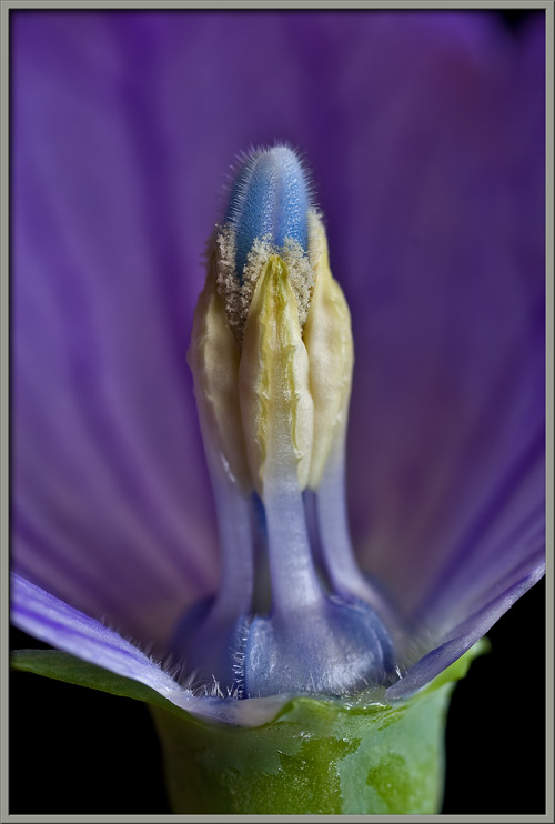
As can be seen below, the flower’s anthers have begun to shed pollen grains which have adhered to the fine hairs on the style’s strikingly blue surface.


In the higher magnification image that follows, the roughly spherical shape of these pollen grains is revealed.


Under the microscope, the stubby, pointed hairs that cover the style’s surface can be seen clearly.


If you take a close look at the following image, you can see that a change has occurred.  The five anthers that were earlier in contact with the style have spread apart, and now rest on the surface of the flower’s petals.


A photomicrograph showing the cellular structure of a petal, and one of its purple veins, follows.


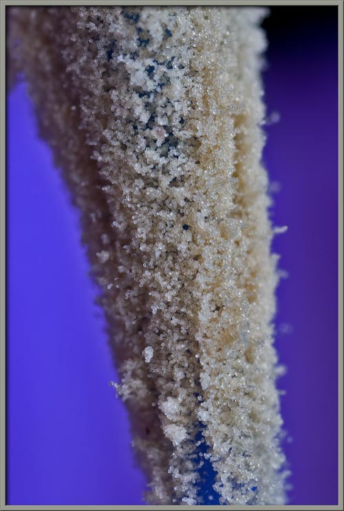
The two images below show the newly revealed surface of the style.  It is liberally coated with pollen grains.


Notice that each anther is connected to the ‘bump’ at the flower’s centre by its flattened filament.  Also note the triangular blue ‘flaps’ in the upper right corner of the image on the right.


These blue flaps can be seen more clearly in the image that follows.  Notice the light coloured hairs that grow between the gaps.  Beneath these flaps is the annular nectary disk which contains the fluid so prized by insects.


The two photomicrographs that follow show the outer, non pollen producing, and inner, pollen producing surfaces of an anther.


As was mentioned earlier, Balloon Flower pollen is roughly spherical in shape.


A higher magnification photomicrograph reveals some of the surface detail.


Close examination of the flower in the image below reveals a significant development.  The stigma has finally appeared!


Notice in the following images, that the stigma is five-lobed, and that the newly exposed receptive surfaces have practically no pollen adhering to them, while the non-receptive back surfaces are abundantly coated.  The Balloon Flower is protandrous; its male reproductive organs (the anthers), mature before the female reproductive organ (the stigma).  This retarded development of the stigma helps to discourage pollination of a flower by its own pollen (a genetically less desirable outcome).  Protandry therefore, diminishes self-pollination.  Ideally, a visiting insect, while obtaining nectar from a flower’s nectary disk, will transfer pollen from another flower of the same species to the receptive surface of the stigma.  As a back-up, in case this doesn’t happen, the stigma lobes curl back in order that their receptive surfaces come in contact with the pollen on the style.  This may result in self-pollination – a process sometimes referred to as ‘selfing’.


The two higher magnification images below show the stigma in side and top views.


Finally, two photomicrographs showing the cellular structure of a Balloon Flower’s leaf can be seen below.


Robert Fortune (1812 – 1880) collected Platycodon grandiflorus plants along China’s coast in 1843 for the RHS garden in Chiswick U.K. (near London).  Their extreme popularity has resulted in the plant’s dissemination worldwide over the past hundred plus years. 


Photographic Equipment

The low magnification, (to 1:1), macro-photographs were taken using a 13 megapixel Canon 5D full frame DSLR, using a Canon EF 180 mm 1:3.5 L Macro lens.

An 8 megapixel Canon 20D DSLR, equipped with a specialized high magnification (1x to 5x) Canon macro lens, the MP-E 65 mm 1:2.8, was used to take the remainder of the images.

The photomicrographs were taken using a Leitz SM-Pol microscope (using a dark ground condenser), and the Coolpix 4500.


A Flower Garden of Macroscopic Delights

A complete graphical index of all of my flower articles can be found here.


The Colourful World of Chemical Crystals

A complete graphical index of all of my crystal articles can be found here.


 All comments to the author Brian Johnston are welcomed.


Microscopy UK Front Page
Micscape Magazine
Article Library


© Microscopy UK or their contributors.

Published in the September 2009 edition of Micscape.
Please report any Web problems or offer general comments to the Micscape Editor.
Micscape is the on-line monthly magazine of the Microscopy UK web
site at Microscopy-UK  


© Onview.net Ltd, Microscopy-UK, and all contributors 1995 onwards. All rights reserved. Main site is at www.microscopy-uk.org.uk with full mirror at www.microscopy-uk.net .