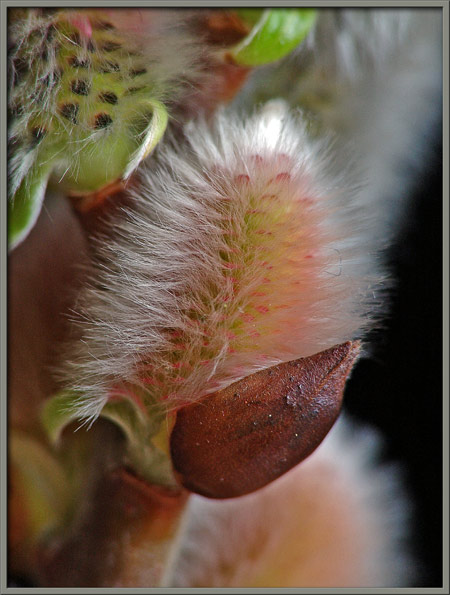
A Close-up View of the

"Pussy Willow"

 
(Male catkins)


Salix caprea


by Brian Johnston   (Canada)


Salix caprea has buds that open in spring before the plant’s leaves make an appearance.  These furry buds, the ‘pussies’ in the name pussy willow, are referred to as catkins.  All willows are dioecious, meaning that male and female flowers occur on separate plants.  In this article, only the more ornamental male flowers have been photographed.  Salix caprea is sometimes referred to as goat willow, pink willow, or florist’s pussy willow.

The image above shows a group of male catkins at different developmental stages.  The pinkish orange colour of the small catkins on the right eventually fades to a light green colour.  Notice that the buds are initially protected by shiny, brown, beetle-shaped structures that fall away as the catkins grow larger.

Compare the branch with blooming catkins on the right, with one about a week before blooming on the left.  (The yellow structures connected to the catkin by thin filaments are the anthers of individual flowers within the catkin.)


Each male catkin is intensely hairy, and these hairs all but obscure the black tips of the bracts (modified leaves) that are associated with each flower.


At the growing tip of a willow branch, three very tiny catkins are completely enclosed by hairy green bracts that in turn, grow out from under brown protective covers.  Note that the brown cover becomes less hairy as time progresses.


Under the microscope, the number of hairs growing from one of the bracts is amazing.  One must focus through the hairs to visualize the cellular structure beneath (right image).


Higher magnification gives a better view of the bract’s hairs.


The two views below show a very immature catkin with its almost completely pink colouration.  At this point the protective cover will detach by the action of the wind or other vibration.


As the catkin matures, the pinkish-orange colour is restricted to the top portion, and a light green colouration creeps up from the base.


The higher magnification used in the three images that follow reveals that the pinkish-orange colouration is due mainly to the triangular bracts associated with the flowers that make up the flower-head.


Eventually, the black-spotted, light green colour is uniformly distributed over the surface of the catkin.


A photomicrograph of a tip of one of the black bracts is shown in the right-hand image.


In the previous macro-photographs, one must deliberately focus deeper into the catkin (using manual focus), in order to see the black bracts.  The camera’s auto-focus, if left to its own devices, will always choose to focus on the closest plane, which in this case is the shroud of fine hairs cocooning the catkin.


If one dissects a catkin by lifting away the overlying bracts, inner structures are revealed.

On the left are two filaments that support anthers (the male pollen producing organs).  (Each individual flower in the catkin has two stamens.)  Notice the interesting shape of the hairs that grow near each filament’s base.)  The right-hand image shows several immature anthers buried beneath the layer of black bracts.


The point of attachment of an anther to its supporting filament can be seen below.


Finally, the catkins begin to bloom, usually in order from the top of the branch to the bottom of the branch.  In contrast, each individual catkin blooms from bottom to top.


Once the catkins begin to bloom, leaves begin to sprout along the length of the twig.  These too grow out from under brown protective covers.


Just below the centre of the image at left below, notice the scar that is left after one of the protective covers falls off.  The image at right shows one of the covers being pushed away from the stem by the emerging hairy leaflet.


Here is a closer view of one of the distinctive scars.


If you look carefully at the image below, you can see the yellow tips of anthers pushing out from under the black bracts in the area near the middle of the catkin.


Higher magnification gives a better view of the process.


Notice in the image that follows, that the stamens may have different lengths on different sides of the catkin, resulting in a non-cylindrical shape.


The mature anthers are banana-yellow in colour.  Notice the white supporting filaments in several of the images.


Each anther appears to be divided into four lobes.


Under the microscope an anther with several attached pollen grains (left), and a supporting filament (right), can be seen.


Higher magnification reveals the ‘bent’ ellipsoidal shape of pollen grains.


As a catkin ages, the anthers begin to shrink to a fraction of their original volume.


During the dissection of a mature catkin, copious sticky nectar coats the fingers.  This nectar attracts bees and other insects to the plant, and when they fly to another female tree, fertilization is achieved.

Most of us, I‘m sure, have seen pussy willow catkins in early spring.  I hope that the images in this article have given you a greater appreciation of these unique and striking structures.


Photographic Equipment

Most of the macro-photographs were taken with an eight megapixel Canon 20D DSLR equipped with a Canon EF 100 mm f 2.8 Macro lens which focuses to 1:1.  A Canon 250D achromatic close-up lens was used to obtain higher magnifications in several images.

A few photographs were taken with an eight megapixel Sony CyberShot DSC-F 828 equipped with achromatic close-up lenses (Canon 250D, Nikon 5T, 6T, Sony VCL-M3358, and shorter focal length achromat) used singly or in combination. The lenses screw into the 58 mm filter threads of the camera lens.

The photomicrographs were taken with a Leitz SM-Pol microscope (using a dark ground condenser), and the Coolpix 4500.


 All comments to the author Brian Johnston are welcomed.

Microscopy UK Front Page
Micscape Magazine
Article Library


© Microscopy UK or their contributors.

Published in the September 2007 edition of Micscape.
Please report any Web problems or offer general comments to the Micscape Editor.
Micscape is the on-line monthly magazine of the Microscopy UK web
site at Microscopy-UK  


© Onview.net Ltd, Microscopy-UK, and all contributors 1995 onwards. All rights reserved. Main site is at www.microscopy-uk.org.uk with full mirror at www.microscopy-uk.net .