|
by Roland Mortimer,
|
Previous Micscape articles by the author on diatoms are here - Part I, Part II, Part III and Part IV.
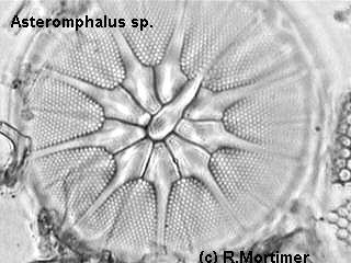
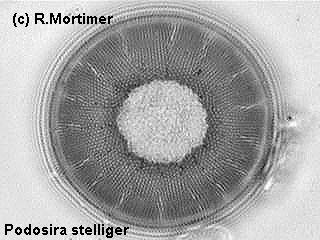
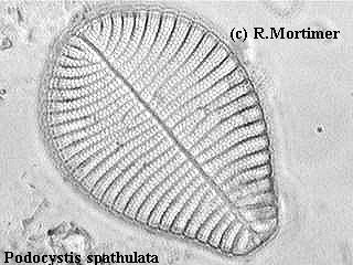
After hearing so much about the versatility of CCD cameras I finally decided to buy one and try it out, but first I installed a video capture board in my old computer to enable me to capture the images of the diatoms from my microscope. The very versatile Heine condenser came into play here too, and once more proved itself to be an extremely useful tool. Moving the condenser element up and down the tube I finally got to a certain position where the condenser threw the light slightly obliquely and the results were really impressive as can be seen from the images in this article. Although they are captured electronic images they have had no further 'treatment' from my computer imaging programmes. As can be seen in some of the images the diatoms look almost 3D. The camera is a digital colour CCD but since diatoms are colourless I switched the camera to black and white mode.
Even with dark field and phase the camera proves
itself as outstanding. Using film and my conventional microscope dedicated
camera I often had disappointing results after spending my money and time.
I also had to choose the diatoms I photographed as I came across hundreds
of species and genera, the cost would be almost ruinous if I used film
to photograph them all. The CCD camera eliminates all of these negatives,
I can instantly see what I'm going to 'photograph' and adjust before capturing,
and, since I don't have to pay for film or developing anymore, I can literally
capture images of every diatom I find and store the images on diskette
then eventually have them all written onto one CD.
 |
 |
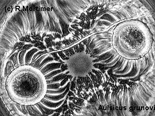
The illumination source used was a 12v 50w halogen
lamp necessary for dark field but the light here is neither dark field
nor bright field, but somewhere in between which gives the nice effect
to the images. Any reasonable dark field condenser can obtain the same
results, it's just a matter of lowering and raising the element and off-centring
until the desired effect is obtained. The image of Auliscus (above
left) was taken in phase, and as you can see most of the detail is shown,
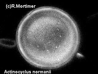
in fact much more than I obtained with a normal camera. The same effects
can even be obtained in dark field as can be seen from the image of Actinocyclus
(above right). The beauty of the CCD camera is it 'searches' for and
increases dim light, so good images can still be had from very fine objects
transmitting little light.
 |
 |
Comments to the author Roland
Mortimer welcomed.
Image gallery by the author.
 |
 |
 |
 |
 |
 |