Well, this is probably not quite what you are expecting from the title; I’m afraid it’s not really salacious or titillating but, nevertheless, interesting and we may be able to actually learn something from Mother Nature about our own nature. As it happens, Mother Nature has been experimenting with reorganizing, refurbishing, and renewing genetic material for probably at least a billion years here on planet Earth. That’s what sex ultimately is all about, recombinations of genetic material to see what succeeds and survives. Humans, of course, often experience exceptional pleasure from sexual activity. Interestingly, we probably need to extrapolate that pleasure principle (or some version of it) to a lot of other organisms as well. Some male mantids and arachnids risk death from attack by the female after sex, so the drive must indeed be intense. One can, I suppose, interpret such behavior as mechanistic, but then that just might translate over to humans as well; a thought that many would find uncomfortable. So, we’ll primarily stay with the invertebrates and there is certainly much of interest to explore here. However, I should warn you that Mrs. Malaprop will intervene with interjections from time to time, but I’ll try very hard to keep her under control. She is simply irrepressible when it comes to certain topics.
Most everyone is familiar with the peculiar mathematics of amoebas which multiply by dividing. On first impression, it would seem that this is a simple process; a single amoeba reaches a stage where it begins to develop a second nucleus and then division starts to occur. A terminological convenience designates one of the cells the “mother” cell and the other the “daughter” cell. [I want to record a reservation here. I am very reluctant to use the term “cell” when talking about protozoa since, in my view, they are complete organisms. If you are interested you can read my article on this topic in Micscape.
Sometimes, they are simply referred to as “sister” cells. As the process proceeds a pinching occurs creating a “waist” which gradually becomes thinner and thinner until only a thin protoplasmic band remains connecting the two. This band is sometimes referred to as an “umbilical cord” which tethers the two cells. This is apparently very strong and tough and the two cells may struggle unsuccessfully to break the tether and finally return to being a single amoeba. The other option would be a successful separation by the two struggling cells. In 2001, some fascinating research revealed that in the process of this struggle to separate, a chemical signal is released as a kind of cry for help. At this point another amoeba, designated as a “midwife”, may respond and squeeze between the two cells and exert enough pressure to snap the “umbilical cord” allowing the two to separate and go their own ways as individual organisms.
Mrs. Malaprop: “And we thought we invented the concept of midwifery!”
Now, in case you think that this is the end of the matter, just remember that some amoebae have hundreds of nuclei, so there’s a nice winter project for you–figure out what’s involved in the reproductive process of multinucleate amoebae.
Mrs. Malaprop: “Moving on, we should consider what happens when a Mecia reproduces–that, of course, is when you get a Pair-o-Mecia or in the modern spelling, Paramecia.”
As it turns out, the reproduction of Paramecia has probably been studied more than any other protozoan with the possible exception of Tetrahymena. There are two main possible processes; asexual reproduction by division and sexual reproduction by conjugation. Before we start looking at the reproducing forms, it is important to understand just how complex an organism Paramecium is. So, with that in mind, I want to give you just a brief overview of a few aspects of this remarkable protist.
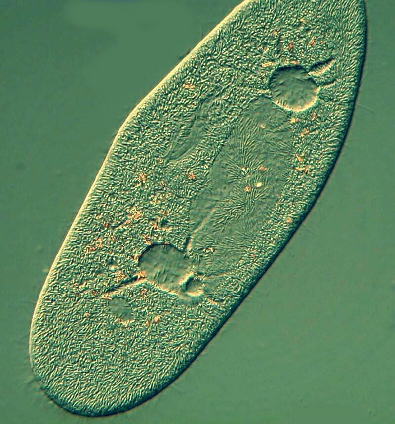
First, I’ll show you 2 images of Paramecium that are the organism as you might encounter it under ordinary circumstances. The first image shows the cytostome or mouth to the upper left and two contractile vacuoles with canals at each end of a large ovate structure which is the macronucleus. The contractile vacuoles regulate the osmotic pressure by expelling excess fluid taken in during feeding.
Mrs. Malaprop: “All of which is just a fancy way of saying that it pumps out liquid to avoid bloating so that it doesn’t have to take die-your-ethics.”
This image was taken using Nomarski Differential Interference Contrast (DIC).
The second image is focused at a slightly different plane and the polarization has been altered. This displays a series of food vacuoles and their contents.
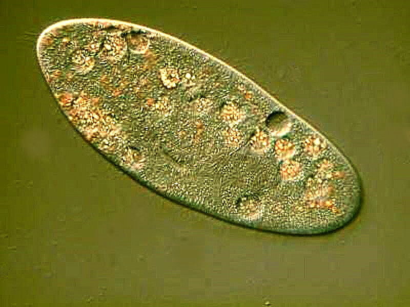
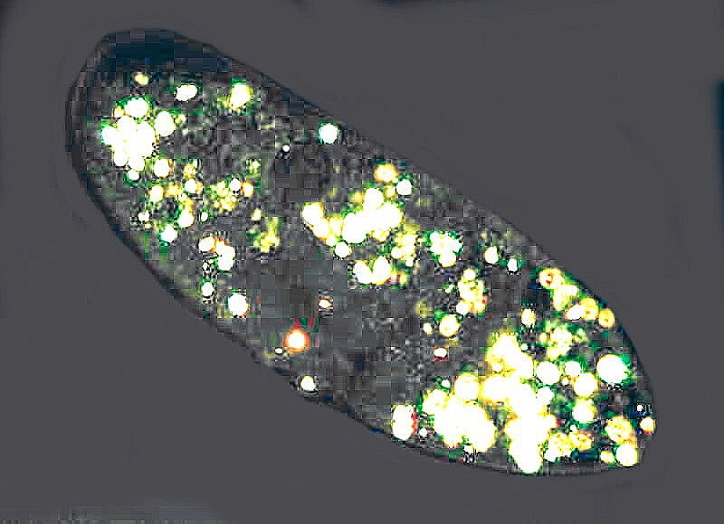
Under polarized light, crystal inclusions become evident and since the late 1800s, researchers have proposed various suggestions regarding their composition ranging from calcium oxalate, calcium carbonate, silica, complex phosphates, etc. Recent research has concluded that the crystals are primarily guanine and hypoxanthine. Not the sort of notion that one would propose without some highly sophisticated technology and extensive laboratory facilities.
Mrs. Malaprop: “Being a primitive amateur, I still like the idea of calcium ox-of-late. Oh well, we don’t always get things our way and as it turns our these animal acids do have interesting appearances under polarized light.”
Anyway, here’s an image of Paramecium under polarization showing crystalline inclusions in vacuoles. When living specimens are examined, they look like miniature motile Christmas trees.
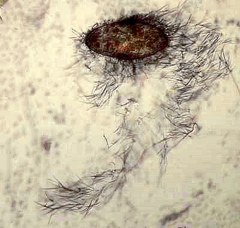
Two more images and then we’ll get to the “sexy” stuff. First, however, I want to show you 2 images of what is largely assumed to be a defense system in Paramecia, namely trichocysts. There are literally hundreds and hundreds of these structures just below the surface of the pellicle or external membrane. Various stimuli trigger the explosive reaction, images of which I will show you in a moment. Here is another revelation of just how complex these organisms are; modern techniques, including electron microscopy, reveal that there are at least 10 components in trichocysts. I use a variety of substances to trigger the trichocysts; methylene blue, iodine, tannic acid, or Harris hematoxylin. This first image was one where the trigger was iodine. Note along the bottom edge of the membrane there are tiny “chambers” which previously housed the trichocysts. It is also important to remind ourselves that this is a 3-dimensional beastie and the entire subsurface top, bottom, sides, possess these structures.
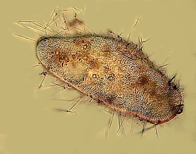
This second image shows the force with which the trichocysts are ejected; the distribution of them exceeds by several times the length of the Paramecium. The trigger in this case was the Harris hematoxylin.
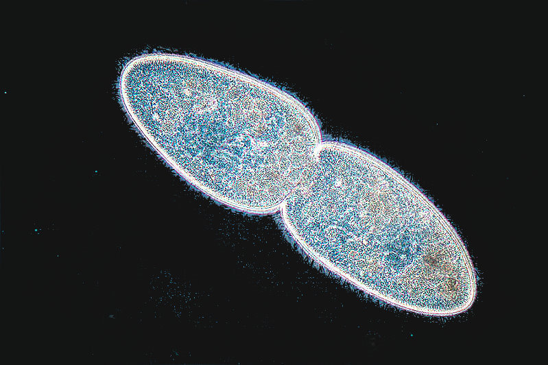
Division or binary fission is the most common form of reproduction in Paramecia. The image below is from Google and was taken by Michael Abbey. I have modified it slightly.
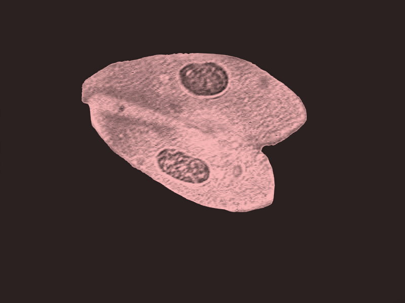
The next image shows 2 Paramecia conjugating.
Mrs. Malaprop: “Ah, now, we’re getting to the good parts.”
The specimens were stained with Harris hematoxylin and you can see the two macronuclei, but it’s the micronuclei that play the crucial role in this process. Here you see the two specimens attached; they will exchange micronuclei, separate and then reorganize their genetic material. This will ideally allow them another 200 binary fission events. This, of course, assumes that the two are compatible “mating types”. This all gets quite confusing as there are “varieties”, “groups” and “types”. Apparently, Paramecium bursaria has 16 mating types; Paramecium aurelia even more and new research keeps suggesting that there are yet more to be discovered.
Mrs. Malaprop: “And you thought human gendender types are complexified. It’s amazing how picky these Paramecia are!”
Well, Mrs. Malaprop’s comment raises a couple of interesting issues (believe it or not). One might construe this complex typology to be a biologically programmed system of eugenics. The other facet is one of a kind of “assertion” of biological dominance. Consider the so-called Kappa strain. It has tiny inclusions which are lethal to non-Kappa strains, so when conjugation occurs, the non-Kappa strain does not succeed in reproducing whereas the Kappa strain does benefitting genetically from the genetic exchange. There is even some suggestion that prions might be involved and also mention of a bacteriophage like particle structure.
Mrs. Malaprop: “Now, if you want some clearifying of these obtuse erotical conceptions, what it boils down to is that you end up with a lot of blonde Paramecia, fewer bruinets, and even fewer ginger rooted hairy beasties.”
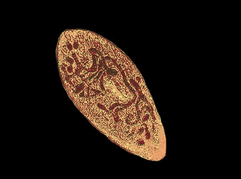
Perhaps the most fascinating aspect of Paramecium reproductive processes is one called autogamy. This can occur when acceptable mating types are not available and/or the conditions are such that conjugation is not a viable alternative. The single Paramecium then remarkably reorganizes its own genetic material in an extraordinarily complex fashion as you can see in the image below. Once again, I stained the specimen with Harris hematoxylin and I have altered the color to create a better contrast. You can see the genetic material scattered in strands throughout the organism.
Reproduction is, of course, a strategy (or a series of strategies) for survival, but there are other processes that are linked to the survival of these organisms. I want to take a look at a very interesting protozoan called Blepharisma and consider 2 other processes.
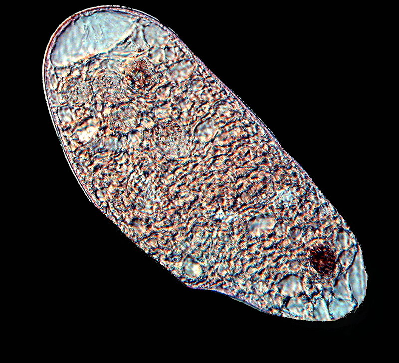
Again, it’s important to first get some general idea of the structures and complexity of the organism. The first image shows a Blepharisma in a slightly contracted state with the posterior end, which has a large contractile vacuole, at the upper part of the image. If you look very carefully, you can see just the hint of the chain or beaded macronucleus. You will also notice that there is a light pink color which is characteristic of this organism and also unique and so the pigment has been named blepharismin.
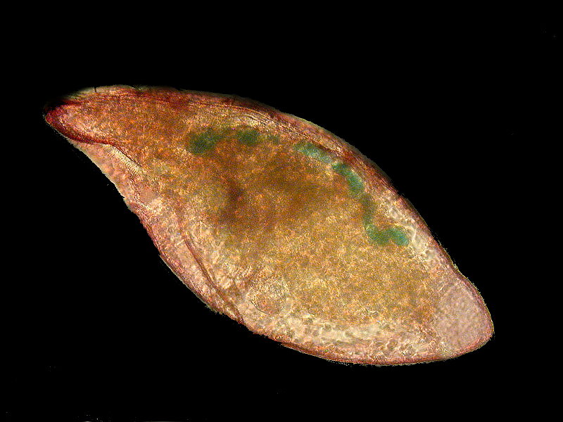
The next image shows a specimen in its more usual form, although still altered by having been stained with Methyl Green–Acetic, a stain which is particularly useful for showing up nuclear elements. Here you can very clearly see the chain macronucleus.
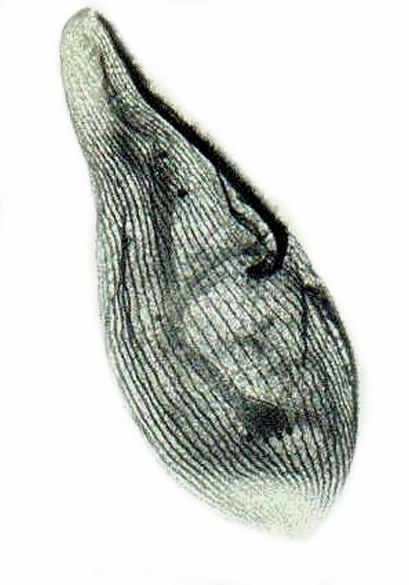
By using a very complex and demanding staining process using Protargol, a silver reagent, one can stain the so-called “silver line system.” This next image shows this elaborate system; also, it appears that this specimen was undergoing autogamy, in other words, deploying another survival system.
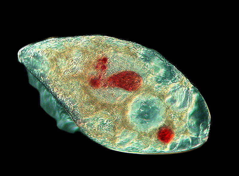
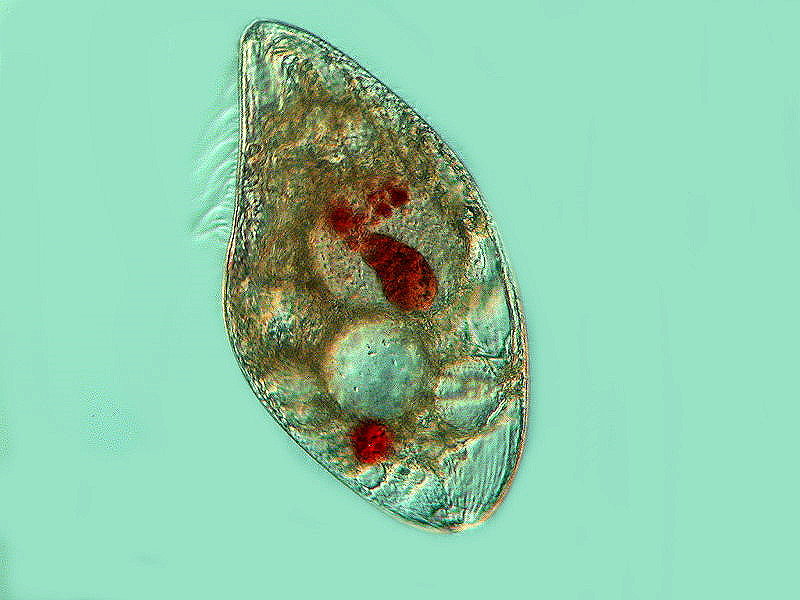
Next, we encounter an incredibly remarkable survival technique; one used in corporate business all the time–destroy your competitors. You want the power and the goodies; in this case, the food and space. Blepharisma is not subtle; it gets rid of its competitors by, quite literally, devouring them. This produces what is know as cannibal giants. This next image is interesting for several reasons; first, it shows us some intensely red areas which are actually other partially digested Blepharisma in vacuoles; second, there are scattered areas which still display the light pink pigment, and third; on the left side we get a view of the undulating membrane around the cytostome. The membrane is somewhat blurred, but it’s length gives us an idea of the size of the cytostome which helps explain how this organism can ingest fellow Blepharisma. The intense red of the ingested specimens is typical and is a result of the compaction of the cell in a vacuole.
In this next image, you can get a clearer idea of the undulating membrane and the numerous vacuoles, including some containing ingested colleagues.
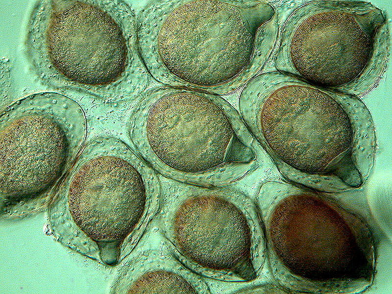
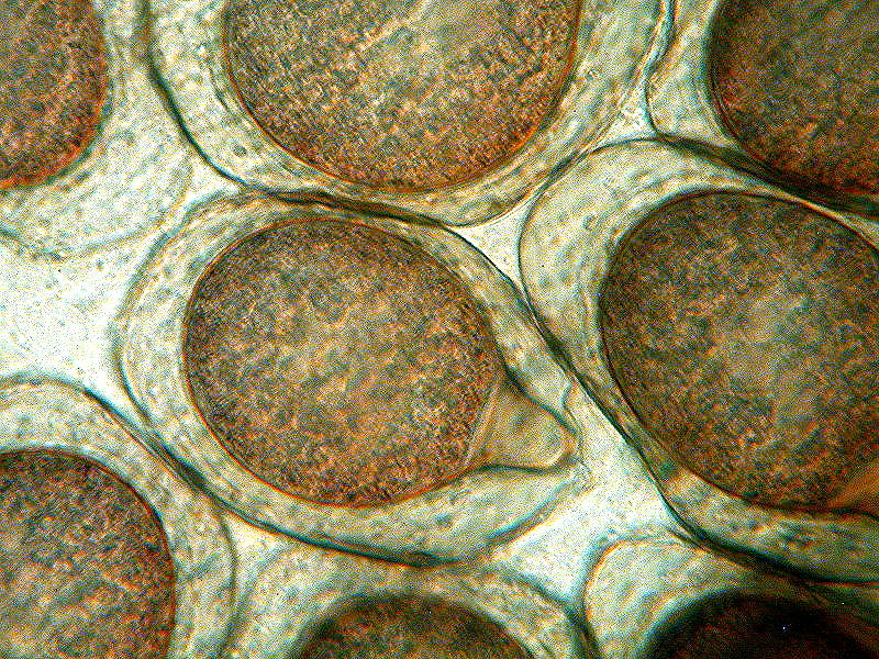
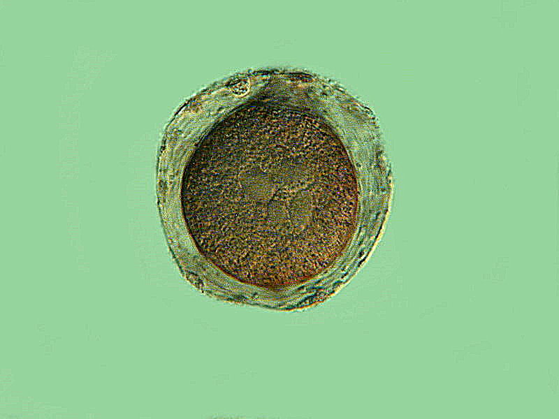
Another remarkable survival strategy is that of encystment. In this fascinating event, the organism dedifferentiates and reduces itself essentially to a highly complex “blob” of protoplasm and its genetic material. The cysts of Blepharisma are highly distinctive in their formative stages as they possess a kind of “nose cone.” First, I’ll show you a grouping of these and then a closeup, followed by a near final stage of encystment where you can see a thickening and toughening of the cyst wall. Some of the nuclear material is also visible. However, note that the vacuoles and the undulating membrane as well as an other organelles have disappeared. This thickening of the cyst wall is to ensure the protection of the content until favorable conditions are once again present so that the process can be reversed and the organism can begin to re-form its organelles. What is so extraordinary here is that the cyst wall must be able to protect against adverse conditions but, at the same time, be able to sense when the conditions are suitable for reconstitution. That is an incredible feat!
Now, let’s move up the phylogenetic scale a bit to some even more complex organisms. Rotifers and Cladocerans (“water fleas”) are both known to practice apomictic female parthenogenesis. This means that the female can go long periods of time without any sexual activity and can produce exclusively female offspring and resting eggs. However, there are species of both rotifers and cladocerans for which no male has been discovered. So much for male dominance in this part of the world; in fact, perhaps males are not even necessary!
Here’s a link if you want to look at some images of rotifers:
And here’s another link for a view of some cladocerans:
And, of course, we mustn’t forget the ostracods also known as “seed shrimp”.
These tiny creatures have been around for millions of years; they are found in both freshwater and predominately in the ocean and the estimates are that there are somewhere between 35 thousand and 70 thousand species. The males have several distinctions: 1) approximately 1/3 of their body is reproductive apparatus, 2) in some species, a single sperm stretched out is 6 times the body length, and 3) the oldest penis known was found in a 425 million year old fossil! Ordinarily, such soft parts are not fossilized and so this is as remarkable as the preservation of the soft parts of certain other invertebrates found in the Burgess Shales.
Mrs. Malaprop: “Oh, now you’ve done it. We’ll have all kinds of braggydoughsy on the part of humans of the male persuasion. I can just imaginate that hulks all over the global will be using small versions of the mediaevil racks that were used to stretch whole people not just immanent parts.”
For the super macho types, there is yet another nightmarish attack on the ego in the male role in the anglerfish. Here we discover what has been described as “sexual parasitism”. The male, who is much smaller, grasps the female and attaches himself to her underside near her genital orifice. However, she retaliates by imprisoning him, growing tissue around him thus enslaving him. He provides sperm for fertilizing her eggs and she provides food for him. She produces a gelatinous string of eggs which are released into the nearby water and the male releases sperm which ferilizes them.
Mrs. Malaprop: “Well, I never! What’s the fun in that? No courtship! No foreplay! Sometimes I think that Mother Nature has a very twisty cents of humor.”
Then there are what might be dubbed the “transmogrifiers”; fish that change sex, sometimes as a result of environmental stimuli, sometimes as a consequence of group dominance, and sometimes due to a programmed developmental process associated with maturation and aging. Moray eels, wrasses, clownfish, and about 500 other species are known to alter sex sometimes permanently but, in other cases, only temporarily. Some, such as gobies, even change back and forth a number of times.
Mrs. Malaprop: “As Alice said “curiouser and curioser”. I wonder if there are any goby sexual purists? It makes it harder to desrimpticate when such a transfourmateion can happen to you at any momentous.”
Well, this is getting to be a bit long and we haven’t touched on regeneration as a survival mechanism nor have we looked at the incredibly complicated and numerous variations in advance vertebrates. So, that will have to wait for some other time.
I hope you weren’t too disappointed by the rather tame images. I guess that old bromide is true that beauty is in the eye of the beholder.