|
Tunicate Tidbits: Ascidian Spicules by Richard L. Howey, Wyoming, USA |
Some people buy real estate, others invest in stocks and bonds–me, I’m an invertebrate spiculator. Yes, yes, as Fred Allen said: “Hanging is too good for a man who makes puns; he should be drawn and quoted.” One of the more delightful aspects of human beings is the eccentric obsessions which we develop; for some, it’s building elaborate model trains, enormously detailed yachts in bottles, collecting ancient coins or medical lithographs or pre-Victorian tea cups. Those with a bent of mind toward natural history and microscopy can also manifest passions for things that seem very odd indeed to the average person: arranging hundreds of diatom shells in a pattern in an area 3/4 x 3/4 of an inch, collecting sand samples containing forams from hundreds of locations around the world, hunting down specimens of dried echinoderms or tunicates to study their skeletal structures (one of my weaknesses, I admit) and the list goes on–passionate curiosity should always drive research, not some crass economic benefit or a student’s need to satisfy some academic whose ambitions have been frustrated.
Some of my earliest microscopic encounters were with calcareous and siliceous structures in dried or pickled specimens of sponges, starfish, sea urchins and tunicates. My first enchantment was pond samples full of protozoa and algae and the second, invertebrate skeletal structures. After 65 years, I am still obsessed by both and am a contender for the title of World Class Bore when some unsuspecting soul gets me started talking about them.
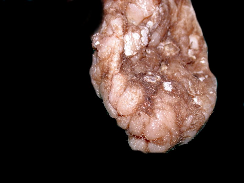
In this essay, I’m going to focus on ascidian tunicates because their alien character captivated my imagination even before I reached puberty—talk about a perverse passion! I obtained a few specimens of Styela plicata from that wonderful, now sadly defunct institution, Turtox and some years later quite a number of specimens of that organism from Gulf Specimen which, unfortunately, but understandably, no longer supplies preserved specimens but only live ones for research. Here is an image of a preserved specimen.
Styela plicata and S. clava, as well as some other species, are by no means endangered and are often classified as fouling organisms and often grow in great numbers on pilings and, to the dismay of yachtsmen, on the bottoms of boats and are thus widely considered a nuisance. They’re the kinds of pests that I much prefer to certain kinds of academic colleagues so, if you want to send me a preserved batch, I’ll gladly pay reasonable shipping costs–for tunicates, not colleagues.
My first attraction to S. plicata was not the tunicates themselves, but all the wonderful things which had been growing on or crawling around on their tunics. The surfaces were heavily populated or colonized with small sponges, bryozoans, hydroids, caprellids, nematodes, small polychaete worms, the occasional miniature brittle star, forams, diatoms, calcareous and non-calcareous algae, barnacles, wee specimens of Mytilus, the blue mussel, sometimes mysterious larvae and, if one gets lucky and gets a specimen that happened to get especially well preserved, one may even find a specimen or two of a colonial peritrich or an errant ciliate.
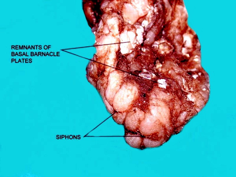
One of my friends once described them as looking like chunks of brain. You’ll notice that they appear quite warty and the tunic is thick and tough; it also seems to be constantly growing because you’ll find little wormtubes down in the folds or bits of sea urchin or sand dollar spines that have simply gotten incorporated into the tunic. It was with this species that I discovered some species of ascidians have spicules; however, getting these to reveal that secret was no easy task. I wasn’t kidding when I said that its tunic is tough and gristly; for sectioning these you need to invest in a micro-chainsaw. That’s only a slight exaggeration and I have another species of Styela whose tunic is so resistant that I had to resort to bone cutters to clip off some small pieces to try to section further. Why bother trying to section this stuff? Rampant curiosity–and as Alice might say, the further one probes ascidians the “curiouser and curiouser” they get. As it turns out, this dense cellulosic armor has some remarkable features, including spicules which we’ll get to in a minute.
However, we need to backtrack a bit before we can make any spiculations. I’m sure many of you know more about tunicates than I do, so you can jump ahead, but others may not know much about them and I’d like to make a brief excursus just to mention a few of the barest basics so that you don’t abandon the joy of tunicates. 1) There are four basic groups of tunicates. The ones that we’re concerned with here are called ascidians; in their larval stage they have the beginning of a backbone (a notochord) which degenerates and disappears after the larva attaches itself to develop into its adult stage. Such attached forms are called sessile; the other 3 types are pelagic or free-swimming. 2) Ascidians can be individual or colonial and some of the colonies are quite large and complex. 3) They are filter feeders and have 2 siphons, one of which, the incurrent siphon, draws water into a large hanging basket called the endostyle which is essentially a filtration system to capture food. This remarkable system is, however, a subject for a future essay. 4) The other siphon, the excurrent one, expels waste, excess water, and sperm and eggs, the latter at different occasions so that they don’t self-fertilize. 5) The heart has 2 “pacemakers” and it will beat in one direction, say for about 200 beats, and then the other pacemaker will take over and reverse the direction of the blood flow while the first pacemaker recharges. 6) Some ascidians have green blood as a consequence of their ability to extract the rather rare element Vanadium from sea water. I could go on, but here we want to concentrate on the spicules.
These are an ancient group of organisms and there are fossilized ascidian spicules, but they are not all that well known even among micro-paleontologists and have often been mistaken for sponge or echinoderm spicules. The organisms themselves are almost never preserved as fossils and the majority of the few samples described as tunicates are dubious, fragmentary, and almost impossible to confirm since there is virtually nothing to compare them with.
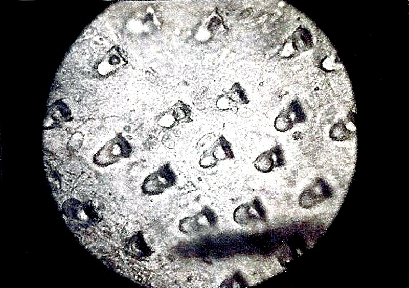
As for modern ascidians, their spicules range from tiny, spiked spheres to small plates. As far as I have been able to determine, they are all calcareous and not siliceous, so they should be easy to isolate, right? Wrong! If we’re lucky enough to get some nice slices or small chunks, we can’t just plunge them into some Clorox, expect the tissue to dissolve, and leave us a nice little collection of clean spicules. Remember, this tissue is largely cellulosic, so if you could get a nice thin slice which you suspect has spicules, you would have to treat it with the enzyme cellulase, which isn’t cheap nor particularly easy to obtain and the results are rather erratic. So, what do amateurs of modest means do? My solution was to buy a box of surgical, disposable #11 scalpels. I’m always compulsively looking for bargains–my sister is the same way and we’re convinced that this is a genetic tendency which we inherited from my mother. I found on eBay an offer of 4 boxes of 100 scalpels each for $40, which means 10 cents each. I suspect the seller may have made a mistake in his listing, but he honored the bid. Even at 10 times this price, it’s worth having a supply of such scalpels. They are indeed “razor” sharp but, they are not designed to do heavy-duty cutting; the blades are brittle and rather fragile, so exercise care in using them (and while they are exceptionally sharp, they are rarely good weapons in drunken brawls in dark alleys). So, now that you’ve got the scalpels, what do you do with them ascidian-wise? Well, we’ve been discussing forms with tough tunics. With a bit of practice, you will soon be getting some nice, very thin slices. I first undertook trying to image these sections in the pre-digital camera days when all I had was an old Polaroid microscope camera which a friend had kindly given to me. Here you can see spicules arranged more or less in rows.
The long tube has a small, ground glass screen which is used to focus the image and as you can see, it is not exactly a precision piece of equipment. Nevertheless, with persistence–since I had a small grant to pay for the film–I was able to learn a few interesting things about this odd creature’s armored house. One of the things that surprised me most was a series of vessels running through the tunic–and odd vessels at that, since they have an internal dividing wall running down the center. I finally figured out that this was part of the circulatory system and because of the eccentric reversible nature of the heart, it was desirable to have 2 vascular channels.
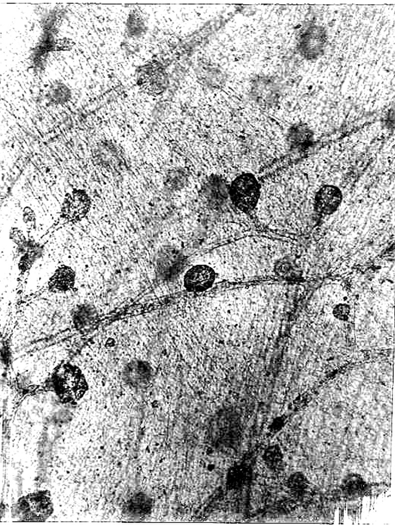
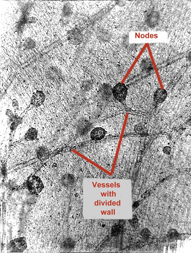
The bulbous nature of the nodes is “not for me to reason why.”
The other images that pleased me were those of tissue sections showing calcareous platelets (or spicules or ossicles as they are also called). Such a combination of a tunic with the tough character of cartilage sprinkled with gritty calcareous bits should be a serious discouragement to most would-be predators. However, there are some reef fish that like to nibble on coral, so there’s no accounting for taste. This production of a defense and then the further production of mechanisms to overcome a particular defense in a series of seemingly endless experiments makes me think less of a Mother Nature figure and more of a perverse little girl with enormous powers and demonic curiosity.
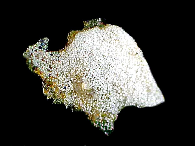
N.J. Berrill’s book on tunicates mentions a significant number of ascidians which have spicules and some of them are even colonial, such as Didemnum. These are a white to yellowish to gray encrusting form which can cover surprisingly large areas. To the casual eye, they look rather drab and insignificant. For the spicule seeker, I once again remind you that you can’t simply place sections of this material in a strong alkali and expect the tissue to dissolve. Nevertheless, with care, persistence (tinged with a bit of fanaticism, and a skillful use of some good micro-tools, you will be able to discover some quite interesting little spicules. In this case, each tiny white spot is a spicule.
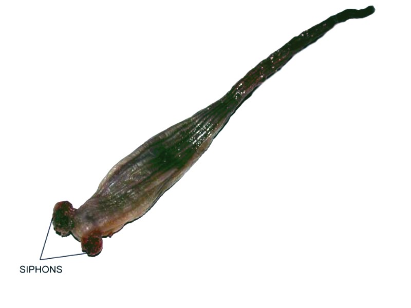
Looking for spicules in tunicates is not the sort of project that is likely to get you any nice, big, juicy research grants. As it happens there isn’t much money in spicules, but there is a very rewarding sense of discovery. There are also plenty of species yet to investigate in this regard. I have some specimens of Styela montereyensis, but I have not yet gotten to checking them for spicules and my searches don’t show any mention of whether or not anyone has found spicules in this species, so here’s a wonderful winter project for some patient soul and just to tempt you, I'll show you a picture of this weird creature.
All comments to the author Richard Howey are welcomed.
Editor's note: Visit Richard Howey's new website at http://rhowey.googlepages.com/home where he plans to share aspects of his wide interests.
Microscopy UK Front
Page
Micscape
Magazine
Article
Library
Published in the October 2016 edition of Micscape Magazine.
Please report any Web problems or offer general comments to the Micscape Editor .
Micscape is the on-line monthly magazine of the Microscopy UK website at Microscopy-UK .
©
Onview.net Ltd, Microscopy-UK, and all contributors 1995
onwards. All rights reserved.
Main site is at
www.microscopy-uk.org.uk .