 Technological change and its impact on our lives proceeds at a breathtaking pace and it has also had a major impact on microscopy at the hobby level, particularly in still and moving image capture. Some of the equipment we almost take for granted now, may have appeared to be almost magical if we time-travelled back a generation
and presented the kit to our younger selves. Below is a summary of key changes in my own hobby level equipment looking back barely 25 years which reflects these changes. It is admittedly a self indulgent review but hopefully it may be of interest.
The slow decline of film photomicrography.
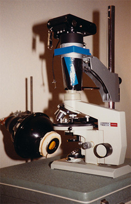
The set-up shown right was the rather basic photomicrography system I used in the mid 80s; professional systems based on 35 mm film had of course been widely used for many years and LOMO had their own range of photo accessories but were beyond my budget as it was for many other hobbyists. This was the best equipment I could afford at the time but had a lot of fun (and frustration) using it and slowly learning by trial and error to circumvent the potential pitfalls of photomicroscopy.
Right: A LOMO Biolam with a Nikkormat FT3 35 mm camera body supported over the vertical photo tube. A stripped Zenith photographic enlarger which fitted in a suitcase served as a camera support and the enlarger lamp as the microscope lamp. Although I had a LOMO Köhler lamp, they were notoriously poor at even lighting despite when a better bulb than supplied was used. The evenly frosted 75W photo-bulb with diaphragm supported in front for critical illumination was preferred, but had to be wary of the lamp overheating.
My success rate, i.e. attaining a sharp, well exposed and colour corrected image was painfully low—I was happy if six or so shots met all these criteria in a 36 roll film. Partly because of this, I rarely used quality film such as Kodak's which was too expensive at my typical failure rate, so mainly opted for the cheaper Boots the Chemist's own brand. I continued to use film as the still recording media into the mid 1990s but rapid developments in relatively affordable video microscopy and computing changed this.
Kodak PhotoCD service - affordable high quality commercial 35 mm slide film scanning.
In the early 1990s, Kodak's PhotoCD service became available in the UK and was offered at Boots the Chemist as part of their film processing and printing services. By 1994 I had a 100 passable 35 mm slides of photomicrographs or potentially so after image editing so decided to try the service (shown right). It wasn't cheap at £65 for 100 slides (£0.60 per slide + £4.95 for the CD), but better value than specialist scanning services. Six scan sizes were embedded into each PCD file from 64 x 96 to 2048 x 3072 (i.e. a max. of 6.3 Mpixels). Having just
read Wikipedia's history of the PhotoCD, apparently it didn't prove that popular or long-lived as desktop scanners became more affordable which offered higher performance. Wikipedia also notes that the proprietary PCD image format was problematical.
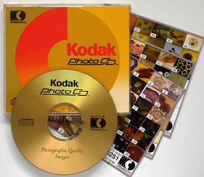 For slides that were sharp but had colour balance or minor exposure problems, the scans enabled more usable images to be derived using image editing software. A number of my 35 mm film slides were of old micro-slides with oxidised mounts where attaining the correct colour balance at point of image taking was tricky. Colour balance corrections of even some unpromising 35 mm slide scans could produce passable images.
Although in 1994 I didn't realise it, a bank of 100 digitised slides was invaluable just a year later when there was an immediate need for digital photomicrographs for a new website, see below.
The rise of video microscopy.
In the mid 1980s, microscopy of moving images was widely practiced in professional circles either by using the long established ciné film technology or the increasingly popular video microscopy such as the 1 inch monochrome Vidicon tube or more recent cameras using CCD sensors. The first edition of S. Inoué's definitive book 'Video Microscopy' was published in 1986. In hobby circles in the mid 80s, capturing moving images was probably limited to a relatively few prepared to use ciné facilities*.*See S Speel, 'An Introduction to Simple Ciné Photography, Quekett Journal of Microscopy, 1985, 35(3), pp.205-207.
Video cameras, especially monochrome Vidicon tube or similar types, were increasingly used in surveillance/security and secondhand cameras from this sector became available after their working life. This availability of used video cameras together with the increasingly commonplace domestic video recorder in many homes brought affordable video microscopy to the hobbyist. I was made aware of these developments by attending Quekett Microscopical Club meetings of the time where the early pioneers were showing its capabilities and also by reading the 1988 seminal paper by Dodge, Dodge and Jones** who discussed both the used and new buying opportunities for the hobbyist keen to explore video for themselves. **A.V. Dodge, S.V.Dodge and K. Jones, 'An Introduction to Video Recording at the Microscope', Quekett Journal of Microscopy, 1988, vol, 36(1), pp.43-53.
I adopted the route with used monochrome Vidicon camera and monitor recommended as a means of assessing the technique on a modest budget and is shown below. I recall this time was one of the most fun I've had with microscopy. As for many hobbyists, the technical aspects appealed and assessing what the equipment could do as well as the thrill of recording live sequences. The burgeoning articles and papers in the magazines and journals were testament to the many enthusiasts exploring the technique.
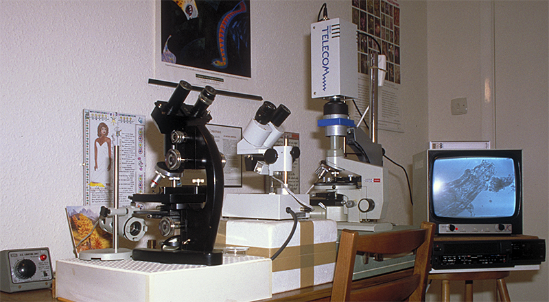
My complete microscopy set-up in the late 1980s. In addition to the LOMO Biolam was a budget Meiji stereo and a Cooke, Troughton and Simms M2000 series phase contrast microscope—the latter was destined for the skip where I worked—formal ownership of scrap was transferred for 50 pence!
The video microscopy set-up on the Biolam is shown, a heavy camera with analogue Vidicon tube and monitor (both ex-security) and a domestic VHS recorder. A live tardigrade is shown. When I moved over to colour (see below) a domestic 14 inch TV with composite video-in was used as better value than a professional colour monitor.(Artwork and calligraphy on wall by my brother Ian.)
 Colour video microscopy beckons.
Demonstrations by some of the early adopters of colour video microscopy, notably the impressive work of Ken Jones and colleagues, made me hanker after a colour system; the used B/W equipment was also likely to have a finite life. Video cameras with colour analogue sensors were available
but were heavy and of modest resolution. After much thought I bought a new high performance colour video camera with digital sensor but with composite video output. These cost high three figure sums in the early 1990s and equivalent to almost my month's salary and of more value than my entire microscopy set-up, but I was very glad I did purchase one of this quality for reasons not fully anticipated at the time, see below.
Colour offered new avenues to explore e.g. retaining colour in live work and polarising microscopy. The first live bdelloid rotifer I videoed, obligingly gave birth to live young and I was delighted to record the happy new arrival.
Although not ideal, hobbyists who already owned a camcorder with fixed zoom lens in the late 80s and 90s could support the camera over a microscope for stills and video capture. Maurice Smith, the co-founder of Microscopy-UK / Micscape successfully carried out all his website imaging needs for some years using this method.
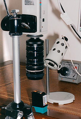
Shown right is the Panasonic WV-CL350 colour video camera purchased. It had a 1/2 inch equivalent sensor (6.4 x 4.8 mm) and 430 TV line (TVL) resolution—almost the best that could be achieved at the time affordably with a single sensor (ca. 460 lines was near the best, hi-res colour models using three RGB CCDs were much more expensive). The monochrome Vidicon 1 inch tube typically achieved e.g. 600 TVL.
The Panasonic is shown here in use for video macroscopy and still image capture, a versatile role I used extensively for the Micscape venture from 1995 onwards. (In the Quekett paper referenced above, Ken Jones showed a similar type of camera with 'C' mount extension tubes attached directly to a microscope objective—very impressive videos of pond life were shown with this set-up at 'Quekett' meets. Ken
kindly shared some of this footage in articles co-authored by Mol Smith in Micscape 1997, see Volvox and Pond Fairies - Plumatella repens.)
Increasingly affordable computers + video capture devices = an early route into digital still image capture.
Timewise we are now in the early 1990s where the IBM PC was becoming more common in the home. I continued to enjoy recording colour video sequences to analogue media (VHS tape) and taking 35 mm film stills—but my dominant imaging requirements were about to change—and very quickly ...
At the end of 1995 a forward looking chap called Maurice (Mol) Smith had already developed and was selling his 'Microscope for the PC' software with a virtual microscope and I was helping with some of the virtual slide sets (we virtually bumped into each other on a pre-Web bulletin board—remember those?). By 1995 the World Wide Web was an established part of the Internet which offered a new route to share our and fellow contributors' passion for enthusiast microscopy—the website www.microscopy-uk.org.uk was born on November 5th 1995 with the first issue of Micscape magazine. I took on the role of voluntary editor after a few issues to allow Mol to develop other site resources.
There was an immediate need for an affordable method of capturing digital imagery from all types of material submitted by contributors for the website—scans of printed matter (before A4 scanners were affordable), digital stills of 35 mm slides, macro and microscopy imagery. Fortunately the video capture device (either external or on an internal PC card) was well established as the demand for capturing stills from domestic camcorders had increased. A notable model and the one I used bought secondhand is shown below, the Snappy by Play Inc.
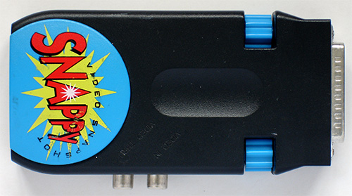
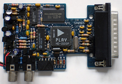
 Above. The Snappy video capture device using a parallel port interface and composite video inputs. Its fun-looking basic exterior and software could be misleading as it was arguably
the best, affordable, external video capture device on the market. Play Inc. had developed their own chip the HD-1500P with high bandwidth and multiple sampling algorithms to extract the maximum possible detail from the analogue video source.
A video through port allowed real time viewing of the video source prior to image capture, i.e. ideal for macro and microscopy image capture.
Micscape articles which made use of the Panasonic camera, macro set-up shown above and Snappy include 'Horsetails, relics from prehistory' and 'Inkjet ingenuity - a tour around an inkjet cartridge'. It was also used extensively to capture images of contributors' submitted images / drawings or diagrams on paper and their 35 mm slides, the latter using a homebrew back projection system (affordable A4 flatbed scanners and
film scanners were some years away).
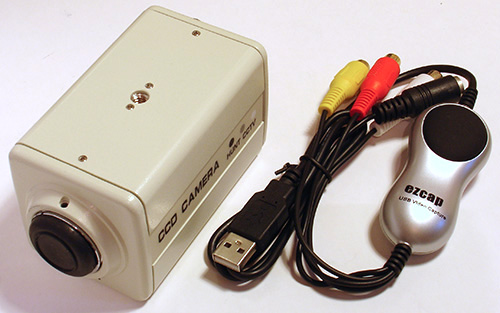
Today, video microscopy with monochrome/colour camera and digital sensor but analogue composite video output is still an interesting and affordable route to explore (shown right). Current hi-res models are typically 520 TVL colour or monochrome 700 TVL both typically <£50 on eBay UK. The night vision capable B/W cameras can have excellent low light capability and can be used for high magnification studies where the low sensor resolution (cf. true digital) is less critical and/or studies outside the visible because of the near IR sensitivity. Modern USB video/still capture tools are also available such as the EZCAP.TV 116 shown (ca. £20).
Exploring outside the visible spectrum.
Although near IR and near UV photomicroscopy with film stock has a long history (with textbooks like Loveland's 'Photomicrography' 1970 having a chapter devoted to it), I suspect it was not an area many hobbyists ventured into. The sensitivity of digital monochrome sensors to these non-visible spectral bands allowed the hobbyist to explore the uses of these bands easily and safely.
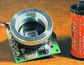 I accidentally stumbled on the monochrome sensor's near IR sensitivity when assessing a miniature CCD camera shown right in 1997 and wondering why it had poor contrast—fortunately knowledgeable Micscape readers pointed out the requirement for a near IR blocking filter for visible light work but the cameras could be used with a visible light block for near IR studies.
The ability to use filters to select a wavelength just within the near UV band also allowed hobbyists interested in 'diatom dotting' (including myself) to push the resolution to the limit with very impressive results obtained by some workers. The rapid development of LEDs in the visible and near UV also aided such studies. They also permitted safer fluorescence studies without the need for mercury lamps
The rise and rise of consumer digital cameras and the increasingly affordable digital SLR or mirrorless equivalents.
Commentators on the rapid progress in consumer digital camera performance often note that a key game changer was the Nikon Coolpix 950; introduced in 1999 at ca. £900 in the UK, it was way outside my budget but prices of the 950 used and of later models new dropped rapidly. Microscopists soon realised that its 28 mm filter thread matched that of some Leica eyepieces and became a firm favourite with professionals and amateur microscopists (although some image artefacts linked to the lens
design were noted in some of the 9xx series and some cameras with similar lens designs). The potential of the Coolpix 950 digital camera for photomicroscopy was discussed in 2000 by Peter Evennett in his landmark article 'The New Photomicrography'.
Like many hobbyists I have been through a few models and styles of digicams now as more appealing features were offered (a more cost effective method is to buy either a used or end of line camera new that is one or two models back from the latest when they can depreciate dramatically). The two camera bodies which I currently own, a Canon 600D and the mirrorless Sony NEX5N offer all the features I can think I require so hopefully have reached equilibrium. For me, key features that have appeared are:
- steady improvement in sensor noise performance, bit depth etc (+ some extra features like HDR bring the dynamic range close to that which the film devotees seek)
- electronic first curtain shutter for essentially near vibration free microscopy (in live view mode) at all speeds (although mirrorless cameras removed the mirror vibration, they can still potentially cause serious vibration if hard coupled to a 'scope from my experiences because of the double shutter action at exposure start, so I regard EFCS to be vital in a mirrorless camera on my scope)
- live view before exposure on reflex mirror DSLR
- articulated and very high resolution screens with zoom in for critical focus
- fully software controlled camera and remote live view
- excellent low light performance with modest ISO and long exposure times for all but the blackest cat in darkest cellar work
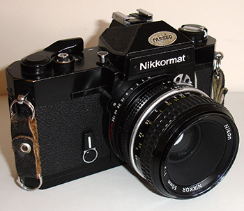
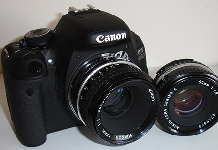
Above. Left - a typical 1980s 35 mm SLR, the Nikkormat FT3 with mirror lock handy for microscopy (but gave an almighty 'thunk' when the focal plane shutter tripped and unsuitable fixed focussing screen). Right a current 'one above budget model' DSLR the Canon 600D. Not dissimilar in appearance but very different in capabilities and convenient features for photomicroscopy.
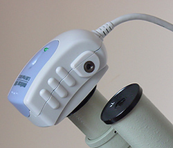 ... with increasingly affordable dedicated digital microscope cameras as an alternative.
The dedicated digital microscope camera has also become more affordable with many hobbyists preferring that route to a consumer digicam. An early version of a more affordable model the Moticam 1000 (ca. 1995) had an inferior sensor to that of a budget DSLR of the time from my own experiences, especially with high contrast subjects, but the sensors have rapidly improved, many now include HD video. Dedicated software for photomicroscopy offer appealing
features such as calibration and on screen measurement, pixel binning etc.
Some users prefer high performance cameras intended for astronomy (and often better value without the big maker's microscopy moniker—the high performance Sony ICX 285 2/3rd inch monochrome sensor can be had in an astronomy camera with cooling for less than £1000, the Zeiss Axiocam MRc with same sensor costs over £7000 albeit with software more suited for microscopy). I still prefer a modest 1.3 Mpixel modern monochrome telescope guide camera for the highest power studies over a DSLR.
Right: Moticam 1000 (1995 model) with 1/2 inch progressive scan CMOS sensor, 1.3 Mpixel colour. Shown here used directly without relay lens for high power monochromatic diatom studies, but an impressive range of couplers and relay lens to support over a microscope eyepiece were included.
In-camera and/or post capture software facilities which include high dynamic range (HDR), stacking multiple images for either increased depth of field and/or better signal noise and stitching to increase field of view have also offer the hobbyist powerful techniques to extend single image capture. Maurice Smith discusses some of these in the October 2013 issue of Micscape.
Taking stock.
As remarked in the introduction, if I time travelled back with my current cameras, a Canon 600D and Sony NEX 5N, to the late 1980s and described their capabilities to my younger self, the cameras' appearance would not be surprising but their capabilities would. It can be easy to take for granted how much progress has been made in such a short time. In the late 80s a few hundred pounds bought a very well used analogue ex-security camera, most modern stills cameras for a not dissimilar price can do
broadcast quality high definition video as well as capture multi-megapixel stills compared with the early video stills (<0.5 Mpixels). The 35 mm film camera of the 80s would be used for microscopy at an ASA typically 100 or lower to maintain quality (although high ASA film and/or push processing was used for where it was required e.g. for low light fluorescence). From my own experiences, the typical modern digital SLR is very capable of low light studies at ISOs of ca 800 and long exposures
of 15 secs.
Hand on heart though, something rather irks me. I recall having way much more fun then than I do now—getting the most out of basic equipment rather than with the ultra capable (but often overly complex) modern cameras. I used to be a technophile, devouring all the latest gadgetry that could afford and explore. Now, I'm more techno-neutral—I adopt the minimum technology that gets a job done and let the continued developments pass me by—maybe there's a moral there somewhere!
Traditional film technologies are of course most certainly not defunct including for photomicroscopy and have their devotees, especially in the large formats. For many hobbyists, including myself seeking digital imagery for the web or modest sized prints, the convenience and instant results of either digital stills or video are too compelling advantages for most work. On my current microscope, a Zeiss Photomicroscope III with built-in film unit I occasionally run B&W film through and process. The digital versus film debates continue but the 'best camera' arguably remains the model that delivers the quality of results desired for each user while balancing factors such as convenience, performance and price—that may be a digital SLR or mirrorless equivalent, a dedicated digital microscope camera or film format / camera model of choice.
Although not really in the hobbyist sphere, the rapid progress in smartphone camera performance has also allowed the phones to have a role in certain branches of photomicroscopy, such as remote diagnosis of disease by sending the images back to experts for assessment. Fluorescence based microscopes for diagnostics using smartphones have been developed.
If my early 20th century hero of photomicrography, Edmund Spitta* who is widely regarded to this day as one of the finest exponents, could have looked 80 years later at a mid 1980s photomicrography system, there would not be much that he would not recognise—silver based film was still in use albeit smaller 35 mm formats were offered, tungsten filament lamps to illuminate would be familiar and electrical principles for exposure calculation coupled to an automatic shutter and film transport could be appreciated. But now in barely a quarter of that time since the 1980s, major changes have taken place in still and video image capture. Is digital technology now starting to mature, or if this rate of progress continues, what 'magic' is around the corner in another generation or two?
*The third edition of his book 'Microscopy ..' pub. 1920 and earlier 1907 edition with extensive full page plates of his photomicrographs are available to read online or download at www.archive.org.
Comments to the author David Walker are welcomed.