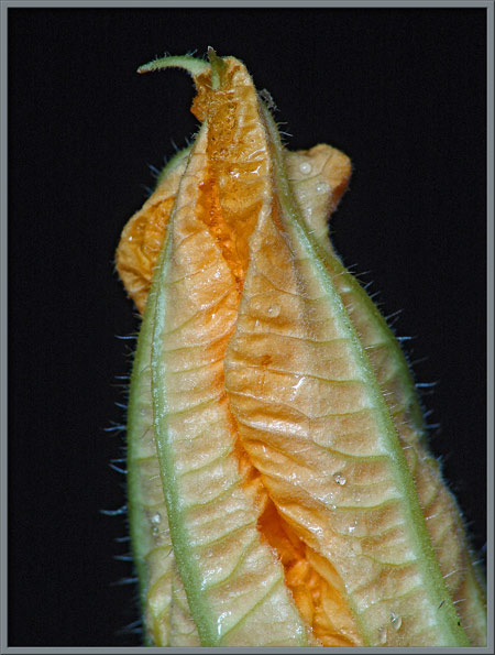
A Close-up View of a Member of the

"Cucurbits"

(Family: Cucurbitaceae       Genus: Cucurbita)


by Brian Johnston   (Canada)


Walking in the steady drizzle on a windy, cold October day,  I certainly didn’t expect to come upon the buds and flowers of this solitary plant.  It was growing quite successfully between the tracks of two railway lines, one seldom used, and one deserted for decades.  How it came to be in that location is a mystery.  Why it was blooming so late in the season, with only a couple of weeks before the first frost, is also a mystery.  Although it had no chance of producing a mature vine crop, the plant was growing vibrantly as if to contest its approaching doom.

Members of the pumpkin, squash, gourd and cucumber family are often referred to as ‘cucurbits’.  This name is derived from their botanical genus classification of Cucurbita.  These were some of the first plants to be cultivated in Europe, Asia and North America.  The family is distinct morphologically and biochemically from other families.  It is therefore called monophyletic.

Most of the cucurbits have separate male and female flowers growing on a long vine which is supported by spiraling tendrils attached to adjacent objects (which may be other plants).  Having flowers of both sexes on the same plant is referred to as monoecious.  Female flowers and buds, such as the one shown above, have a clearly visible ovary which eventually develops into the cucumber, squash, etc.  This makes them easy to identify.  Male flowers and buds have no such miniature fruit attached to their base, and look like the two shown below.  Notice the many extremely fine hairs that cover both bud and stems.


Male Flower

The first flowers to open on the long stem are almost always male.  Bees are alerted to the location of the vine by the pollen on these male flowers.  By the time the first female flowers open, the bees have an established route to the plant, and they transfer pollen from male to female flower resulting in fertilization.

The buds of male flowers are quite beautiful, with their yellow-orange furled tops and ring of five green sepals (modified leaves) at the base.  Notice in the second image, the green lines that radiate out from the stem.  There is one aligned with each sepal, and a fainter one aligned with each primary vein of a petal.


If one of the buds is cut in half longitudinally, the main veins of the petals are clearly visible in the ‘outside’ view.


The ‘inside’ view reveals a dense mass of folded orange tissue with many very fine hairs covering all surfaces.


The bud feels strangely spongy when handled.  The microscopic views which follow explain why.  Between the surfaces of the petals, there can be seen to be many tubular structures which bend and collapse as the bud is compressed.


The photomicrograph which follows shows the granular cellular material of the unopened bud’s petals.


At higher magnification, the base of one of the tubes seen earlier seems to be composed of a series of squashed spheres.


Again at high magnification, there are many reddish chain-like structures which seem to bridge the gaps between the layers of tissue.


As can be seen below, the green sepals just above the bulbous base of the bud, are covered by many fine hairs.  (The water droplets seen in these and other images are the result of the photography of the specimens obtained during a fine rain.  Images were obtained immediately because of the tendency of the specimens to wilt, even when placed in water.)


In my opinion, the most photogenic stage in the development of this plant, is the bud form.  Some of my favourite images follow.


Eventually, the bud opens to reveal the bright yellow-orange flower, which unfortunately lasts for only a day or less.  The flower consists of five triangular petals which are fused together at the base.  (The ring of petals is called the corolla.)  At the tip of each petal there is a tiny, green, needle-like spike which is about 10 mm long (right hand image).


The two closer views that follow reveal the stamen at the center of the flower.  The cone-shaped yellow anther is connected to the rest of the flower by a sturdy white filament.  The upper surface of the petals is covered by a multitude of fine hairs, many of which have clumps of pollen stuck to them.


If the petals are removed, the stamen, consisting of an anther, (male pollen producing organ) and supporting filament, can be seen more clearly.  (There is just a hint in the first image that there may be more than one stamen, but if so, the multiple stamens have fused to form one structure.)  If you look closely, individual spherical pollen grains are just visible in the images.


The pollen grains are clearly visible in the image on the right below.  On the left you can see the hairs which help distribute these grains to investigating bees.


Photomicrographs of pollen taken using dark-ground illumination reveal that the surface of each grain is covered with many tiny bumps, and a few larger diameter, higher protuberances.


The use of a phase contrast condenser with a non-phase objective provides a different view of a pollen grain.


Two ‘side-view’ photographs of a partially open flower follow.


Notice how the tiny hairs on a sepal collect a variety of different diameter rain droplets.


The outer edges of the petals of a flower are always curled inwards.  (The left image was taken from the outside of a flower, and the right from the inside.)  As with all of the surfaces of the plant, the petals are intensely hairy.  


A much higher magnification shows some of these tiny hairs.


Female Flower

As was mentioned earlier, the female flowers begin development after the male flowers.  In the case of my plant, the female buds failed to open; they were killed by the first hard frost of the season.  Two images of a very early stage bud can be seen below.  The spherical ovary is beneath the developing petals and is therefore referred to as inferior.


At a later stage the developing petals take on a characteristic orange tint.  Although the bud lacks the colour of the open flower, it more than makes up for this deficiency with its striking architectural form.


When the petals of a female flower are cut away, three yellow, unusually shaped stigma lobes, (female pollen accepting organs), are visible.  In the second image, notice the ring of tiny green hairy protuberances that surround the base of the petals.  The last image reveals the furry nature of the three stigma lobes.


The ovary beneath the female flower is almost perfectly spherical, and is held by a thick ribbed stalk called a peduncle.  One of the main characteristics of the cucurbit family is how the fruit is formed.  The ovary (fruit) is fused with the outer protective covering of the ovary (receptacle tissue) to form a hard rind. Botanically, this type of fruit is called a pepo.   In the right-hand photograph, the fleshy white tissue that will develop into the inner portion of the fruit, (if the flower is successfully pollinated), can be seen.  Surrounding this is the dark green protective tissue that will form the rind.


Leaves

The front and back surface of a leaf can be seen below.  The leaves are positioned alternately on the stem and are palmately shaped with five irregular lobes.  The back surface of each leaf is prominently veined.


Close-ups of the front surface of a leaf are shown below.  The photomicrograph on the right shows the hairs at the edges of the chlorophyll filled blocks.


The back of the leaf has a series of protruding light green veins.  Under the microscope, some of the hairs covering a vein are visible.


A much higher magnification reveals the cellular structure of the vein, and the fact that some of the protuberances are not hair-like, but round in shape (glandular).


The edge of a leaf can be seen below.  The image at right shows the variation that exists in the size of the attached hairs.


Occasionally a pollen grain, or clump of grains becomes attached to one of the hairs on the leaf’s surface.


The base of a hair can be seen to be segmented in the image below.


Stem

Immature, small diameter stalks, (left image), are almost round in cross-section, and have pale yellow striations.  Mature stalks, (right image), are darker green, five sided, and hollow.


Tendrils

The heavy plant is supported by many coiled tendrils, (modified shoots), like the one below.  They grow quickly, and entangle any other plant or object in the vicinity.


Under the microscope, the tendrils can be seen to have hairs growing from their surfaces. The occasional adhering pollen grain is visible as well.


What is it?


Is the solitary cucurbit that I found on that rainy October day a cucumber, gourd, muskmelon, pumpkin, squash or watermelon?  I’m not certain!  Since the frost killed the plant before the fruit had a chance to fully mature, identification is difficult.  The largest fruit, (seen above), was 3.25 cm in diameter.  Perhaps the final answer is not important.  This article is about an entire botanical family, not just one member!


Photographic Equipment

An eight megapixel Sony CyberShot DSC-F 828 equipped with achromatic close-up lenses, (Nikon 5T, 6T, Sony VCL-M3358, and shorter focal length achromat used singly, or in combination), was used to take all of the macro images. The lenses screw into the 58 mm filter threads of the camera lens. Still higher magnifications were obtained by using a macro coupler (which has two male threads) to attach a reversed 50 mm focal length f 1.4 Olympus SLR lens to the F 828. The photomicrographs were taken with a Leitz SM-Pol microscope (using a dark-ground condenser), and a Nikon Coolpix 4500 camera.   


Additional Information

Pumpkins, squashes and gourds - Cucurbitaceae


The Cucurbit Network

   
Cucurbitaceae Information


Cucurbitaceae Information


 All comments to the author Brian Johnston are welcomed.


Microscopy UK Front Page
Micscape Magazine
Article Library


© Microscopy UK or their contributors.

Published in the October 2006 edition of Micscape.
Please report any Web problems or offer general comments to the Micscape Editor.
Micscape is the on-line monthly magazine of the Microscopy UK web
site at Microscopy-UK  


© Onview.net Ltd, Microscopy-UK, and all contributors 1995 onwards. All rights reserved. Main site is at www.microscopy-uk.org.uk with full mirror at www.microscopy-uk.net .