I was asked to look at a pond which had a 'scum' on the surface. Using a filter paper I scooped out from the surface and was left with a brown deposit on the paper. Looking at the deposit in certain lights, there was a definite sheen.
Taking some of this deposit onto a microscope slide
it was evident that this was made of individual cells. Using high power,
the cells resembled those of Porphyridium with a stellate chloroplast.
This is a member of the Rhodophyceae which do appear as brown in colour
in freshwater.
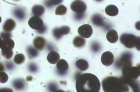
Overall appearance of the pond
scum
|
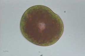
An individual cell of the algae, ca. 10µm in diameter (objective 40x) |
I extracted the filter paper with acetone and obtained a bright green solution. Using this extract I performed a one way chromatogram using acetone and petroleum ether (10/90) as the solvent. The resulting chromatograph revealed the positions of two chlorophylls (green and blue-green), xanthophyll (yellow-brown) and a carotene (yellow).
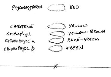
Chromatogram of algal extract. The colours do fade rapidly so only their positions are noted. |
Most interestingly, at the top of the chromatogram, was a bright red spot. I am inclined to believe that this red spot is phycoerythrin. I would be interested in receiving some feedback on these observations. I thought it would be of interest to report how the two disciplines were utilised in this study, i.e. the original microscopic appearance and then showing the presence of the red pigment, using chromatography.
Comments to the author Mike
Morgan are welcomed.
Safety note:
The acetone/petroleum ether solvents used are highly flammable. Such solvents
must only be used by skilled personnel with a knowledge of the safety hazards
under suitable laboratory conditions.