Tunicates
With Salad On The Side
by
Richard L. Howey, Wyoming, US
Images by Jan Parmentier and Wim van
Egmond, Holland
NO! I'm not suggesting that tunicates are a succulent delicacy
and I have never read about any culture which uses them as food,
but human beings do consume some very odd things and regard them
as delicacies—fish, grasshoppers, chocolate-covered ants,
rattlesnake meat, beche de mer, okra, "thousand
year old" eggs, and Big Macs. Nonetheless, there are several
factors that mitigate against regarding tunicates as potential
comestibles. Some species have calcareous spicules deposited in
the tunic which would make them rather like eating gristle with
sand embedded in it.
Many years ago, when reading a general textbook on invertebrate
zoology, I became fascinated with these odd creatures and I began
collecting specimens. At first, I bought preserved specimens from
biological supply houses, then later I was able to collect
specimens for myself on the coasts of Maine and later on the
coasts of Oregon. My passion for tunicates was such that I wrote
the directors of various marine institutes asking for any spare
specimens they might have. As a consequence, I now have
specimens, not only from Maine and Oregon, but Florida, Alabama,
Mississippi, California, Hawaii, Alaska, North Carolina, and
Antarctica.
| Tunicates are intriguing for a number of
reasons. First of all, there is good evidence that the
first description of these organisms dates back to
Aristotle who thought they were sponges and, for a casual
observer, this is still an easy mistake to make. The
sessile (or attached) species of tunicates, the
ascidians, are mostly an undistinguished looking lot. Styela
plicata is a form which I got in abundance in 5
gallon preserved "fouling assemblage"
collections from a company in Florida which,
unfortunately, no longer provides preserved specimens.
Having numerous examples of this particular form led me
to concentrate my tunicate investigations on this
species. One day I was showing a friend some specimens of
Styela plicata and his response was: "Good
Lord, those are ugly. They look like chunks of
brain." That baptismal rite produced a description
which has stuck to this day and I still use his
description when I show this tunicate to new,
unsuspecting visitors. However, I also took his remark as
a challenge to show him how fascinating and idiosyncratic
Styela plicata is. The ascidians produce a
"tunic" surrounding their delicate internal
structures. The tunic is a real oddity in the animal
world, for it consists largely of cellulose compounds. It
is tough, gristly, warty and ridged. Interestingly, there
are vessels for the transport of "blood"
running through this tunic. The vessels have a membrane
dividing them which allows fluid to flow up one side and
down the other. |
This is a critical innovation, since the heart of the
ascidians is controlled by two "pacemaker"-like
structures. The heart beats, pumping the blood in one direction
for about 100 pulses, until the controlling pacemaker begins to
lose its "charge". Then, the other pacemaker takes over
and pumps the blood in the opposite direction while the first
pacemaker "recharges" and this alternation continues.
Tunicates have an incurrent siphon and an excurrent siphon. Water
containing minute food particles is admitted through the
incurrent siphon into the pharyngeal basket. This basket is an
elegant structure designed rather like a net or lacy lattice. The
"fibers" of this net are covered with cilia which
create currents directing the particles to the
endostyle—stream of mucous only a micron or two thick, which
flows constantly down toward the stomach. There the food is
digested and the waste is passed on into the anus which opens
into the excurrent siphon and is there ejected. The ascidians are
somewhat contractile and when disturbed, expel water, thus giving
them their popular name of "sea squirts". Some
ascidians concentrate vanadium compounds, which are relatively
rare in sea water, and the vanadium salts impart a greenish color
to the "blood".
It is difficult to generalize about tunicates, because they show
such an amazing diversity. They belong to the phylum Urochordata.
Though sometimes mistaken for sponges, they are at the
"top" of the invertebrate phylogenetic tree. The
ascidians have a "notochord" or primitive backbone in
the larval or "tadpole" stage. In the adult forms, this
structure disappears. In addition to the sessile forms, there are
the Salps, the Doliolids, and the Appendicularia. These are
pelagic groups and often look as though they were made of glass.
The salps are lovely creatures; tubular, streamlined and
jet-propelled. The incurrent and excurrent siphons are at
opposite ends of the body and if one looks carefully, one can see
a series of from 4 to 9 transverse muscle bands which run around
the body. The incurrent siphon takes in water and food and when
the muscle bands contract, the body of the salp is propelled
forward by means of the expulsion of the water through the
excurrent siphon. The much-touted ingenuity and inventiveness of
humans is often overshadowed by the creativity of nature. At
least, with the development of some of the new disciplines in
biotechnology, we are beginning to realize that we can learn an
enormous amount by studying the remarkable strategies of
so-called primitive organisms.
An individual salp can
vary in size from about 1½ centimeters to nearly 20
centimeters. The salps have two forms of reproduction.
There are the asexual forms, the so-called nursemaids,
which reproduce by budding and can form
"chains" up to 25 meters in length! The other
form reproduces sexually and produces its offspring
within the body and then ejects it through the excurrent
siphon. The first person to demonstrate the generational
change between these two forms was the 19th Century
German poet-scientist Adelbert von Chamisso.
The much smaller doliolids are a delight to behold, for
they look like tiny glass barrels containing a delicate
spring.
|
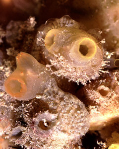
Image: Ciona with 'salad'
© Jan Parmentier 1998
|
The pyrosomes ("fire bodies") are colonial and range
in size from 10 centimeters to as much as 10 meters in length!
They appear as a thick tube closed at one end. The individual
animals that compose the colony measure only a few millimeters.
The name "fire body" derives from their peculiar
luminescent ability. When subjected to certain stimuli, pyrosoma
glow with a bluish-green luminescence. There is an account of an
early expedition during which a 3 or 4 foot pyrosoma colony was
hauled up in a net and dumped on deck at night. A crew member
traced his name on the surface of the organism and watched with
amazement as it appeared luminescently.
Perhaps the most remarkable group of the tunicates is the
Appendicularia. They are the only pelagic forms that have a
notochord and they retain it throughout their lives. This group
is sometimes also know as the Larvaceae; in part, because there
is a resemblance to the larval stages of the ascidians. The most
intriguing feature of the appendicularians is that they are like
tiny tadpoles and occupy a gelatinous "house" which
they secrete. This house has a filter to screen out debris and
food particles which are too large. When this filter gets blocked
or if the organisms senses a threat, it can escape through a
"trap door" in the house. But most amazing of all, the
tadpole has another house ready to be unfurled. These organisms
are another demonstration of the endless inventiveness of nature.
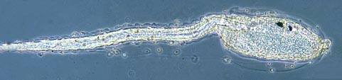
Image: Tunicate larval stage or 'tadpole'. © Wim van Egmond
1998
| Another fascinating group is the colonial
ascidians. These are sometimes described as "sea
pork" as they have both the texture and appearance
of salt pork. Never having indulged in that particular
delicacy, I cannot vouch for the accuracy of the
description. Some of the preserved samples which I have
are the size of a basketball and have either a pinkish of
purplish tint. They consist of a gelatinous mass
containing thousands of small tunicates. Some of the
colonial ascidians, such as Botryllus have a
very distinctive arrangement of zooids and look like
petals of a flower arranged around a common center into
which the excurrent siphons expel their waste products. |
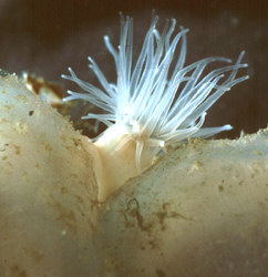
Image: Ascidian with anemone
© Jan Parmentier 1998
|
After this long-winded description, you may well have
forgotten that the title of my essay is "Tunicates With
Salad On The Side", so now it's time to get to the
"salad" part. The sessile tunicates or solitary
ascidians are often an important part of what are called
"fouling communities" and can be found in great numbers
on the pilings of piers, cliff faces, in reef communities,
occasionally in clumps on the sea bottom, and even on the bottoms
of boats. Styela plicata, the "chunk of brain"
tunicate, and Styela montereyensis, which looks like an
elongated flattened okra pod, except that it is dark brown, have
thick leathery tunics which are almost always heavily colonized
by other organisms. This is also true of Ciona intestinalis
which, when living, looks like a semi-transparent vase, but
nonetheless possesses a tough, gristly tunic. On Styela
plicata and Ciona intestinalis, I have found an
abundance of "salad". For the microscopist, collections
of tunicates from fouling communities are a treasure trove. Most
of the associated organisms which one finds are quite small and
are ideal for the microscopist who wants to make small mounts
without a great deal of special preparation and/or sectioning. On
just Styela plicata and Ciona intestinalis
alone, I have found small brittle stars, small chitons, tiny
mussels, sponges, polychaete worms, serpulid worms, small
barnacles, diatoms, sponge spicules, coralline algae, eggs and
cysts that one has virtually no hope of identifying, bryozoans,
ostracods, nematodes, tiny anemones, copepods, mites, caprellids,
other tunicates, such as, the ubiquitous colonial "sea
pork", and even the tubes of the ciliate protozoan Folliculina
which, with its gray-green tube, is highly distinctive.
Filamentous algae, dinoflagellates, and other small flagellates
are also to be found.
The tunicates themselves are well worth many hours of study and
some of the ascidians even have elegant spicules embedded in the
tunic. To examine these spicules is a challenge, because one must
cut thin sections of the tunic and then if one finds spicules,
dissolve out the surrounding cellulose tissue using the enzyme
cellulase. Every investigation of these bizarre creatures is a
voyage of discovery. There are even specialized parasitic
copepods that live inside the salps. These can be isolated and
mounted separately for study and the same can be done with many
of the small organisms associated with the ascidians. The chitons
and brittle stars can be placed in a dilute solution of potassium
hydroxide or sodium hypochlorite. Commercial Chlorox can be used,
if diluted—one can stand the smell. Personally, I detest the
odor and prefer to use potassium hydroxide, but whichever one
uses, caution must be exercised, since these are powerful
caustics and will burn the skin. The plates of the chiton can be
arranged in order on a slide and provide a handsome display. The
numerous bits of the calcareous skeleton of the brittle star can
be selected for variety and mounted either according to variation
in form or in patterns. One can use these caustics on polychaete
worms to isolate the setae (bristles) for mounting and on sponges
to separate out the spicules. Obviously the diatoms can also be
isolated and mounted and I have found round, triangular, and
rod-shaped forms. Clearly, the fouling communities are miniature
zoos and I heartily recommend tunicates with salad on the side.
Comments to the author Richard Howey are welcomed.
Editor's note:
Read the Micscape article Tunicates by Jan Parmentier and Wim van Egmond where some of the
species described are illustrated.
© Microscopy UK or their
contributors.
Published in November 1998
Micscape Magazine.
Please report any Web problems
or offer general comments to the Micscape Editor,
via the contact on current Micscape Index.
Micscape is the on-line monthly
magazine of the Microscopy UK web
site at Microscopy-UK
WIDTH=1
© Onview.net Ltd, Microscopy-UK, and all contributors 1995 onwards. All rights
reserved. Main site is at www.microscopy-uk.org.uk with full mirror at www.microscopy-uk.net.