A Close-up View of a Member of the
 
Protea Family -

a Grevillea (or Spider Flower)


by Brian Johnston   (Canada)


Documentaries about Australia usually focus on the unique and fascinating animal species that inhabit the landscape.  The subject of this article is just as unusual, but it belongs to the plant world.  The genus Grevillea is one of the most popular and widely cultivated of the country’s plant genera.  Grevillea species of all shapes and sizes are to be found in various natural locations throughout Australia.  They are all members of the Protea family (Proteaceae).  A colourful example of the same family, the “Pincushion Protea” was the subject of an earlier article.

Since the various Grevillea species hybridize readily, it is likely that the example studied here was either a chance hybridization or a cultivar produced in a nursery.  The plant was imported to Canada from Australia for cut-flower use in exotic flower arrangements. 

As can be seen below, the flowers of the plant are quite small, and occur in a cluster called an inflorescence at the tip of a branch.  The inflorescence can contain up to 100 individual flowers.  In some cultivated plants (like this one),  the flowers are clustered in a cylindrical brush shape (or raceme).   The raceme in this case is about 10 centimetres long, with a diameter of about 2.5 centimetres.  Several racemes in bud stage are visible.  I was surprised to find that the stem was extremely strong and very hard.  An ordinary pair of scissors or common kitchen knife was no match for this sturdy branch. 


The leaves of the plant occur in V-shaped “sprays” of leaflets, and are unusual in structure.


Closer views reveal that the brown central vein of each leaflet is wrapped, sandwich-like, by green edging.


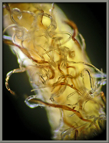
Higher magnification shows that both the central, and outer structures are covered by very fine hairs.  The photomicrograph at right shows some of the hairs in the central area of a leaflet.


In the bud stage, the flower looks like a column composed of furry brown spheres, with the occasional tiny green leaflet poking out here and there.  As time passes, the spheres move away from the stem and it is evident that they are held on the end of short brown stalks.


Just how hairy the buds are can be seen in the higher magnification images below.


Some plant inflorescences bloom from top to bottom, others from bottom to top.  In this species, one side of the flower-head blooms before the other.  As you can see from the images, the concept of “blooming” as is normally recognized in other plants has to be redefined for this flower!


Below, you can see the tip of the inflorescence with a couple of flower stems off to the left, and a circular dark ‘wound’ produced when one of the flowers fell off.  Notice the intense hairiness of the stem and flower stalks.


As the inflorescence begins to ‘bloom’, the stalks holding the spherical buds elongate and become curved.  Eventually, the upper, almost white surface of the stalk splits, revealing the light green style which supports the hidden female reproductive structure.  The image on the right shows a stalk before it splits longitudinally.


Three additional images show this stage.


Over time, the flower’s stalk stops lengthening, but the style continues to grow in length.  This forces the style to erupt out of the stem and form an arc above the stem.  As this process continues, the split in the style widens (right hand image).


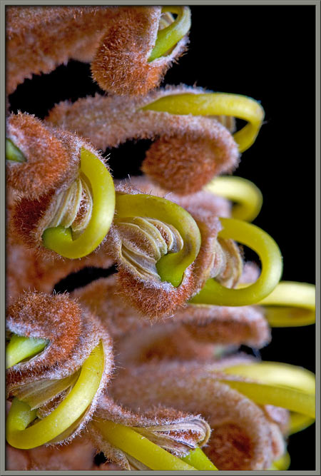
Eventually,  the inflorescence looks like the one shown below.  The many looped green styles give the flower-head a very strange appearance!


Notice that the style ends in a green enlargement – the stigma (female pollen accepting organ) – which is buried within the furry spherical ‘bud’.


The three light coloured ribbons that are exposed by the split in the style are the surfaces of the flower’s anthers (male pollen producing organs).


A single flower, dissected from the rest of the flower-head, can be seen below.


The back surface of an anther is covered with numerous tangled, hair-like filaments as can be seen in the photomicrographs below.


Eventually, the anthers open and the stigma uncurls.  Unfortunately, my cut-flower expired before this occurred.  Instead, I pulled the stigma from the anther chamber to reveal the chamber’s interior.


The receptive surface of the stigma can be seen in the image at left.  On the right is a photomicrograph of the flower’s triangular pollen adhering to the surface. The knobby bump at each corner can be seen more clearly in the higher magnification phase-contrast image (last).


One would expect that if the flower’s stigma is in contact with the pollen covered anthers, self-fertilization would occur. This is usually not the case.  Proteas are protandrous; the male organs mature before the female ones.  When the immature stigma picks up pollen from the stamens, fertilization doesn’t occur.  The stigma simply serves as an organ of pollen transfer, and is therefore called the “pollen presenter”.  A few hours after being exposed to the air, the pollen falls from the structure.  From 24 to 36 hours after the flower blooms, grooves (called stigmatic grooves) open in the tip of the stigma which can accept pollen from another plant.  Fertilization can then occur.


Photographic Equipment

Almost all of the macro- photographs were taken with an eight megapixel Canon 20D DSLR equipped with a Canon EF 100 mm f 2.8 Macro lens which focuses to 1:1.  A Canon 250D achromatic close-up lens was used to obtain higher magnifications in several images.

A few were taken with an eight megapixel Sony CyberShot DSC-F 828 equipped with achromatic close-up lenses (Canon 250D, Nikon 5T, 6T, Sony VCL-M3358, and shorter focal length achromat) used singly or in combination. The lenses screw into the 58 mm filter threads of the camera lens.  (These produce a magnification of from 0.5X to 10X for a 4x6 inch image.)

The photomicrographs were taken with a Leitz SM-Pol microscope (using dark ground and phase contrast condensers), and the Coolpix 4500.


A Flower Garden of Macroscopic Delights

A complete graphical index of all of my flower articles can be found here.


The Colourful World of Chemical Crystals

A complete graphical index of all of my crystal articles can be found here.


 All comments to the author Brian Johnston are welcomed.


Microscopy UK Front Page
Micscape Magazine
Article Library


© Microscopy UK or their contributors.

Published in the November 2008 edition of Micscape.
Please report any Web problems or offer general comments to the Micscape Editor.
Micscape is the on-line monthly magazine of the Microscopy UK web
site at Microscopy-UK  


© Onview.net Ltd, Microscopy-UK, and all contributors 1995 onwards. All rights reserved. Main site is at www.microscopy-uk.org.uk with full mirror at www.microscopy-uk.net .