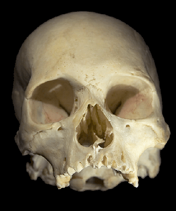
The Human Skull
 |
The Human Skull Photographs and Article by: Jessica Peterson
|
The skull is the part of the skeleton that covers and protects the brain and other sense organs. The human skull is composed of 22 types of bones. These bones are separated into two categories, the bones the form the cranium and facial bones The section of the brain that holds the brain is known as the cranium. The cranium is made up of eight flat bones that are connected. The cranium bones form the forehead and eye socket and the back of the skull. The facial bones include fourteen bones, the majority of them symmetrical to give the face symmetry. The facial bones include bones such as the maxilla, which is the upper jaw and the nasal bone. Joints known as sutures hold the bones together. At birth, the bones are not fully fused together, making the skull flexible. As a person grows, the bones of the cranium are fully fused together. |
|
The small holes in the skull are known as foramina. They allow nerves and blood vessels to pass through the skull. Processes are parts of the skeleton that hold extra tissue for muscles and ligaments to attach to. The features of the bone give the head and face form physical features and characteristics. |
|
Inferior View of the Maxilla Bone
The Maxilla Bone is located in the center of the face, help structure part of the eye socket, and holds the upper teeth |
Anterior View of the Maxilla Bone and The Infraorbital Foramen transmits the |
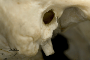 |
Anterior view of the External Auditory Meatus and the Styloid Process Both the Auditory Meatus and the Styloid Process are located on the Temporal Bone. The Auditory Meatus allows sound to enter the inner ear. The Styloid Process |
|
Foramen Magnum and Occtipal Bone (left)
The Foramen Magnum allows the spinal cord to pass through. The Occtipal Bone is the cranial bone located at the back of the head, and covers the occtipal lobe of the brain Coronal Sutures (right)
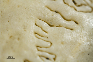
The suture connects the frontal and parietal cranial bones |
 |
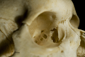 Zygamtic Bone, Eye Cavity, Sphenoid Bone, and Lacrimal Bone
The Zygamtic Bone is one of the major bones for forming the cheek. The Lacrimal Bone also forms a cavity for the tear gland. |
Equipment Used:
There are various problems that come across when photographing a skull. It is fragile, and good care has to be taken so that it will not crack or break. This becomes difficult when wanting to show the interior view, for the top of the skull is rounded and it will not stay in one spot by itself. The skull is generally one color and has very little contrast. Until you get close to it, the texture is hard to notice.
|
The Sphenoid Bone helps form the eye cavity. |
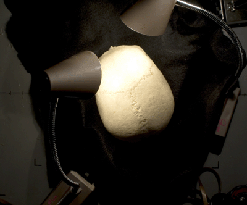 |
The directional lights showed the features of the skull by creating contrast. Having highlights and shadows help to show the texture and formations of the skull.
|
Squamosal suture and Coronal Suture |
Lighting Set Up |
The squamosal suture connects the parietal and temporal cranial bones |
Nasal Bone The Nasal Bone structures the upper part of the nose and the nasal bridge |
Frontal View . |
The Saggital Sutures connect the two paritial bones |
Contact Information: |
|
Refrences: |
|
Best Viewed in Firefox at resolution 1024x768 |
Return to index of articles
by students on the 'Principles and techniques of photomacrography'
course, November 2007, Article hosted on Micscape Magazine (Microscopy-UK). |