Recently, Frank van Campen and Herman Snoeck, from the Antwerp Royal Society for Micrography ( Koninklijk Antwerps Genootschap voor Micrografie) visited our Dutch Society for Microscopy (NVVM). Frank and Herman delivered a one evening course in making plant sections, and colouring and embedding them.
After a delightful evening, each of the Dutch members returned home with a self made slide. Our Belgian friends are experts in making slides of botanical and zoological samples and were very willing, on our request, to teach us some of their craft. They presented, as a gift, some twenty very fine slides to our club, to show us what is possible with exercise and patience.
One of the slides showed diatoms from Port Townsend, situated a bit north of Seattle, USA, on the Pacific coast. As I had never seen diatoms from the Pacific, I studied that slide in more detail, at the same time trying my new digital camera (Nikon Coolpix 995) on my microscope. I was very much helped by the article in the Micscape issue of September, in which Vishnu V.B. Reddy, USA, described his experiences.
A friend of mine, much better in using his hands, copied the adapter described by Vishnu, and I immediately took to work with it. It worked excellently, and as a result I am glad to show several of the diatoms.
Altogether, a nice cooperation between several international partners!
Comments to the author Jan
Parmentier are welcomed.
Pictures
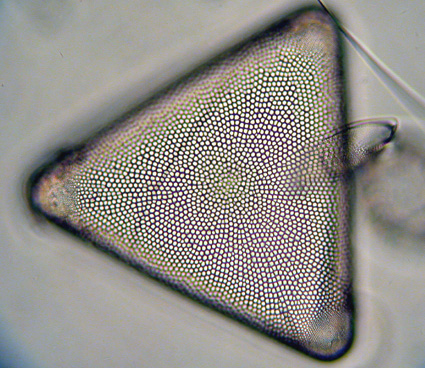
Picture 1 : Trigonium arcticum, (objective 40x) |
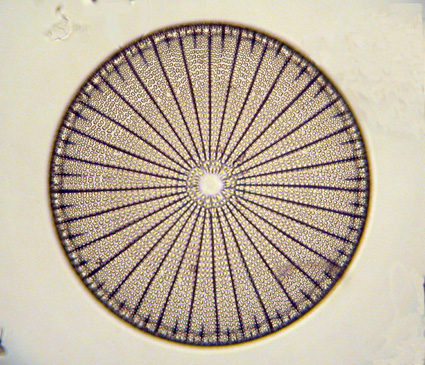
Picture 2 : Arachnoidiscus sp., (objective 40x) |
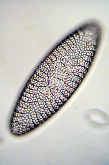
Picture 3 : Isthmia nervosa, (valve view, objective 40x) |
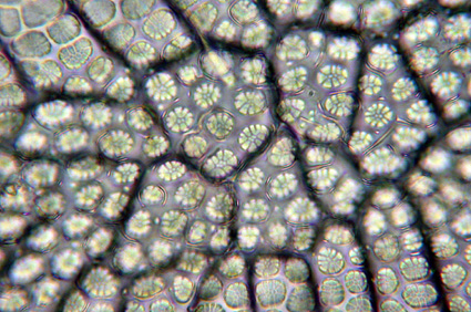
Picture 4 : a detail of Isthmia nervosa, (oil immersion, obj. 100x) |
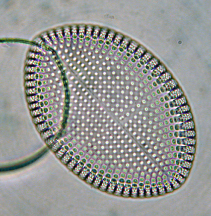
Picture 5 : Cocconeis scutellum, (obj, 100x oil immersion) |
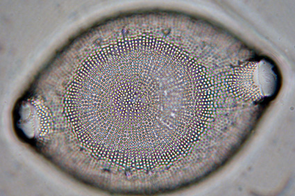
Picture 6 : Probably an Odontella sp., (obj. 100x, oil immersion) |