|
Canadian pondweed by Dave Walker, UK |
Still from video below |
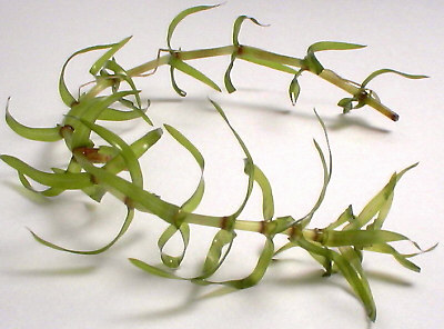
Canadian pondweed (Elodea canadensis) is often mentioned in biology and freshwater life books as one of the best subjects to demonstrate the phenomenom called cyclosis or cytoplasmic streaming. The plant thrives in my garden pond, so the onset of the long winter evenings seemed a good opportunity to try and capture some video clips ....
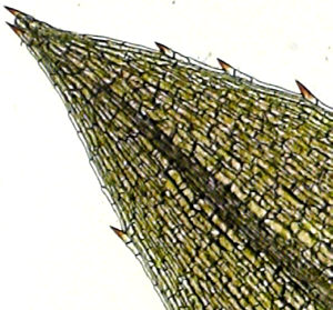
Preparing a slide for study: A leaf can be peeled off the stem and a small piece cut off and mounted in a little pond water under a coverslip. The leaves are only two cells thick so permits easy viewing of the cyclosis. Younger paler leaves (e.g. in buds) may show the cyclosis more clearly, especially for video capture.
| Canadian pondweed.
The long strands of dark green leaves in whorls of three are easily spotted
if the weed is present in a pond. If the plant doesn't occur locally, a
garden centre or aquarist may sell it.
Elodea is very common in the UK, although it's not a native plant and was introduced from North America (ca. middle 19th c.). |
 |
| Tip of a young leaf,
objective 3.5x.
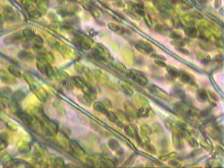
The weed the author took from his garden pond in November initially showed only a small amount of cyclosis in the long thin cells of the central leaf vein. But after a few minutes of study under the microscope, the bright light and warmth of the lamp encouraged cyclosis and the chloroplasts migrated to the cell walls and started their cyclic motion. |
 |
| A real time video
clip in brightfield using a Russian achromatic 20x (NA 0.40) objective
and 10x eyepiece.
The slight pink hue is not an artefact, parts of the leaf do have this colour. The videos are in avi format and use the common Cinepak Codec compression. If avis can't be viewed, click here for an animated greyscale gif (6 sec. real time autolooped, 2 frame/sec, 572kbytes). |
The window may remain blank until file is downloaded, (6 second clip autolooped, 5 frames/sec, 585kbytes). If interested in viewing a longer 12 second clip, 1.12Mbytes, click here. |
The cyclic movement of the chloroplasts is caused by the streaming of the cytoplasm. A hint of this streaming is evident as the bright band along the cell wall in the centre of the video clip above. This is clearer in visual studies, where the cytoplasm can be observed moving along the cell walls and in strands across the cells. The cells at the leaf edge of Elodea are only one cell thick and have fewer chloroplasts, so these cells may show the streaming more clearly, particularly with enhanced contrast techniques. The author's attempts to video the cytoplasm itself streaming under phase contrast weren't very successful, as the depth of field is tiny and the cytoplasm strands tended to move in and out of focus.
A number of web sites give good descriptions of the phenomenom, the underlying mechanisms and which other plant cells can be studied to show cyclosis. For example:
Cytoplasmic streaming - a short summary on the Encyclopaedia Britannica site.
Cell biology. Cytoplasmic streaming - an article by Bob Ridge summarising aspects of various 'streaming' studies in different plant cells, including horsetail and clover root hairs. (International Christian University, Mitaka, Tokyo). Thank you to the author who informs me that this page is an extract from a book on root hair biology. To see the contents of the chapter go to http://mac122.icu.ac.jp/rls/contents.html.
Experiments in pollen and seed germination for pre-college teachers - a resource page by Dr Mary Lee Sparling (California State University) describing how to view cyclosis in both Elodea and the pollen tubes of Tradescantia.Comments to the author Dave Walker are welcomed.The plant cell - a variety of interesting experiments to study aspects of plant cells under the microscope. Includes a splendid image of a Tradescantia stamen hair cell which clearly shows the cytoplasm strands. (Univ. of Wisconsin Botany course.)