Batrachospermum
by Gary Baird, US
 When it comes to describing algae,
the colors of the rainbow do just fine. Green, Yellow,
Blue-green, Red...each has its place. But when it comes to
freshwater algae, Red is rare. Fortunately, the Red algae, or
Rhodophyta, are well represented by a very common genus, Batrachospermum.
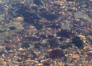
I discovered this specimen on stones at the bottom of a swiftly
flowing intermittent stream in late winter. Batrachospermum
seems to flourish in this fast water environment, for it was not
growing in more quiescent stretches of the same stream. From
shore, the Batrachospermum thallus looks almost black. I
stepped out into the cold water to recover this specimen. What a
beautiful sight it was! Like little glass beads strung on a
thread, the delicate strands felt slimy, gelatinous, to the
touch. The filament branched profusely, like a tree (arbuscular
is the term botanists use).
When it comes to describing algae,
the colors of the rainbow do just fine. Green, Yellow,
Blue-green, Red...each has its place. But when it comes to
freshwater algae, Red is rare. Fortunately, the Red algae, or
Rhodophyta, are well represented by a very common genus, Batrachospermum.
I discovered this specimen on stones at the bottom of a swiftly
flowing intermittent stream in late winter. Batrachospermum
seems to flourish in this fast water environment, for it was not
growing in more quiescent stretches of the same stream. From
shore, the Batrachospermum thallus looks almost black. I
stepped out into the cold water to recover this specimen. What a
beautiful sight it was! Like little glass beads strung on a
thread, the delicate strands felt slimy, gelatinous, to the
touch. The filament branched profusely, like a tree (arbuscular
is the term botanists use).
 Even though Batrachospermum
is classified with the Rhodophyta, it really does not look Red at
all! One of my reference books1 says that it contains
phycoerythrin and other chemical components peculiar to
Rhodophyta.
Even though Batrachospermum
is classified with the Rhodophyta, it really does not look Red at
all! One of my reference books1 says that it contains
phycoerythrin and other chemical components peculiar to
Rhodophyta.
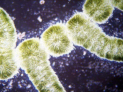
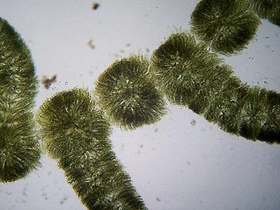
Here shown in darkfield illumination, the thallus is resplendent
in its beauty of organization and detail and color. The tiny
branches are in whorls around the functional "stem" of
the thallus. This "stem" is actually a chain of large,
single cells. Rhizoid like structures make it look more complex.
Magnification shows the considerable complexity of Batrachospermum's
spatial organization, an organization even further complicated by
the reproductive structures of the plant.
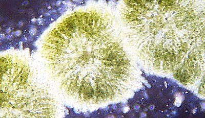 Each of the
"beads" or whorls is composed of countless individual
cells, each so small as to make characterization a real
challenge. Within this complexity develops the female sex organ
(carpogonium) and the male sex organs (antheridia cells). The
male cells do not swim, meeting the female carpogonium simply by
chance in drifting with the current.
Each of the
"beads" or whorls is composed of countless individual
cells, each so small as to make characterization a real
challenge. Within this complexity develops the female sex organ
(carpogonium) and the male sex organs (antheridia cells). The
male cells do not swim, meeting the female carpogonium simply by
chance in drifting with the current.
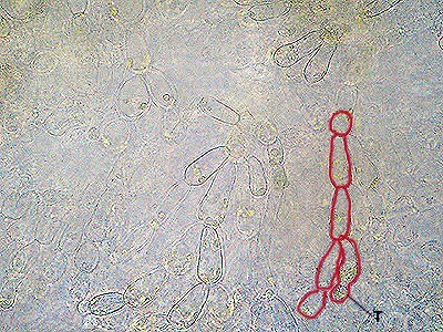
 Batrachospermum's
reproductive complexity is difficult to follow, both
microscopically and in the written accounts of experts. The
literature describes the formation of a 'trichogyne' as a
percursor to carpogonium development. This illustration and the
one following show what I believe to be a trichogyne. It is
marked in the outlined photograph with a "T". It
appears there at the terminal end of a lateral branch of cells.
Batrachospermum's
reproductive complexity is difficult to follow, both
microscopically and in the written accounts of experts. The
literature describes the formation of a 'trichogyne' as a
percursor to carpogonium development. This illustration and the
one following show what I believe to be a trichogyne. It is
marked in the outlined photograph with a "T". It
appears there at the terminal end of a lateral branch of cells.
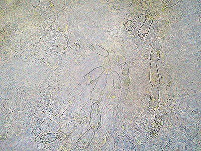
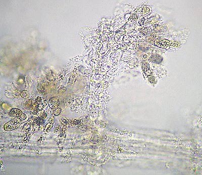 This photo of an old culture of Batrachospermum
shows a branch laden with carposporangia, the reproductive spores
ready to be released. The life history of Batrachospermum
is complex, and is best seen piecemeal, looking at different
stages of growth at different times. This particular culture sat
six weeks on my shelf! I was ready to throw it out and checked it
on a whim, to see if anything different was extant. How
serendipitous!
This photo of an old culture of Batrachospermum
shows a branch laden with carposporangia, the reproductive spores
ready to be released. The life history of Batrachospermum
is complex, and is best seen piecemeal, looking at different
stages of growth at different times. This particular culture sat
six weeks on my shelf! I was ready to throw it out and checked it
on a whim, to see if anything different was extant. How
serendipitous!
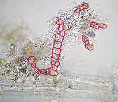 Here is the same photo with some
of the cells of the carposporangia outlined in red. These cells
are basically ready to cast off into the current, out to seek
their own place in the world. These structures are easier to see
in old, bleached out tissue than in the fresh green growth.
Here is the same photo with some
of the cells of the carposporangia outlined in red. These cells
are basically ready to cast off into the current, out to seek
their own place in the world. These structures are easier to see
in old, bleached out tissue than in the fresh green growth.
Another reference work2 states that ten species of Batrachospermum
are known from the U.S., albeit this reference is quite old. B.
moniliforme is said to be very common, and likely is the
sample here depicted. I lack sufficient taxonomic data to
completely key it out, however.
 So next time you go
stream-dipping, do not pass up the plain black growth at the
bottom of springs and freshets. Great beauty awaits your efforts!
So next time you go
stream-dipping, do not pass up the plain black growth at the
bottom of springs and freshets. Great beauty awaits your efforts!
For more information about Batrachospermum, I personally
found these references3,4 to be most useful.
Comments to the author Gary Baird welcomed.
References
1How
to Know the Freshwater Algae, by G.W. Prescott. Copyright 1970 by
Wm. C. Brown Company Publishers.
2The Freshwater Algae of the United States, by Gilbert
M. Smith. Copyright 1933 by McGraw-Hill Book Company, Inc.
3The Structure and Reproduction of the Algae, Volume
II, by F.E. Fritsch. Cambridge University Press, 1965.
4Freshwater Microscopy, Copyright 1965 by W.J.
Garnett. Constable and Company, Ltd, London, and Dover
Publications, Inc. New York.
Editor's
note. The editor thanks Gary for permission to
mirror this article from his home pages. Visit Gary Baird's home pages where his
'Naturalists Notebook' illustrates and describes aspects of the
natural history in his home area of Carthage, Missouri, US.
Text and Photos © 1999. Gary Baird. All
rights reserved.
©
Microscopy UK or their contributors.
Published
in the May 1999 edition of Micscape Magazine.
Please
report any Web problems or offer general comments to the Micscape Editor,
via the contact on current Micscape Index.
Micscape is
the on-line monthly magazine of the Microscopy UK web
site at Microscopy-UK
WIDTH=1
© Onview.net Ltd, Microscopy-UK, and all contributors 1995 onwards. All rights
reserved. Main site is at www.microscopy-uk.org.uk with full mirror at www.microscopy-uk.net.