DESMIDS & OTHER ALGAE -
PRIMARY PRODUCERS in the FOOD CHAIN
by William Ells
Coniferae, Walnut Tree Lane, Loose, Maidstone, Kent. ME15 9RG. UK.
 I
suggest invertebrates feeding on algae is a good field of study for amateur
microscopists, even those of us with little experience. As you read 'MICSCAPE'
I assume you already have a microscope or are about to acquire one. With
a 10:1 objective to search a slide, (do get a mechanical stage if you can
afford it, it makes searching much easier) a 40:1 for detailed examination,
a 10:1 eyepiece, a few slides and cover glasses, a live sample from a pond
etc., boggy places and moss squeezings are best for desmids, and you are
in business.
I
suggest invertebrates feeding on algae is a good field of study for amateur
microscopists, even those of us with little experience. As you read 'MICSCAPE'
I assume you already have a microscope or are about to acquire one. With
a 10:1 objective to search a slide, (do get a mechanical stage if you can
afford it, it makes searching much easier) a 40:1 for detailed examination,
a 10:1 eyepiece, a few slides and cover glasses, a live sample from a pond
etc., boggy places and moss squeezings are best for desmids, and you are
in business.
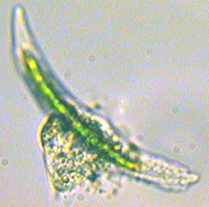
Figure 1. Amoeba eating Closterium.
Take notes of your observations, make sketches of anything
you do not recognise to look up later with measurements if possible (how
this done will be shown later). It is not too difficult to record your
observations with a camera (see earlier article
on photomicrography). Although my main interest is in the desmids I
take note of other algae and its consumers in a sample.
 In
1986 I found amoebae feeding on desmids, I sent some photomicrographs to
Alan Brook Professor Emeritus at the University of Buckingham, he made
similar observations of a different amoeba and desmid species, for details
see Brook & Ells (1987). A good example how the amateur can get involved.
Figs 1 & 2 are from the same period.
In
1986 I found amoebae feeding on desmids, I sent some photomicrographs to
Alan Brook Professor Emeritus at the University of Buckingham, he made
similar observations of a different amoeba and desmid species, for details
see Brook & Ells (1987). A good example how the amateur can get involved.
Figs 1 & 2 are from the same period.
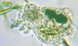
Figure 2. Amoeba with ingested desmids
 You
may need to study a sample at intervals over a day or two in order to see
what is being ingested, digested and excreted. The method I use is :- Put
a drop of the sample on a slide; spread a thin smear of petroleum jelly
(Vaseline ™) on a spare slide, then drag the edge of a cover
glass along this slide to pick up a continuous strip of the jelly, repeat
with the other three sides of the cover glass, then lower the cover glass
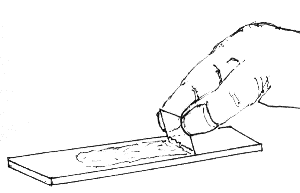
over the sample drop, see Fig. 3 right.
You
may need to study a sample at intervals over a day or two in order to see
what is being ingested, digested and excreted. The method I use is :- Put
a drop of the sample on a slide; spread a thin smear of petroleum jelly
(Vaseline ™) on a spare slide, then drag the edge of a cover
glass along this slide to pick up a continuous strip of the jelly, repeat
with the other three sides of the cover glass, then lower the cover glass
over the sample drop, see Fig. 3 right.
You will need to experiment to get the right size drop of water containing
the specimens and the right amount of jelly on the cover glass. If you
use oil immersion objectives you will find it easier with the cover glass
sealed than with the cover glass floating on a film of water.
After the first examination the slide should be kept in
a cool place, a north facing window sill is ideal, protected from dust
with a watch glass or something similar. You will find algae and protozoa
etc. will keep alive for a week or more using this method. Or you may find
you have bacterial soup or fungal attack renders the sample useless.
I have also kept samples in jam jars (nothing fancy) for
several months dipping into them from time to time. In one such sample
collected from a tarn near the Drunken Duck Inn in the English Lake District
in the company of a group of sober desmid enthusiast on a foray from the
Freshwater Biological Association. The small tarn is not named on my Ordnance
Survey map.
Each mount I examined from the tarn had a few small rods
of compressed debris I knew to be faecal pellets of a small crustacean
or possibly insect larvae. Macerating the pellets on one of these mounts
by applying pressure and a twisting motion to the cover glass, I identified
large numbers of empty semi cells of the desmid; Staurodesmus triangularis
(Lagerh) Teiling 1967, and two semi cells of Closterium costatum
Corda ex Ralfs 1848.
In the jar containing the tarn sample were two freshwater
shrimps of the genus Gammarus, I removed and examined these under
a low power (35:1) stereo microscope and found one had a pellet in its
anal cavity. After fixing, I removed the pellet, placed it on a slide and
macerated it as before. From this one pellet I found no less than 80 empty
Staurodesmus semi cells, one Euastrum sp. and one Staurastrum.
Note all the ingested Staurodesmus cells were broken at the isthmus
- this agrees with the laboratory experiments carried out by Coesel (1997).
However the desmids ingested by the amoeba were excreted with the cell
wall intact, the protoplasm apparently being taken up by the digestive
process through the pores in the cell walls.
The shrimps having been in the jar six months had little
choice of diet. I could not find any living
Staurodesmus at this
time, possibly they had all been eaten by the shrimps. Other desmids were
still thriving, mostly Closterium spp. and a few of that handsome
desmid Euastrum verrocusum alatum.
Aquatic oligocheate worms are inveterate feeders on algae,
in water collected from Thursley Common, Surrey on the 21.5.88 and examined
on the 18.7.88 there were several oligocheatae that had been feeding on
two species of
Trachelmonas. One of the worms had ten Trachelmonas
which could be clearly seen in its digestive tract, and there appeared
to be even more in a dense mass within which individual cells could not
be easily distinguished. Trachelmonas is one of several micro-organisms
claimed by both the botanist and the zoologist, the smaller of the two
species consumed, about 21 x 11 µm. and the one in greatest abundance
in the sample, had an ochre coloured lorica with no obvious markings and
a distinct collar. The other was larger 36 x 19 µm. with a paler
colour lorica, a granular surface and a distinct collar. More than a hundred
species have been described in texts on algae and protozoa.
 Further
observations made in September 1990 in a sample collected from a boggy
pool near Blair Atholl, Perthshire, oligocheate worms were found that had
been feeding on diatoms and desmids. In one could be seen a diatom of the
genus Surriella, and in another three cells of the desmid Tetmemorus
granulatus. The worms were approximately 1.25mm. long, their head 219µm
broad. The Tetmemorus were approximately 200µm. long by 35µm
broad, so the three were nearly half the length of the worm. The cells
were unbroken, one empty, one nearly so, the other with shrunken chloroplast.
Further
observations made in September 1990 in a sample collected from a boggy
pool near Blair Atholl, Perthshire, oligocheate worms were found that had
been feeding on diatoms and desmids. In one could be seen a diatom of the
genus Surriella, and in another three cells of the desmid Tetmemorus
granulatus. The worms were approximately 1.25mm. long, their head 219µm
broad. The Tetmemorus were approximately 200µm. long by 35µm
broad, so the three were nearly half the length of the worm. The cells
were unbroken, one empty, one nearly so, the other with shrunken chloroplast.
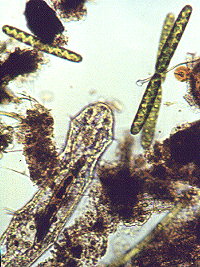
Later in the same year an aquatic oligocheate worm with
a desmid of the genus Closterium in its gut was found in a sample
from Sutherland, Scotland. (Fig. 4. right - note the recently divided cells
of the desmid Spirotaenia condensata). Both these samples were freshly
collected material without the restrictions on diet of the earlier samples.
Lund & Lund (1995) show part of an oligocheate worm with a desmid,
genus Euastrum in its gut (page 275).
In 1997 during examination of a sample of water from Dartmoor,
I found a rotifer that I now know to be Tetrasiphon hydracora, in
its stomach could clearly be seen among a green mass a desmid of the genus
Micrasterias. I sent a drawing of the rotifer to Mr. Eric Hollowday
who identified the animal for me, I quote from his letter :-
' This animal is a real "desmid cruncher" to be sure!
Rus Shiel of Australia showed a remarkable video in Poland three years
ago showing it crunching and munching its way through one desmid after
another with astonishing speed, the trophus seems to be excellently adapted
for this purpose, so keep it out of your cultures.'
This rotifer is probably rare, fortunately for the desmids,
it is the only time I have seen it, Eric Hollowday a rotifer specialist
tells me he has seen it alive only once, from Thursley Common.
I understand that in Japan a species of algae is cultivated
to feed the specially bred rotifers of the genus Brachionus which
in turn are fed to fish fry for the ornamental fish market.
I have seen in a video by Mike Dingley a Heliozoan with a desmid
, genus Cosmarium impaled on one of its spines (axopodia) we do
not know if it is ingested or not.
In spite of all these observations there is still plenty
of work for the amateur. Are there other rotifers that actually take in
alga cells, we know there are some that feed by sucking out the protoplasm?
Do protozoa feed on algae they chance upon, or are these primitive animals
able to select their prey? Do some species of aquatic worms prefer diatoms
to desmids? Why not share anything you discover with us? Send a description
of your observations with a photomicrograph if possible to the Micscape
Editor for possible publication.
Acknowledgements:-
The author is indebted to Eric Hollowday for identifying
the rotifer and for his helpful comments. Thanks to Faith Coates for the
excellent sample from a bog on Dartmoor.
References:-
Brook A.J. & Ells W. (1987) The Feeding of Amoebae
on Desmids. Microscopy Vol. 35 Part 7. pages 537-540. The Quekett Microscopical
Club.
Brinkhurst R.O. (1963 revised 1971) Aquatic Oligocheata.
F.B.A. Pub. No. 22.
Coesel P.F.M. (1997) The edibility of Staurastrum chaetoceras
and Cosmarium abbreviatum (Desmidiaceae) for Daphnia galeata/hyalina
and the role of desmids in the aquatic food chain. Aquatic Ecology 31:
pages 73-78. Kluwer Academic Publishers.
Jahn T.L., Bovee E.C. & Jahn F.F. (1949) How to know
the Protozoa. Wm. C. Brown Pub. Dubuque Iowa.
Lund H.C. & Lund J.W.G. (1995) Freshwater Algae. Biopress
Ltd. Bristol. England.
Editor's note: Comments and feedback via email
to Bill Ells
are welcomed.
Please report any Web problems to the Micscape
Editor, via the contact on current magazine index.
First published in Micscape Magazine, May 1998 ( ISSN 1365 - 070x )
Micscape is the on-line monthly magazine of the Microscopy UK Web
site at http://www.microscopy-uk.net/mag/indexmag.html
WIDTH=1
© Onview.net Ltd, Microscopy-UK, and all contributors 1995 onwards. All rights
reserved. Main site is at www.microscopy-uk.org.uk with full mirror at www.microscopy-uk.net.