by Wim van Egmond
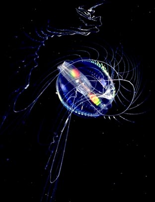
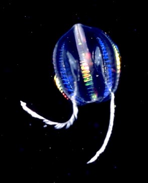
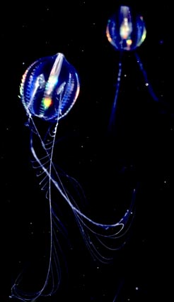
Comb-jellies, or sea gooseberries as they are also known, are one of the most beautiful animals the ocean has to offer. They are not related to jellyfish but form a group of their own: the Ctenophores.

The Ctenophore depicted in this article is Pleurobrachia pileus. It grows not much bigger than two centimetres. Therefore you can't really call it a microscopic organism. But it has many features that are interesting to study under the microscope. Ctenophores can be found in every ocean. Some even reach considerable sizes.

 
The sea gooseberries can be easily collected. They often swim close to the surface. With a little net or even a glass jar they can be scooped from the water. When put in a glass container they can be kept alive for many days.

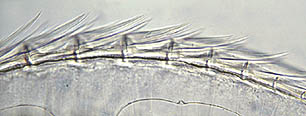
One of the most obvious characteristics are the locomotory organs, eight rows of comb-like structures that show irridescent colours when they reflect direct sunlight.

They are real predators. With it's long tentacles it captures food like copepods, the larvae of many organisms and other small creatures floating in the plankton. It brings the food to it's mouth which is shown in the pictures at the head of this article. Most textbooks however show the organism with it's mouth facing downward. I always see them with the tentacles pointing down.
 

One row of combs under the microscope.
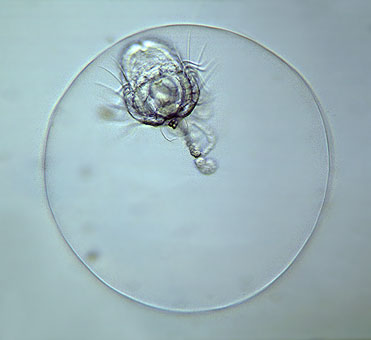

  
When I was recently studying some plankton samples I saw this very tiny egg. It was no more than half a millimetre but to my surprise contained a larva of a ctenophore. All body organs were already there. Also the comb plates can be seen. I didn't see eight as in the adult but four. Even the tentacles are visible. (This larval development is so different from that of jellyfish that it is clear that they are not closely related animals)

Most striking is the aboral sense organ. In the pictures of the adult organism it is difficult to see because it is so small compared to the size of the animal. The picture of the larva clearly shows the sense organ. A small dome contains a little round structure that is connected to the eight combs. With this the organism can control it's position in the water.


This picture was taken with oblique illumination to give some contrast to the very transparent organism. By adding a few dots of vaseline under each corner of the coverslip I prevented the egg capsule from being squashed by it. Because of the shallow depth of field you see only a circle of what is in fact a perfect sphere.
    Comments to the author Wim van Egmond are welcomed.

Footnote: Thank you to Claudia Mills for kindly telling us the species name of the Pleurobrachia illustrated above. Visit Claudia's web page on Ctenophores at http://weber.u.washington.edu/~cemills/Ctenophores.html


Microscopy UK Front Page
Micscape Magazine
Article Library


Micscape is the on-line monthly magazine of the Microscopy UK web
site at Microscopy-UK

Please report Web errors to the Micscape Editor,
via the contact on current Micscape Index.  All Material Copyright: © Wim van Egmond

WIDTH=1


© Onview.net Ltd, Microscopy-UK, and all contributors 1995 onwards. All rights reserved. Main site is at www.microscopy-uk.org.uk with full mirror at www.microscopy-uk.net.