
|
The Discovery of the Structure of DNA - Seventy Years on by David Walker, UK |
In the 25th April 1953 issue of the prestigious journal Nature, three papers were published describing and illustrating the structure of DNA. Proposed structures had been suggested before; Linus Pauling had published in February 1953 but the Crick and Watson team breathed a sigh of relief when they realised it was incorrect and Linus had not 'pipped them at the post'. Later extensive studies confirmed the new structure proposed, the famous double helix with paired bases inside the two anti-parallel phosphate-sugar backbones.
Nature in the April 27th 2023 issue on the 70th anniversary published an 'Opinion Comment' article What Rosalind Franklin truly contributed to the discovery of DNA’s structure by Matthew Cobb and Nathaniel Comfort reassessing the still controversial topic of how Rosalind Franklin and her key X-Ray crystallography work was treated. This article and an Editorial How Rosalind Franklin was let down by DNA’s dysfunctional team are open access. <Minor rant mode on> I was astounded though when visiting the 1953 issue in their archive that the three landmark papers are still behind a paywall - $10 for 48h rent, $40 to buy pdf for each! Tut tut Nature; regrettably as a non-affiliated researcher my nickname for some journal publishers is 'The Information Police'. Fortunately an increasing number of publishers are more enlightened for older resources in their publications. A Google reveals institutions that share all three papers as they regard them of historical value that they should be available to all. <Minor rant mode off>
One of the most balanced 'Historical perspective' of the discovery of the DNA structure that have read recently was by Aaron Klug, The Discovery of the DNA Double Helix published in the J. Molecular Biology 2004, 335, 3-26. Sadly behind a paywall but a legitimate student educational resource site www.studocu.com displays online to read. Franklin later worked in a very successful and productive collaboration with Klug.
Models of various complexity can be downloaded in seconds and admired in 3D on a screen but it is only when you build a model where its beauty can really be appreciated. As a self-confessed archetypal geek I admire the structure every morning, it sits hanging from a ceiling at the foot of my bed. Early sunshine casts wonderful shadows on the wall. This Cochranes of Oxford Minit model (right) is straightforward to build with clear instructions, the tricky multi-point suspension with key atoms on the strings have been pre-assembled. The kit was reviewed in the Micscape April 2021 issue. Simpler smaller models are also instructive, two are shown above and were reviewed in Micscape here.
I enjoy collecting stamps on narrow thematic subjects related to my interests. Unsurprisingly those depicting aspects of the DNA structure are more numerous so only select examples that present more detailed chemical aspects rather than the more stylised examples. A selection of these are shown below. I have a great admiration for the skills of the invariably unsung stamp designers but the right handed DNA is not always presented correctly. In the splendid A Philatelic Ramble through Chemistry (pub. Wiley-VCH, 1998) by E. Heilbronner and the late F. A. Miller they have a section Comical Errors on Stamps where four DNA related stamps are illustrated. I don't necessarily share their ''malicious joy of catching the artist and the post office with egg on their face" particularly as DNA has more potential design minefields.
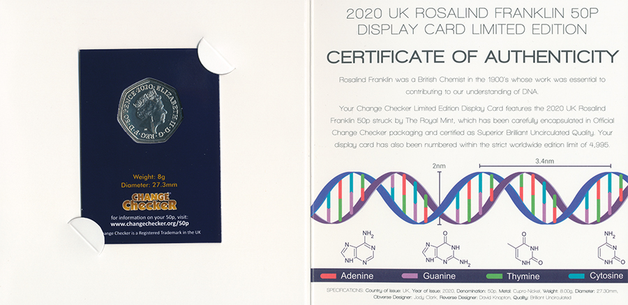
The British Royal Mint in 2020 issued a 50p coin celebrating Franklin's work. The reverse of the coin shows a stylised XRD pattern of the double helix, see cover below which uses the same coin.
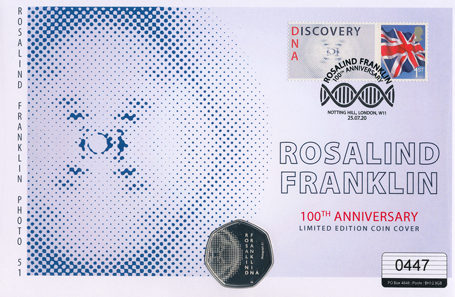
This coin cover in the Westminster Collection series celebrating the 100th birth anniversary of Franklin again shows a stylised XRD pattern on both coin and cover. It is not always easy to spot if the DNA helix has been correctly presented depending on the 3D perspective of the design. The rule of thumb have seen suggested is that imagine each strand of the backbone is a spiral staircase and if walk down each should always be turning to the right i.e. a right handed helix. (I'm not certain if the previous set above is correct). But this example's postmark is clearly incorrect, it is just two wavy lines one laid on the other.
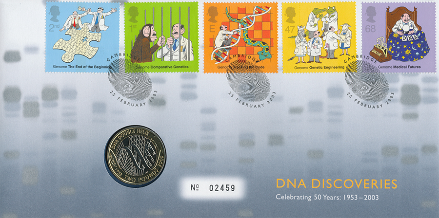
Great Britain issued a set of 'DNA Discoveries' stamps in 2003 on the 50th anniversary. GB publish some splendid designs on issues but am not a fan of this set.
With a science background I would have preferred the less cartoonish design themes and to show the personnel involved. The Royal Mint issued a Ł2 coin with the helix on both a face and edge.
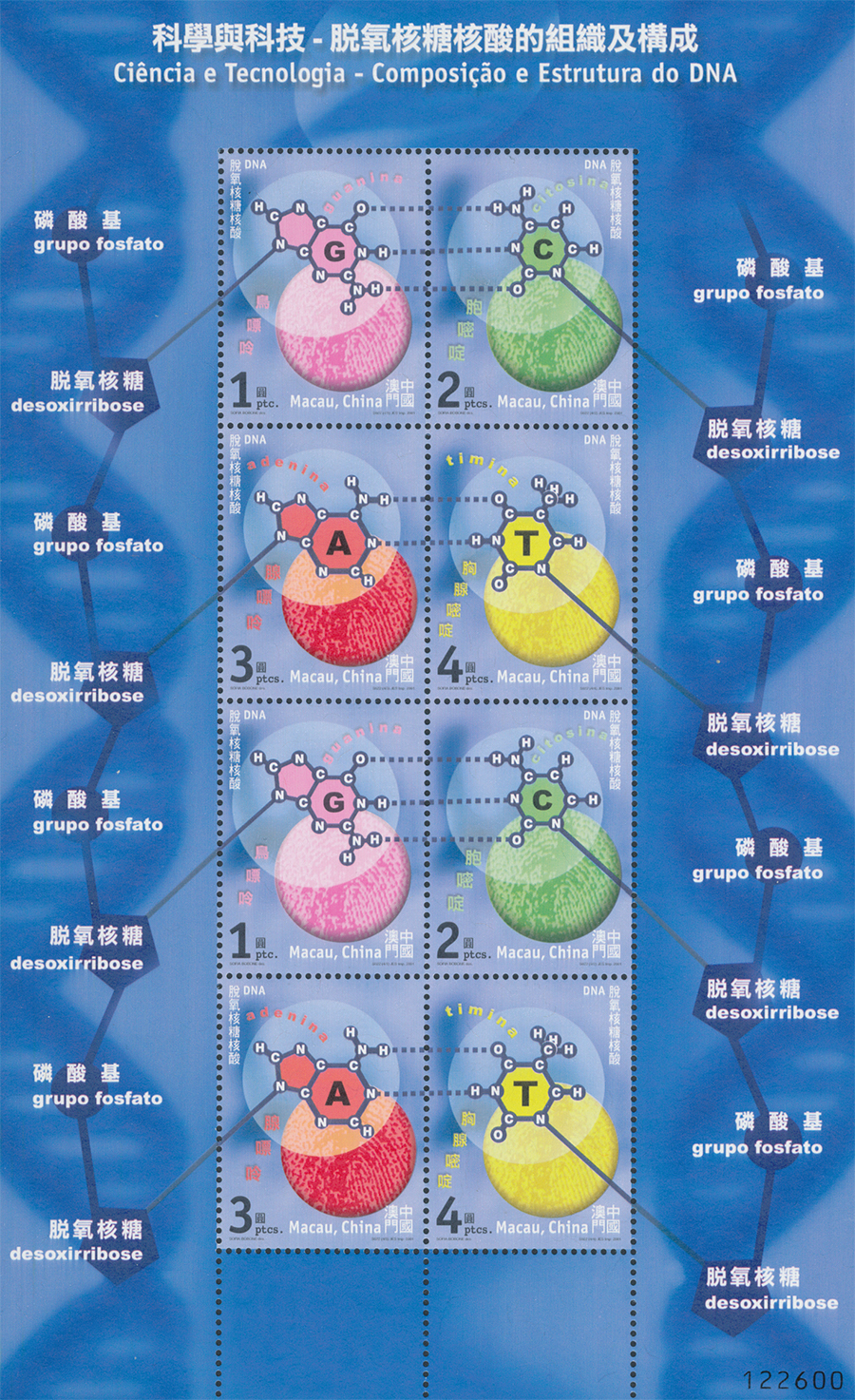
One of my favourites issued by Macau depicting the base pairs and how G-C and A-T hydrogen bond. One of the key insights of Crick and Watson to finally build the correct model was realising that the two base pairs occupy the same physical size to each fit within the outer phosphate-sugar backbones. Other key inputs were the published work of Erwin Chargaff who determined that the number of units of
the bases G and C were in the ratio 1:1 in DNA as were those for A and T, suggesting the preferential pairing. Franklin's famous XRD photograph 51 of the hydrated B form had a pattern that was indicative of the characteristic double helix and also her work that showed DNA had C2 symmetry which meant the phosphate-sugar backgrounds were anti-parallel. While model building, fortuitously an organic chemist Jerry Donahue who shared an office with them pointed out they were using the wrong tautomeric keto-enol
form of the bases and only hydrogen bond correctly with the correct tautomer.
(I have fond memories of keto-enol tautomers, my undergraduate third year physical chemistry project at Nottingham University was studying the effect of solvents on the keto-enol tautomers of an organic molecule using a venerable, even in the late 70s, NMR machine.)
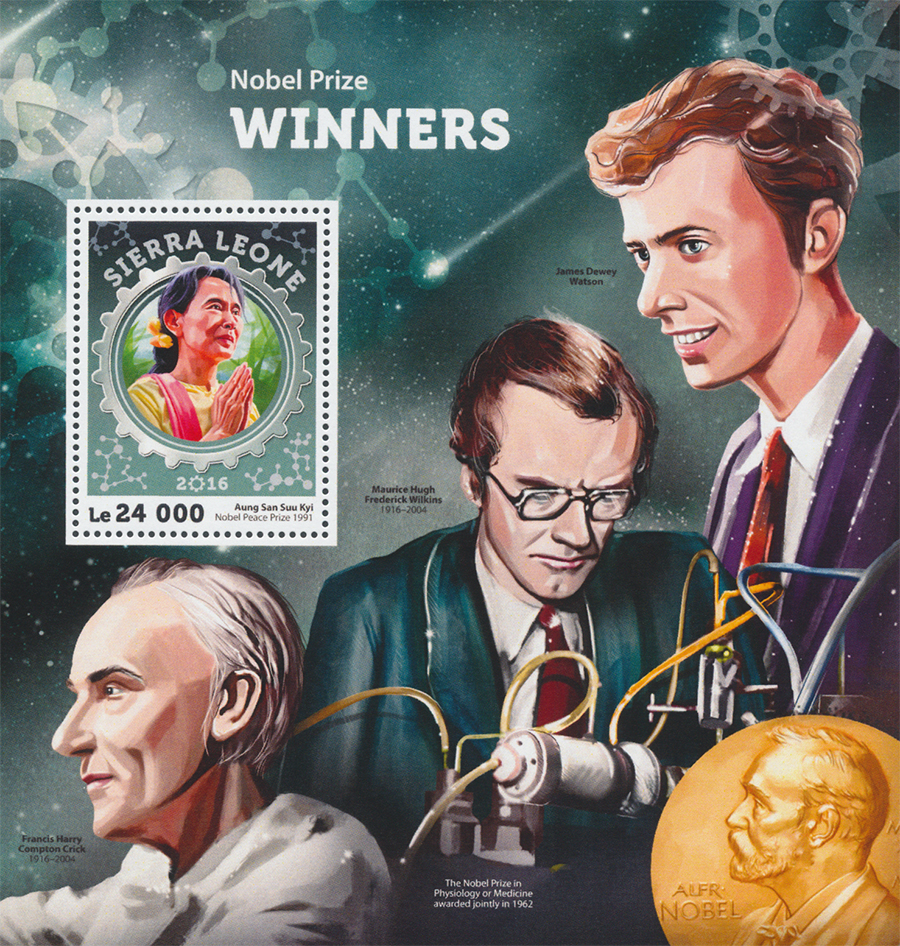
This Sierra Leone sheet shows the three 1962 Nobel laureates; Maurice Wilkins using some of the equipment at King's College, London and Francis Crick and James Watson who worked at the Cavendish Laboratory, Cambridge. Regrettably Nobel prizes were not generally awarded posthumously before 1974 when the rule not to was formalised. The Wikipedia entry for 'Photo 51' cites the Nobel Prize website 'Facts' page which notes that the prize had been twice awarded posthumously before 1962 (the year before for the Peace Prize in 1961) when the DNA prizes were awarded. Sadly Franklin died of ovarian cancer and associated illnesses in 1958. As writers since have remarked, deciding which of a maximum of three people were to be awarded the prize if Franklin was considered would have been tricky.

Another of my favourites. A molecular model and a table of the three base sequences known as codons that code for each amino acid. Uracil 'U' is seen here as this amino acid replaces thymine in RNA.
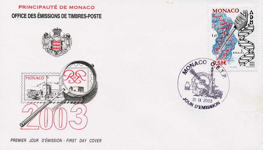
Monaco 2003, 50th anniversary. Engraving designs seem less common nowadays but do like the crisp delineation of features that this printing method provides. The diagrammatic right handed double helix is clearly shown.
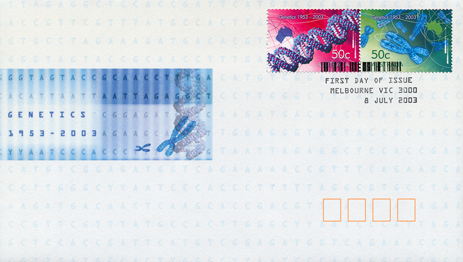
An Australian 2003 issue, 50th anniversary showing a splendid molecular model and also a base sequence and chromosomes.
Comments to the author David Walker are welcomed.
Published in the May 2023 edition of Micscape.
Please report any Web problems or offer general comments to the Micscape Editor .
Micscape is the on-line monthly magazine of the Microscopy UK web site at Microscopy-UK
© Onview.net Ltd, Microscopy-UK, and all contributors 1995
onwards. All rights reserved.
Main site is
at www.microscopy-uk.org.uk.