Get ahold of a piece of chalk, real chalk, not this modern, compressed, something or other that they pass off as chalk, but a good old-fashioned piece of blackboard chalk. What you are now holding in your hand is mostly calcium carbonate and there might even be some shells of foraminifera (forams), they were once marine amoebae. It is astonishing how often nature uses calcium carbonate in constructing things and the extraordinary variety of forms and organisms in which it occurs. Here, I want to give you a taste of that variety. It can occur in structures that are tiny and delicate and in shells that are, relatively speaking, massive, such as, Tridacna, the giant clam, which can weigh over 450 pounds and be 4 and a half feet in length. At the other end of the scale are the coccolithophores which sometimes “bloom” in extraordinary numbers. They are spherical cells ranging from 5 to 100 microns in diameter with calcareous platelets ranging in size from 2 to 25 microns. Nonetheless, during a really active bloom, they can be detected on satellite images.
Just to show you how beautiful chalk can be, we’ll start with a modest-sized example from the tropics.

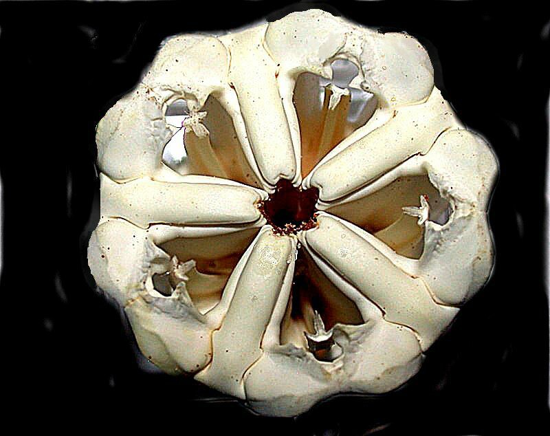
This is the test or “shell” of the sea urchin Coelopleurus mailiardii from the Philippines. Every spot where there is a little protuberance or “ball” is the base for the ball-and-socket joint for the attachment of a spine. The base of the spine has a socket which fits over the ball and is attached by muscle tissue which can rotate the spine. The spines can extend 3 or 4 times in length out beyond the diameter of the test. If you take a second careful look, you will see rows of tiny holes in the test. These are where the tube feet come through and, in some species of echinoids (sea urchins), the tube feet can be extended out even beyond the tips of the spines. An amazing bit of chalk, what? But the surprises aren’t over. These organisms have an amazing bit of dental architecture which is generally comprised of about 40 calcareous structures fused into an amazing device called “Aristotle’s lantern and, rightly so, since he was the first to describe it. I will show you 2 images of an especially large and very elegant one. First a side view and the a top view.


The teeth are at the bottom and not visible in either of these views, but if you take an intact dried urchin and turn it over, you will see 5 small white chisel-like bits protruding. These are the tips of the teeth. An elaborate array of muscles attached to the teeth slide back and forth on the lantern, moving the teeth, allowing the urchin to have a nosh by scraping algae off the rocks and substrate.
Something a bit larger is the shell of a Nautilus, a marine artistic marvel.

Quite properly attired for almost any formal occasion, I would say. Now, what do you think would happen if we were to cut it in half? “Why on earth would anybody want to do that?”, you might ask. Well, some of us were cursed with insatiable curiosity which can lead to beautiful discoveries and sometime unpleasant consequences. In this case, the answer is quite simply; I’ll show you an image.

Look at those chambers! Look at that spiral! Hail Fibonacci. Now, what if we were to take another Nautilus and do a center cut of the shell? Well, we get something even more visually remarkable. Pretty impressive for chalk.

I won’t ramble on about it: I’ll just ask you to look at it and think about it and then look at it again.
A bit ago, we mentioned sea urchin spines and what one usually encounters are sharp nail- or needle-like structures, but these are certain types that are so strange that if you encounter one in isolation, you might be hard put to even recognize it as an echinoid spine. The tropical cidaroids provide numberous examples. The “slate pencil” urchin, Heterocentrotus mamillatus (link to Google Images) has thick distinctive spines.
Psychocidaris oshimai (link to Google Images) has bizarre spines that hardly seem describable as spines.
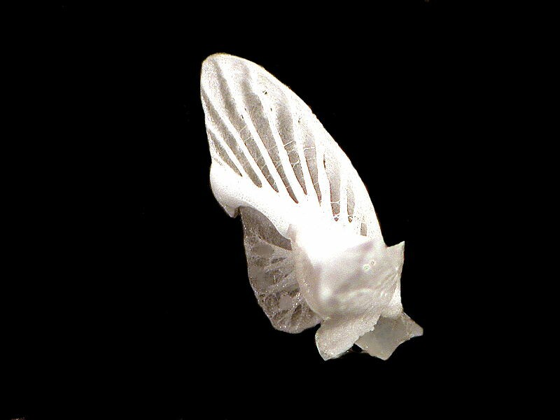
However, for my money the most un-urchin-like spine comes from Cidaris mikado. The image I’m going to show you is an upside down view, just to make things more confusing. What looks like a little hat up at the top of the spine is the ball structure that fits into the joint aperture on the surface of the sea urchin test. The fenestrated, lacy part (the facing-down side) is what you would see when you look at an intact urchin. Seeing this in isolation, who would think that this is a spine?

Several years ago, I came across a branching piece of what looked to be coral, but turned out to be a sponge. I think. Or maybe it’s a hybrid (those are all the rage these days)–perhaps a Corong or a Sporal. In any case, it has some acicular spicules, but also some spheres–geometry is everywhere in nature. Here are some along with a single acicular (needle-like) one.

And here’s a close up of a couple of the spheres.

Corals are repositories of calcium carbonate and soft corals have some very distinct spicules. These include the whip corals, fan corals, and forms like the sea pansy (Renilla). The image below shows a common type that can occur in staggering numbers in a single specimen.

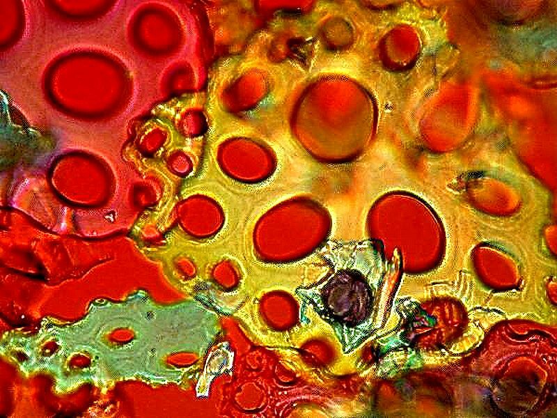
Spines can also be found in extraordinary numbers in certain species of sea cucumbers (holothuroids), Eupentacta, for example, whose dermis is a dense mat of spicules. Other species have very few and some seem to have none at all. An interesting little tropical worm-like form Lissothuria has abundant fenestrated spicules which under polarized light plus a rotary compensator are very colorful. I”ll show you 3 views.


Lovely examples of calcareous architecture can be found among the foraminifera. Computer graphics make it possible to enhance images so that one gets different perspectives and even increase contrast and detail. Here is a very nice foram specimen.

Then with a shift of color contrast, we get a quite different image.

Then, I took a different, but closely related specimen, created a white background, enhanced focus and color contrast and then used the “Invert” function in the graphics program. Here is the result.

You can clearly see the internal chambers and spirals they form. It is almost like having an X-ray image.
Sometimes, it is even tempting to attribute a sense of humor to Mother Nature.
Here is a foram funny-face showing one eye, the mouth, and a huge nose.

Now, let’s bounce back to the echinoderms: first the sea cucumbers. There is a genus of these which was of special interest to 19th Century mounters, Synapta, because it has both fenestrated plates and anchors.

These are extraordinary creatures; certain species can exceed 10 feet in length! and are sometimes known as the “snake” sea cucumbers.
Next, let’s go back to sea urchins, only this time to flattened ones. Any one interested in marine life is very likely to have seen or even possess specimens of sand dollars and, of course, the entire shell is calcareous. However, here we’re going to look at some incredible “mouth parts”. As mentioned above, the regular urchins have the elaborate Aristotle lantern as “mouth parts” . These are also very elaborate but, as one would expect, flattened.

Even more fascinating, however, is that when one disassociates these structures into their components, one discovers “birds” and “wings”.


Clearly this topic is much too extensive to cover in one essay, so I’ll stop here and hope to continue soon with more remarkable examples of the beauty of chalk.
All comments to the author Richard Howey are welcomed.
Editor's note: Visit Richard Howey's new website at http://rhowey.googlepages.com/home where he plans to share aspects of his wide interests.
Microscopy UK Front
Page
Micscape
Magazine
Article
Library
© Microscopy UK or their contributors.
Published in the May 2020 edition of Micscape Magazine.
Please report any Web problems or offer general comments to the Micscape Editor .
Micscape is the on-line monthly magazine of the Microscopy UK website at Microscopy-UK .
©
Onview.net Ltd, Microscopy-UK, and all contributors 1995
onwards. All rights reserved.
Main site is at
www.microscopy-uk.org.uk .