|
Exploring the use of Peltier plates for a microscope cold stage.
Further trials: Water freezing under a coverslip and frost crystal formation on uncovered surfaces.
by David Walker,
UK
|
In the August 2015 and April 2018 issues of Micscape I shared my trials of using Peltier plates, both solid and with a central hole to make cooling (or heating) stages for hobbyist microscopy. Trials of the annular plate have continued and various aspects are shared below which hope may be of interest. My particular goal
was to be able to study water freezing under a coverslip with indoor comfort.
My interest in ice formation was piqued by the harder winter than normal in the north of England with daytime temps down to -5°C during snowfalls. I enjoyed studying with a hand lens the flakes collected in the garden on black velvet. Although local conditions only gave the large agglomerates of snow crystals i.e. the true snowflakes and not the striking individual crystals that have been shared, e.g. by Micscape
contributor Ted Kinsman.
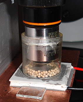
Condensation The formation of dew if chilling the stage below the dew point needs to be prevented if studying coverslipped subjects, otherwise the image is quickly corrupted. After some trials, the setup used to stop condensation is shown below. Also required is a mini-drybox under the copper plate to prevent condensation on the underside of the glass slide. A connecting
side hole drilled in the copper plate with a few pellets
of drying agent were used and sealed with a slip, this was shown in the April 2018 article.
|
A mini drybox was enclosed around the low magnification objectives used. The collar was an old objective case, with bottom drilled out for a snug but loose fit (to allow focussing) and case length reduced to suit.
An inner acetate collar was added with base an annulus of Sellotape.
Molecular Sieve 3A drying agent was the desiccant, this is readily available pre-activated on eBay. Unlike self indicating silica gel, there is no indication when saturated so regular replacement is needed.
The collar sits within the edge of the large 22 mm square coverslip for the water layer so as not to affect the drying agent.
When left for a few hours to form a dry environment it worked well with no condensation seen on the slip upper surface even when plate temps as low as -15°C were used.
The Peltier plates tend to skate around on the thermal paste, especially in warmer weather so small pieces of glass slide were glued onto the copper plate to keep them in place.
The glass slide for the water layer was actually a coverslip to allow the best heat transfer to the cold plate.
|

|
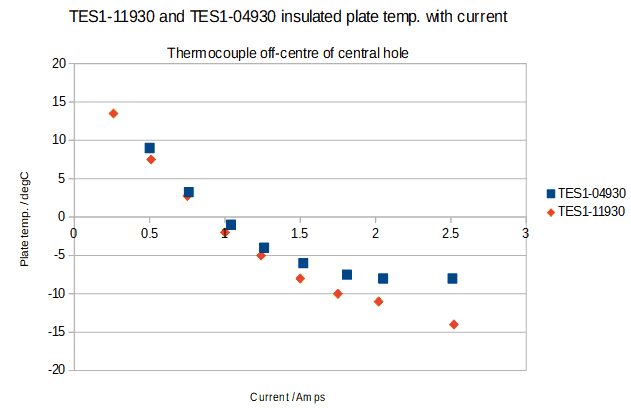
Assessing the plate temperatures Important lesson learnt, plates with higher power ratings are not required for microscopy studies. For the two plates with central holes, it was useful to know the optimal plate temp. that could be achieved for a given current.
These are plotted below.
|
Plate ratings with central hole:
TES1-04930 5.8V, 3A, 10W, ΔT ca. 64°C,
26 mm diameter, 10 mm central hole.
(This low wattage plate together with a solid equivalent will be reported on in a later article as they may be the most ideal for microscopy cold stages.)
TES1-11930 14V, 3A, 24W, ΔT ca. 66°C,
3 x 3 cm, 5 mm central hole.
ΔT is the maximum differential temperature across the plate under optimum conditions. If the hot side cannot be efficiently cooled this is reflected in poorer performance for cooling.
For a given current the plate temp. achieved was similar. But the wattage of one was twice that of the other (i.e. the volts required for a given current were about double) and thus twice the heat dissipated.
As remarked in earlier articles, effective heat removal is a key challenge in devising compact cold stages suited for microscopy. The area to be chilled is small for microscopy field of views, so plates with the lowest wattage capable of achieving the temps. required is the ideal.
Unfortunately, in this regard I initially ventured along the least ideal path for Peltier studies shared in August 2015. The cheapest and most readily available plate is the TEC1-12706 rated at 52W so there is a real challenge as I found trying to dissipate the heat. Fan assisted heatsinks were unable to maintain the key ΔT across the plate and the cold side starts to warm because of this.
The plateauing out of the temp. for the TES1-04930 was because a small fan and thin copper plate was used and at higher currents was unable to dissipate the heat. When an extra fan was added temps dropped further from -8°C to -11°C.
|

|
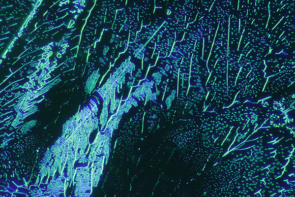
Studies of water freezing under a coverslip The TES1-11930 has been used to date for these studies using the cold stage described in the April 2018 article. With a current of ca. 2.6A the water freezes in seconds. The video below is 4X real time with the Zeiss 6.5X planachro objective and green
/ blue quarter Rheinberg using a 60 mm 12 LED ring on the Photomicroscope III base. The use of LED rings instead of a condenser is a technique described by Dr Kevin F Webb.
Ice is not birefringent for polarised light studies so variations on darkfield were best. Note added Sept. 13th 2018. Later learnt that ice is weakly birefringent so the thin ice layers under the coverslip would not show the typical colours. A later article will explore the effect of ice thickness on cross polar studies. The thickness of ice on its appearance under crossed polars is explored in this October 2018 article.

A still from ice crystal formation under the same conditions as the video above.
Studies of frost crystal formation on uncovered cold surfaces In the article last month I remarked that watching dew condense on a cold surface and later freezing to amorphous ice was fascinating to study. If the cold plate temperature is low enough, frost crystal formation directly from the vapour
phase can also be studied, this with no attempt to insulate the plate from a room at temps of typically 20°C.
I recently treated myself to a secondhand copy of the classic work 'Snow Crystals: Natural and Artificial' by Ukichiro Nakaya (1954 English edition, reprint 2013). Chapter 4 is devoted to 'Artificial Production of Frost Crystals'. Nakaya studied both natural and manmade frost and often found crystal habits for frost that were equivalent to that of snow crystals albeit the symmetry is not seen. Frost
formation thus offered as he noted a more accessible way of studying the nature of snow crystal formation but he was also a pioneer of artificial snow crystal formation and their study.
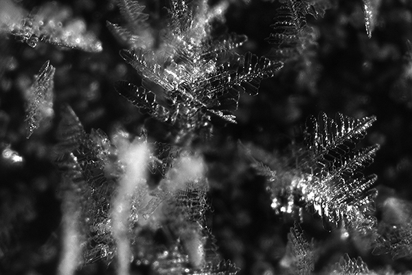
The qualitative studies I've made to date with frost formation on uncovered Peltier plates did give rise to different crystal habits. With low enough plate temps., even dendritic forms typically seen in snow crystals in winter were seen growing rapidly on the non-insulated plate. There seems to be plenty of scope for the hobbyist to vary parameters such as plate temp, surrounding temp. and humidity to see how it affects frostcrystal formations.
Frost crystals grow more readily on sharp edges so encouraged this by studying their growth on both the edge of a razor blade and a pinhead; a video for the latter is shown below. Frost crystals were a challenge to video well because of the limited depth of field of the typical 6.5X compound microscope objective, stereo optics didn't have the numerical aperture to capture crisply.
Frost formation on a needle point. Zeiss 6.5X objective, 2X real time, 1 minute duration. Initially dew condenses and freezes to amorphous ice but as the needle
cools further, frost crystal formation direct from vapour to solid phase occurs.

Dendritic frost crystal formation on a TEC1-12706 plate at 10V / 2A. This plate only drops to low temperatures for short periods due to difficulties dissipating the heat.
Comments to the author David Walker are welcomed.
©
Microscopy UK or their contributors.
Published
in the May 2018 edition of Micscape.
Please
report any Web problems or offer general comments to the
Micscape
Editor .
Micscape
is the on-line monthly magazine of the Microscopy UK web site at
Microscopy-UK
© Onview.net
Ltd, Microscopy-UK, and all contributors 1995 onwards. All rights
reserved.
Main site is at www.microscopy-uk.org.uk
with full mirror at
www.microscopy-uk.net
.


