|
What Has 8 Dorsal Plates, One Foot, 11,500 Eyes And Is Edible? by Richard L. Howey, Wyoming, USA |
Well, O.K., not all species have that many eyes; in fact, some don’t have any at all. These extraordinary creatures are members of the class Polyplacophora and of the family Chitonidae. Interestingly, they’re fairly common around tide pools, yet they have not been extensively studied. It’s hard to get grants unless you can convince the panel that your research can produce some rather immediate practical results and preferably ones which will be very profitable. Well, apparently that is not so with chitons. Chitons are fascinating mollusks and I have been intrigued by them for many years and only now am I getting around to really investigating how fascinatingly weird they are. It turns out that some species have eyes or more precisely ocelli; that is, they consist of a single lens with some nerve cells connected to each of the ocelli. In addition to the lens, there is a pigment layer and a retina. However, even more remarkable is the composition of these eyes; most ocelli are composed of complex proteins, but these are aragonite, a form of crystallization of calcite. They have been described as “rock” eyes and “ceramic” eyes. Many species live in tidal areas where there is the pounding of surf and then a withdrawal of water. Such actions would be very hard on protein-based eyes and so these aragonite eyes are an amazingly effective adaptation. There are a few other examples of mineral-based eyes, but the ones in chitons are astonishing in that they definitely provide images of objects, although very primitive ones. There have been experiments that show that they are able to discriminate between shapes and mere changes in light intensity. These lenses have another astonishing property; they allow the chiton to see both in air and underwater as the tides move in and out. This works out for the chiton because of an unusual property of aragonite, namely, that it has 2 refractive indices: one which functions for aquatic vision and the other for atmospheric vision.
The eyes, according to Libbie Hyman in her classic work The Invertebrates, Volume VI, pp. 99-100, were first described by H. Moseley in 1885 on a specimen of Acanthopleura echinata, with an estimate of 3,000 eyes on the cephalic valve and 8,500 on the remaining 7 valves. (I suspect that the actual counting was done by an assistant and not Moseley himself. After all, no research laboratory can survive without slave labor.) This is a species with a number of small spines extending up from the mantle. Since that time, the eyes have captured the attention of a number of researchers and these aragonite structures remain nearly unique in the animal kingdom. The valves are also composed of aragonite and they are embedded in a thick cuticle called the mantle which extends around them and runs all along the outer edge of the organism.
Many chitons appear rather drab and thus are easily ignored by a casual observer. The dried and preserved ones are usually even less striking due to the fact that many lose their color when dried or preserved in fluid. However, some forms are dazzling as you can see here.
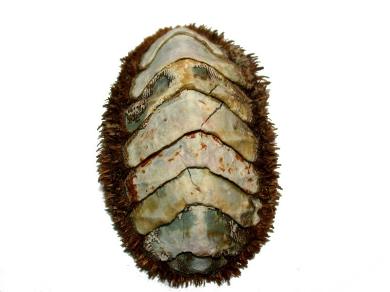
The body plan of chitons is astonishing in terms of its armor. The plates or valves are often interlocking and fused and yet can flex. The valves are arched and this allows a chiton to elevate the central part of its body above a rock surface to which it is attached and this action allows for a sort of pumping action which provides access to oxygen for the gills. However, the chiton can flatten itself and adhere tenaciously to the rock surface helping to protect it from would-be predators. The rock-hard valves and spines and spicules are also discouraging to predators since the only edible parts are underneath all of this heavy plating. There are, though, some species which have a thick and very tough mantle which covers the plates as in the large gumboot chiton, Cryptochiton stelleri, and according to Ed Ricketts in his classic book Between Pacific Tides, the foot would be edible only in emergencies as it has a strong, unpleasant odor. This species can get up to 14 inches in length and weigh over 4 pounds.
Most chitons are rather small and we’ll look at a variety of specimens ranging from ones that are less than an inch in length to a large gumboot chiton.
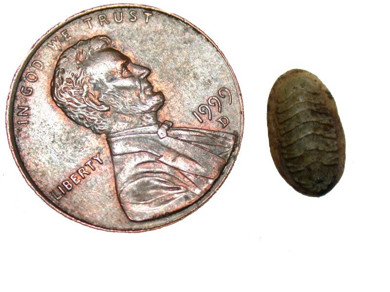
First of all, let’s look at a Callistochiton which is very wee indeed. The penny gives you an idea of its relative size.
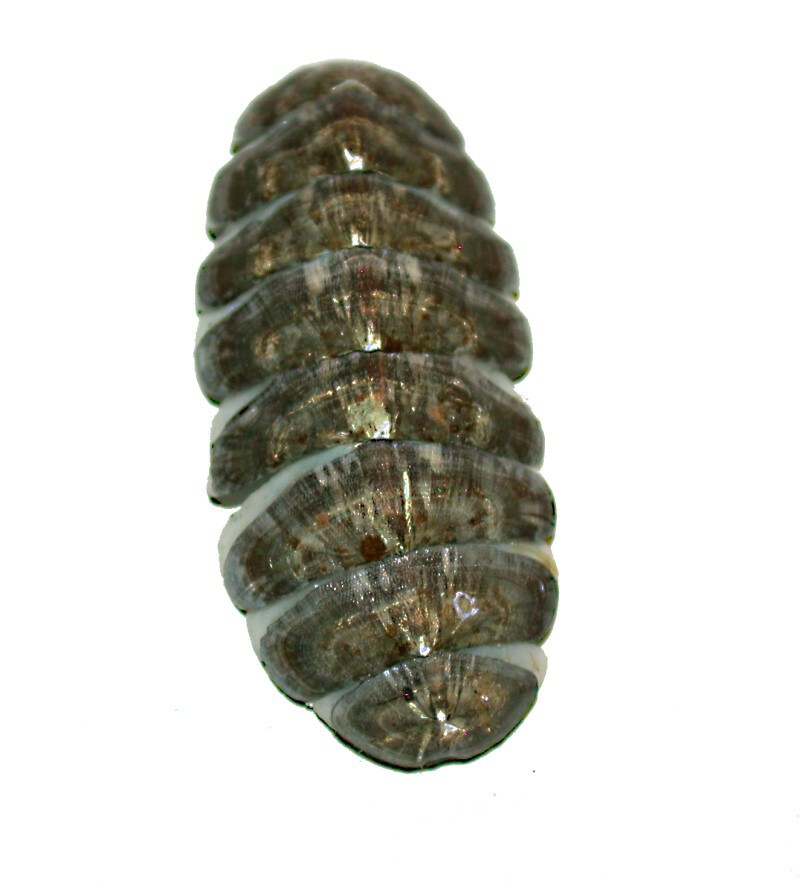
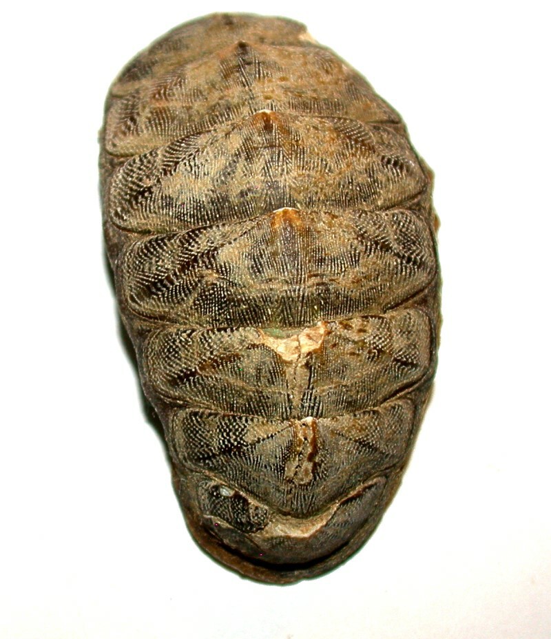
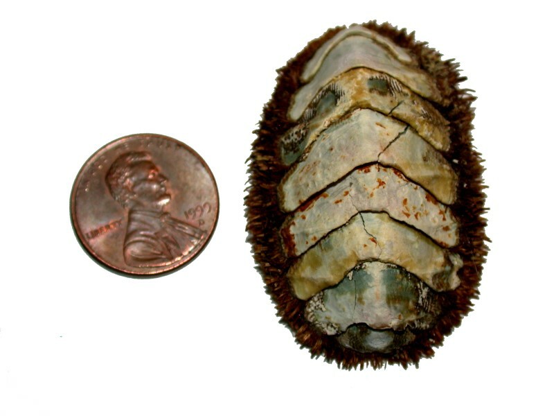
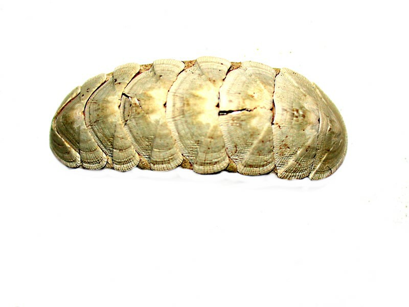
If you look closely you can see the 8 separate plates, but they are much more readily observed in other species. In the next example, which is an unidentified species, the valves are readily visible. The taxonomy of chitons is complex and tricky, but this image shows the plates clearly and is rather anomalous in that part of the mantle is usually visible around the outer edge, but not so in this specimen.
Again, an image with a penny is helpful to give us an idea of the size.
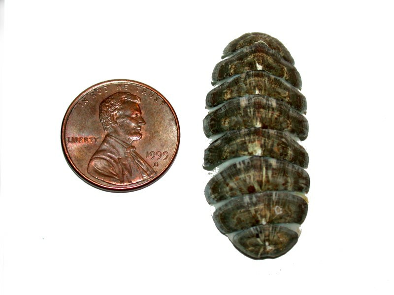
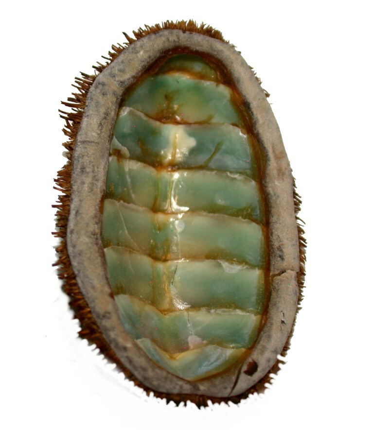
You will notice a series of narrow areas between the valves which have a slight greenish tinge. The foot has been removed and, as a consequence, we have just the “shell”. This might account for why there is no fringe of mantle surrounding the outer edge. When we turn this shell over, we are met with a colorful sight–the inside of the valves have a lovely light green color.
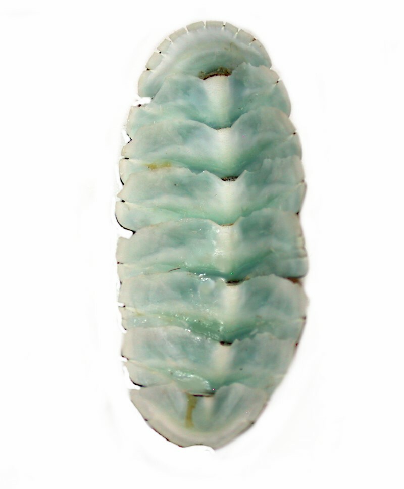
We also encounter an interesting optical illusion here. It appears that the valves or plate arch up toward us but, in fact, they curve downward forming an elongated oval cup which previously contained the foot.
I mentioned earlier that the classification of these organisms is tricky and that, in significant part, is due to the wide range of variation in patterning and other adornments such as spines and even the color of the mantle. Interestingly the term “chiton” comes from ancient Greek; in Greece and Rome a loose garment made of wool or linen, a loose tunic or a kind of mantle, was called a chiton.
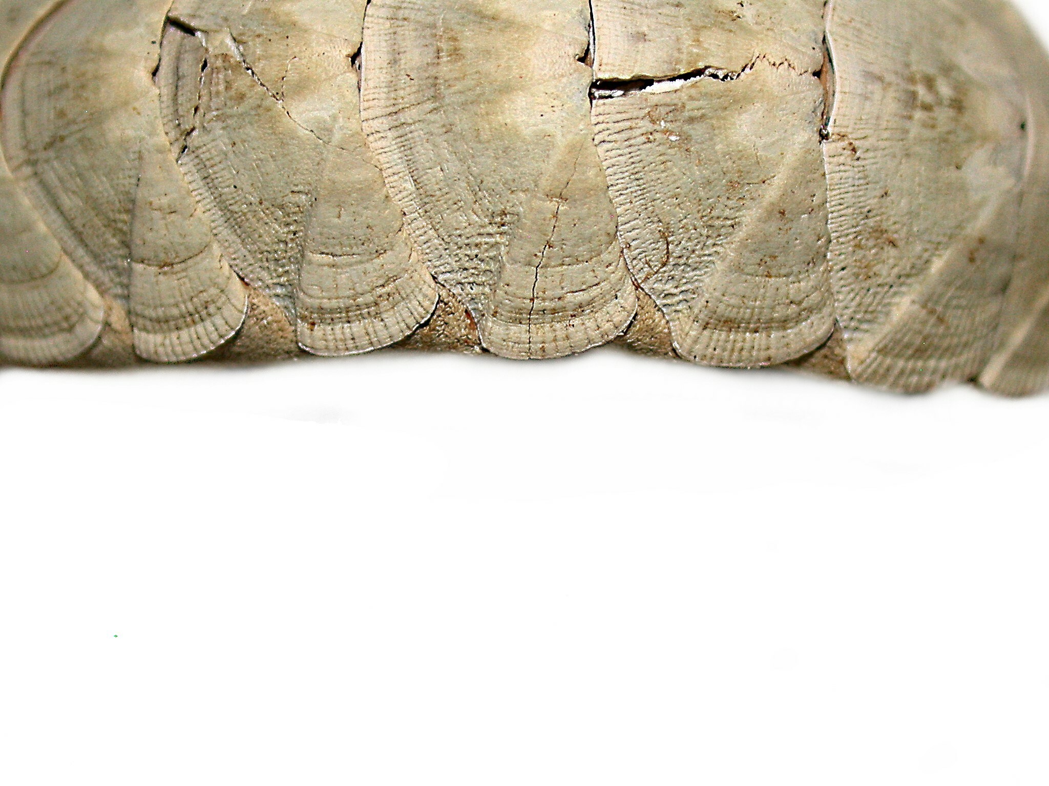
Let’s take a look at some of the patterning on the dorsal surfaces of a few chitons beginning with another unidentified species. The outer “wings” of the valves almost look like they were intricately etched.
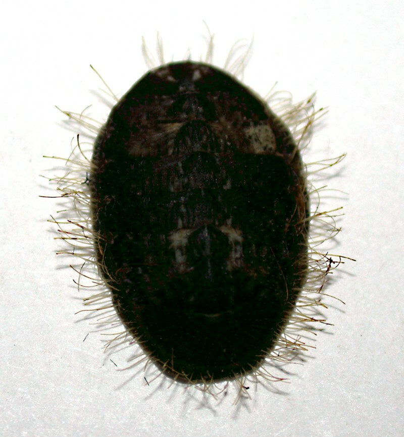
In others, the mantle has encroached in such a way that the valves are almost invisible, but this specimen of Chaetopleura lurida has a distinctive feature of a different sort–it’s a hairy chiton.
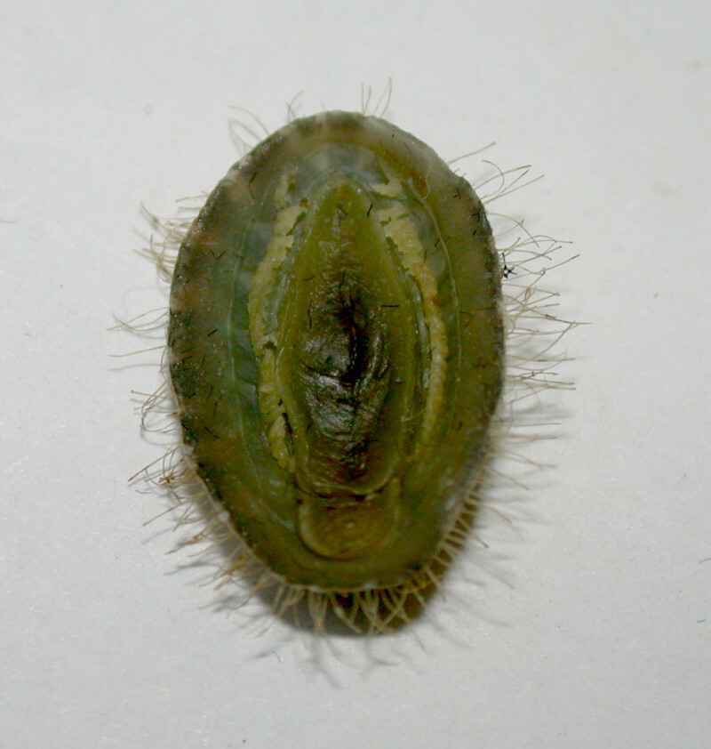
And this specimen, we can turn over and see the foot and again be surprised by the contrast of color between the dorsal and ventral surfaces.
And then there are fuzzy ones such as this specimen of Mopalia which also has a greenish-colored underside and once again presents us with an optical illusion because of the absence of the foot.
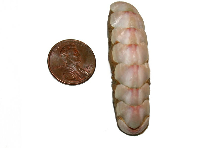
Some forms are veriform or elongated and consequently rather worm-like. I’ll show you 2 examples. The first is unidentified and also shows us yet another coloration pattern, this time pink.
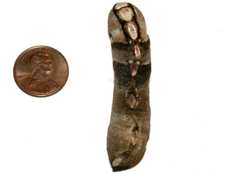
The second is a specimen of Cryptoplax and here we have an example of a chiton where some of the valves are very distinctly separated by the cuticle of the mantle.
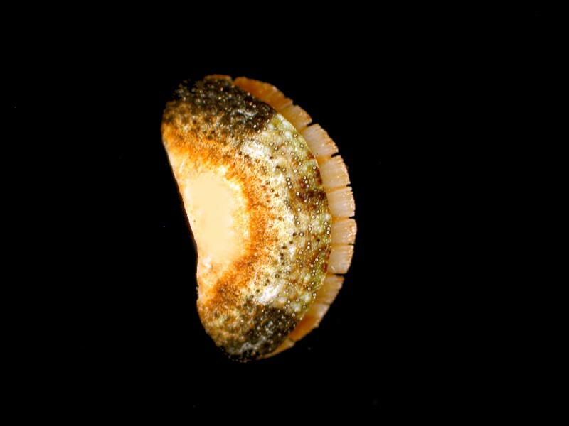
The next specimen looks almost like a shard of a Southwestern American Indian piece of pottery with its rich earth tones. Here the valves are closely in contact and well-embedded in the mantle; nonetheless, they remain distinct. This is a specimen of the genus Ischnochiton.
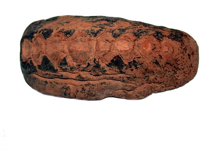
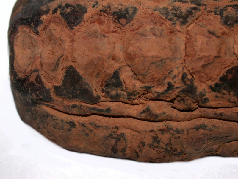
This is a specimen of considerable interest and I’m going to show you several closeups. The first is a closeup of the dorsal surface showing the shape of five of the valves, the smallest being the posterior valve.
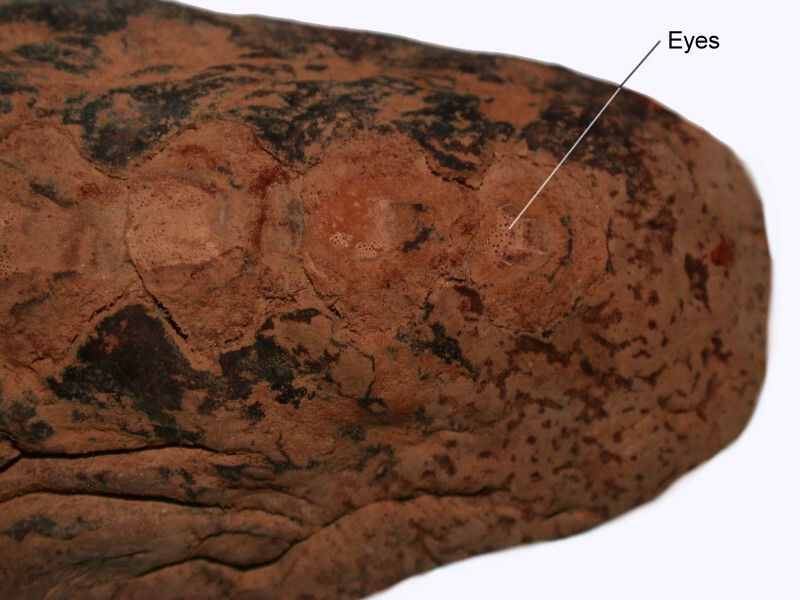
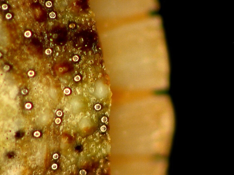
Next is an image showing you some of the eyes in the cephalic or anterior valve. I can hear you thinking: “You’ve got to be kidding!” Nope, they’re those little black dots. But, have no fear, I have another species in which the eyes are much clearer and I’ll show you that in a bit. (That is what’s known as a “teaser” to keep you reading.)
Now, we have a view of the underside and can observe the mouth.
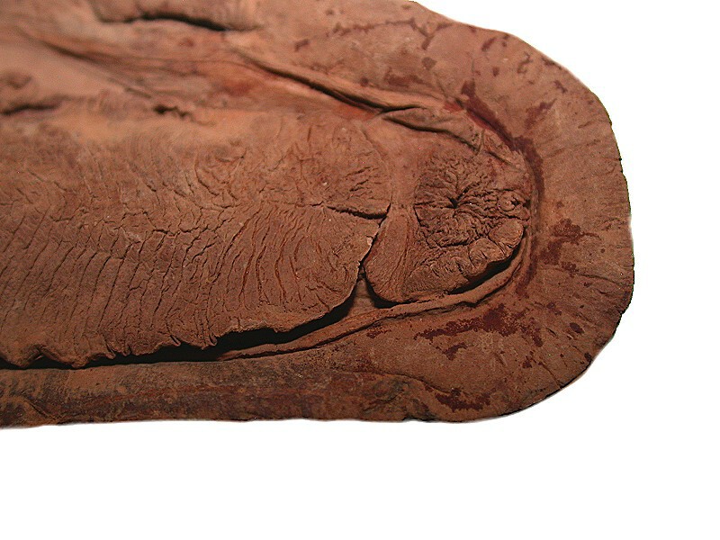
Behind that ring of tissue is a series of powerful muscles which control the opening and closing of the mouth, but also a set of muscles which move the radula, a narrow band of very sharp teeth. Recent research has revealed that these teeth are the hardest ones known in any animal. Amazingly, they are composed of not only protein and mineral material, but metallic magnetite, a form of iron oxide. You can find out more here.
Here we have a view of the foot and a part of the mantle and in the groove between them are tube-like structures which are gills.

Some chitons like this Amphineura have artistic inclinations and produce elaborate and pleasing patterning on the valves. First, I’ll show you the whole organism and then a closeup of the patterning.
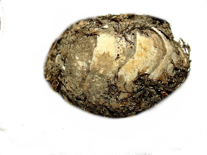
Next, let’s take a look at a really mossy chiton on which it is difficult to discern much more than the presence of the valves. It is probably a species of Mopalia. First, a dorsal view.
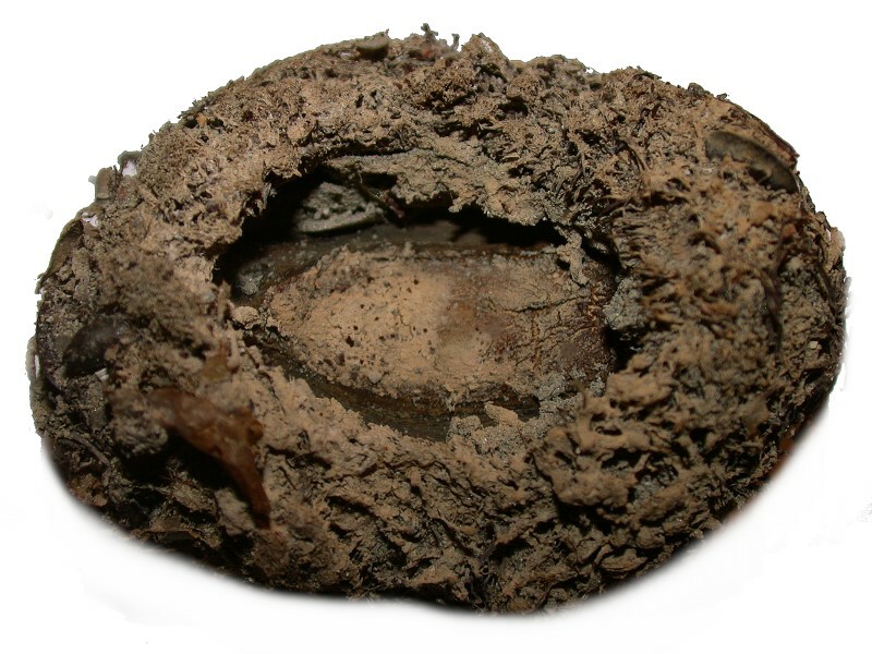
Then, a ventral view and you can see the foot which appears to be recessed in a bowl-like configuration; in part, this is due to the fact that during preservation, the chiton arched and produced extreme curvature, thus pulling the foot inward and partially covering it over.
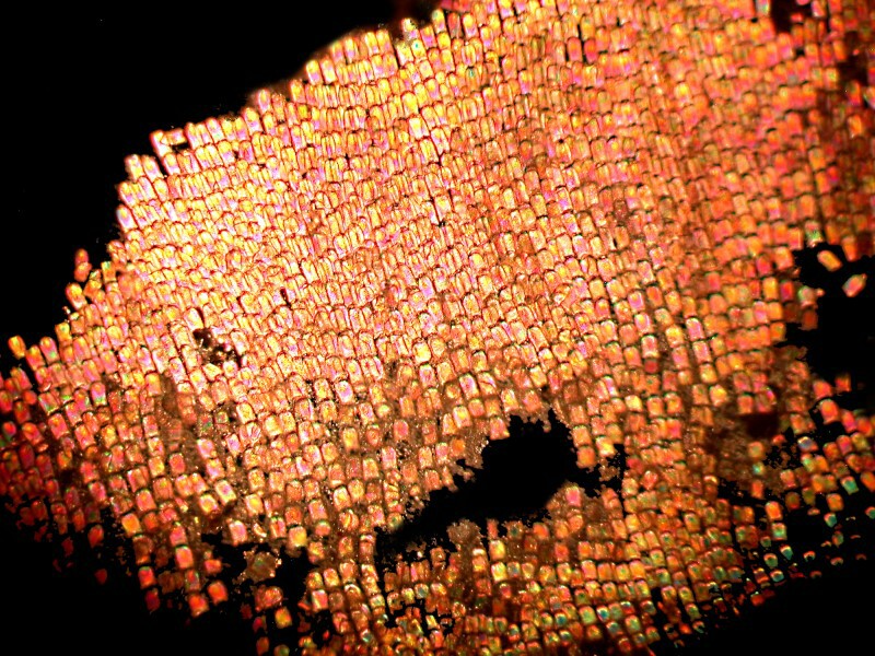
Well, this has gotten rather long, so we’d better turn to the issue of the eyes. I first noticed them in some valves which had been isolated for craft projects. It was a small packet of 25 pieces and I had ordered them on a whim. The organism from which they came was not identified, but from the general shape, size, and patterning, it is fairly certain the they are from a species of Amphineura. As I said at the beginning of this essay, I have long been curious about chitons and particularly the valves, but I was always too lazy to spend the time and effort to isolate them, so being able to buy a small packet of them was splendid. When I put one under my stereo-zoom microscope, the first view was this.
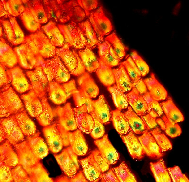
I increased the magnification and this is what I saw.
It immediately flashed through my mind: “Those look like ocelli–eyes!” But, could they be? And, could there be so many? Yet, I was almost certain that they were indeed eyes. About 15 years ago, I read about some remarkable calcite eyes in ophuiroids (brittle stars) which cover virtually the entire upper surface of the organism, so I knew that some very strange receptors were indeed possible.
Because the valves are arched or curved, they are difficult to photograph unless one uses a stacking program and that’s not so easy with an ordinary stereo-zoom that has only a coarse focus. So, I decided to take a pair of bone shears and cut some pieces of the valve until I got something that would allow me to take a careful look. Here is one example.
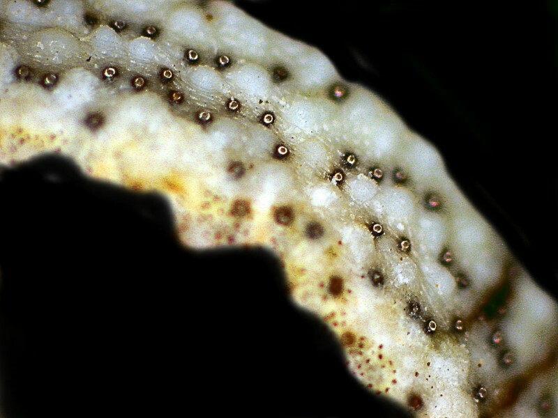
I also noticed some brownish pits which were no longer eyes. Over time, some are damaged and the organism is constantly producing new ones. Some chitons live to be over 20 years old and in their environment, there is a good deal of abrasion which takes place.
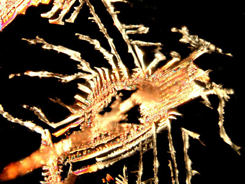
In the next essay, I hope to show you some additional types of chitons, but also extend the examination of some of the micro-structures. There are some curious and mysterious forms hidden away in these creatures. Just to whet your appetite: Some crystalline structures, possibly spicules (polarized light):
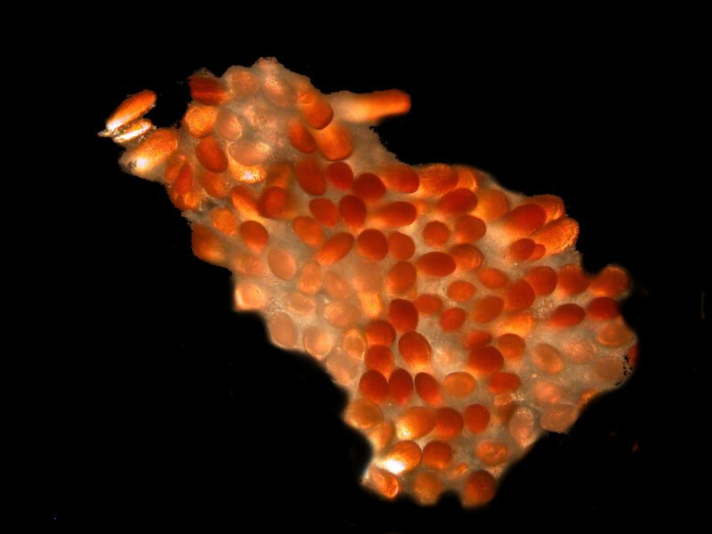
some ovoid spherules, possibly eggs:
and masses of tiny highly refractive structures (possible spicules) embedded in a thin membrane and which look almost like butterfly scales.
My brief excursion into the world of chitons has, once again, made me aware of the vast range of my ignorance and I look forward to learning much more about these remarkable creatures.
All comments to the author Richard Howey are welcomed.
Editor's note: Visit Richard Howey's new website at http://rhowey.googlepages.com/home where he plans to share aspects of his wide interests.
Microscopy UK Front
Page
Micscape
Magazine
Article
Library
Published in the May 2016 edition of Micscape Magazine.
Please report any Web problems or offer general comments to the Micscape Editor .
Micscape is the on-line monthly magazine of the Microscopy UK website at Microscopy-UK .
©
Onview.net Ltd, Microscopy-UK, and all contributors 1995
onwards. All rights reserved.
Main site is at
www.microscopy-uk.org.uk .