A Close-up View of a Prairie Mallow Hybrid


A Close-up View of a

Prairie Mallow Hybrid


Sidalcea malviflora 'Little Princess'


by Brian Johnston   (Canada)


The Mallow Family, (Malvaceae), is best known for its spectacular blooms, as illustrated by the Hibiscus, Hollyhock, and Rose-of-Sharon plants.  Even the lowly Common Mallow (Malva neglecta), considered by many to be a pestilential weed, is a family member.

Sidalcea malviflora, Prairie Mallow, is a wildflower native to high coastal meadows, and open woodlands from Oregon to northern Baja.  It is often called ‘Checkers’, ‘Checkerbloom’, ‘Dwarf Checker-mallow’ or ‘Dwarf Checkerbloom’ because the flowers are approximately the size of a checker game piece.  The plants have round, lobed basal leaves, upright stems with deeply cut upper leaves, and a spike of funnel-shaped flowers that possess a prominent pistil.  ‘Little Princess’, the cultivar studied in this article, resembles a miniature Hollyhock with striking pink flowers.

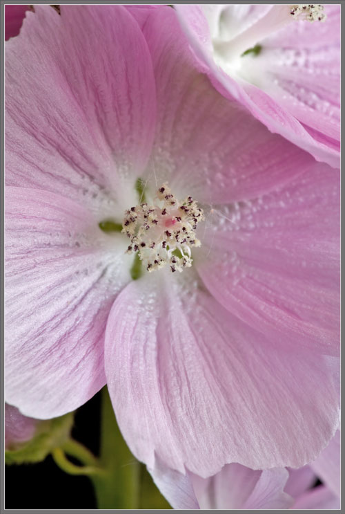
The first image in the article, and those that follow show why this cultivar is so highly valued by gardeners.  Each flower spike is simply ‘pretty as a picture’!


This profusion of pink blooms begins as a group of green, ridged buds with pointed tips.  There is no hint at this point of the flowers’ eventual colour.  In many bud spikes, the upper-most members are difficult to distinguish as buds at all, and have a white or very faint pink colouration.


If we move closer to the early-stage buds, we see that their coating of fine hairs is particularly dense along the ridges.


The ‘rogue’ bud spike shown in the three images that follow has some buds with an unusual orange-pink colouration.  This is certainly not typical, and while normal buds are enclosed by a ring of green sepals (modified leaves), the rogue ones possess these strangely coloured ones.


A typical bud begins to bloom when the ring of sepals (called the flower’s calyx) splits apart near the top, and the flower’s petals become visible.


The green sepals have stopped growing, but the tightly packed whorl of petals (called the flower’s corolla) continues to lengthen.  Four images follow that show examples of this process.


Eventually the five overlapping petals that form the corolla open out to form a roughly cup-shaped bloom.  Notice that each pink petal is attached to the flower’s stalk by a stubby, narrow, white base.  This adaptation results in five distinctive oval ‘gaps’ that add visual interest to the opening flower.


Over the period of a couple of days, a flower transitions from closed bud, to cup-shaped young bloom, to a final mature flower with an ‘almost’ flat surface.  Examples of this transition can be seen in the two images that follow.


Projecting from the flower’s centre is a sturdy, pale-pink stalk which supports its reproductive structures.  At this point however, only a mass of dark, almost black anthers coated with beige pollen grains is visible.  The pistil has yet to put in an appearance.


This situation is short-lived however.  Soon, the flower’s bright red, multi-lobed stigma lobes are pushed out from beneath the forest of anthers by the lengthening style.


In the cup-shaped young flower shown in the image on the left, the pistil is not visible, while in the mature, flat flower shown on the right, the stigma lobes are discernable.


As the style continues to lengthen, the red stigma lobes are pushed farther away from the group of anthers.


Sidalcea malviflora’s petals are rough in appearance due to the large number of three-dimensional longitudinal veins that cover their surfaces.


Photomicrographs of a petal’s surface show the wide range of colouration of its constituent cells (left image), and their jigsaw-puzzle-like shape (higher magnification right image).


Similar photomicrographs showing a different area on the petal are shown below.


To me, the shape of the flower’s dark anthers resembles an assortment of chromosomes!


The sequence of images that follows, (taken with increasing magnification), reveals the structure of the flower’s pistils, and the spherical shape of Prairie Mallow pollen grains.


For comparison, here is a view of the jumbled mass of anthers and their supporting filaments taken using macrophotographic equipment (left), and photomicrographic equipment (right).


Most Prairie Mallow pollen grains are perfectly spherical, and are covered with short, spike-like projections.  Strangely, several grains (in the first two images) look like deflated beach balls!


Notice that when the stigma is pushed by the lengthening style up through the mass of pollen covered anthers, its lobes are packed tightly together in a column (left image).  If the stigma of a flower comes into intimate contact with its own pollen during its passage up through the anther mass, why doesn’t self-pollination occur?  (Self-pollination is detrimental to the long term viability of the species.)  In order to discourage self-pollination, the receptive surfaces of the stigma lobes are facing one another, and are in close contact until the stigma has passed through the danger zone.  Only later do the lobes separate to reveal fresh receptive surfaces to visiting insects (right image).  Thus cross-pollination is favoured.


A number of images follow that show views of the separated stigma lobes of two Prairie Mallow flowers.  In one, there appear to be 8 lobes, while in the other 9 lobes are present.


The two photomicrographs below show the body of a stigma lobe.  Notice that the red receptive surface is covered with hair-like projections that help to acquire and retain pollen grains.


Additional photomicrographs reveal the tip of a stigma lobe.  It’s easy to see that if the ‘hairy’ receptive surfaces of all of the lobes were facing one another in a columnar structure, pollen grains could not adhere to the hairs, thus largely preventing self-pollination.


Here are two additional photomicrographs that show pollen grains adhering to a stigma lobe tip (left image), and the surface of an anther (right image).


Once pollination has occurred, the column that supports the stamens and pistil dries up and falls off.  All that is left of the flower is the ring of sepals and the green ovary.


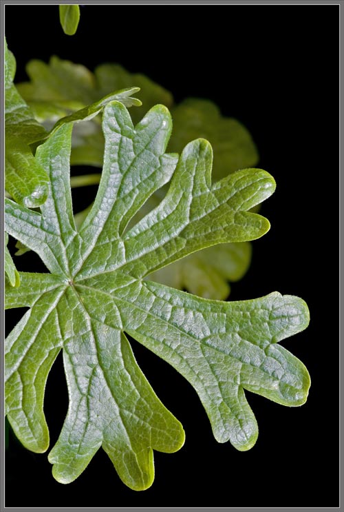
Now let’s look more closely at the plant’s leaves.  Near the top of the stem, the leaf has a palmate shape with extremely deep lobes.


A little lower on the stem, the tip of each lobe is itself lobed.


Farther down the stem, the base leaf shape is still palmate, but the base lobes are less deep.


As we continue to move down the stem, the depth of the base lobes continues to decrease.


At the base of the stem, a leaf’s shape is very different when compared to one positioned just beneath the flower spike.


The two images below show higher magnification views of the glossy upper surface of a leaf.


A photomicrograph of its under-surface follows.  Note the many oval stomata and guard cells that control gas entry into and out of the leaf.


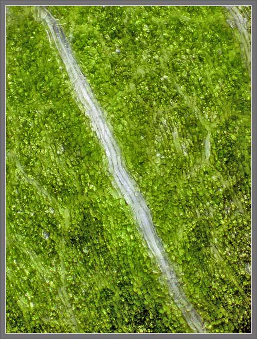
The image on the left reveals the cellular structure of the upper surface of a leaf.  On the right is a photomicrograph showing one of the spike-like hairs that grows from the leaf’s edge.


Prominent veining is visible on the underside of one of the plant’s lower leaves.


Higher magnification views of this vein structure follow.


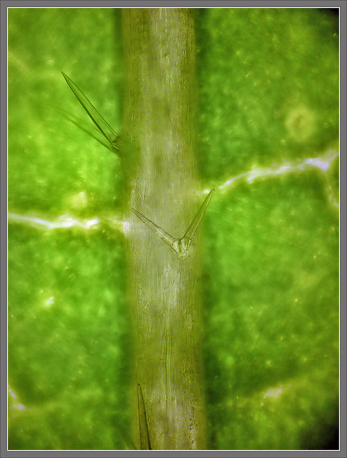
At low magnification the fine hairs that grow from a leaf’s surface are not visible.  The higher magnification view on the right shows them clearly.


Extremely sharp spikes grow in pairs from the surfaces of a leaf’s veins.


Not only is Sidalcea malviflora ‘Little Princess’ a visual feast for the eyes, but it is also easy to grow.  In fact, if grown in moist soil with full sunlight, it may bloom through the summer, and into the fall!


Photographic Equipment

The low magnification, (to 1:1), macro-photographs were taken using a 13 megapixel Canon 5D full frame DSLR, using a Canon EF 180 mm 1:3.5 L Macro lens.

A 10 megapixel Canon 40D DSLR, equipped with a specialized high magnification (1x to 5x) Canon macro lens, the MP-E 65 mm 1:2.8, was used to take the remainder of the images.

The photomicrographs were taken using a Leitz SM-Pol microscope (using a dark ground condenser), and the Coolpix 4500.


A Flower Garden of Macroscopic Delights

A complete graphical index of all of my flower articles can be found here.


The Colourful World of Chemical Crystals

A complete graphical index of all of my crystal articles can be found here.


 All comments to the author Brian Johnston are welcomed.

Microscopy UK Front Page
Micscape Magazine
Article Library


© Microscopy UK or their contributors.

Published in the May 2011 edition of Micscape.
Please report any Web problems or offer general comments to the Micscape Editor.
Micscape is the on-line monthly magazine of the Microscopy UK web
site at Microscopy-UK  


© Onview.net Ltd, Microscopy-UK, and all contributors 1995 onwards. All rights reserved. Main site is at www.microscopy-uk.org.uk with full mirror at www.microscopy-uk.net .